Ulnar Nerve
Historical document, translated for reference. It reflects medical knowledge of the 1920s–30s and is not medical advice.
Summary
This article from the 1928–1936 Soviet Great Medical Encyclopedia details the anatomy, innervation, and pathology of the ulnar nerve. It describes the nerve's course through the arm, its clinical significance in trauma, and the characteristic symptoms of its paralysis, such as the 'claw hand' deformity.
Encyclopedia article (1928–1936)
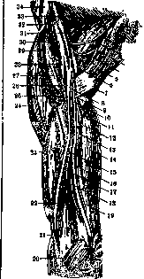
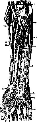
ULNARIS NERVUS, syn. n. cubitalis, ulnar nerve, one of the large nerves of the upper limb, a mixed nerve. It originates in the brachial plexus, runs initially together with the a. brachialis medial to it (Fig. 1), then passes behind the septum intermusculare mediale in the direction of the sulcus nervi ulnaris humeri, where it is located directly on the bone between the epicondylus medialis humeri and the olecranon; from here it passes between the initial bundles of the m. flexor carpi ulnaris to the anterior surface of the m. flexor digitorum profundus, where the Martin-Gruber anastomosis exists between the ulnar nerve and the n. medianus; distally, the ulnar nerve heads alongside the a. ulnaris along the radial edge of the m. flexor carpi ulnaris and in the middle of the forearm, and sometimes closer to the hand, it divides into the ramus dorsalis manus and the ramus volaris manus (Fig. 2). For the branches of the ulnar nerve, their topography, and anastomoses, see Nerves of Man. The ulnar nerve innervates the m. flexor carpi ulnaris, m. flexor digitorum profundus, m. abductor digiti quinti, m. flexor brevis digiti
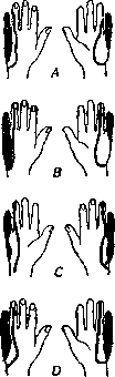
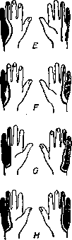
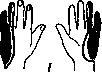
quinti, m. opponens digiti quinti, m. adductor pollicis, m. flexor pollicis brevis (deep head), mm. lumbricales IV et III, and all the mm. interossei; the ulnar nerve conducts sensory impulses from the volar surface of the hand corresponding to the skin of the palm of the V and the middle of the IV finger, from the little finger and the ulnar half of the ring finger, and on the back of the hand from its ulnar half, from the back of the V and IV fingers, and the ulnar side of the proximal phalanx of the III finger. The following variations are encountered: the ulnar nerve innervates only the m. lumbricalis IV or, in addition, the m. lumbricalis III; the m. flexor digitorum sublimis may receive innervation from the ulnar nerve, and the m. adductor pollicis from the n. medianus. In rare cases, all muscles of the thenar receive innervation from the ulnar nerve. On the other hand, cases have been described where the mm. interossei and lumbricales were not affected during paralysis of the ulnar nerve and contracted upon stimulation of the n. medianus with an electric current. For variants of cutaneous innervation, see Fig. 3.
B. M. E. Vol. XXXIII.
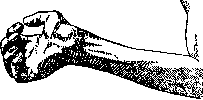
Pathology. The ulnar nerve becomes diseased more often than the n. medianus, but less often than the n. radialis. In cases of wounds and fractures on the arm and forearm, the ulnar nerve is often affected together with the n. medianus. In non-critical changes occurring in both indicated nerves simultaneously, the hand sometimes assumes a position called
the "main en predicateur" (Fig. 4). With the preservation of muscles innervated by the n. radialis, it is characterized by hyperextension of the hand, extension of the fingers at the proximal phalanges, and flexion at the middle and distal phalanges. Isolated damage to the ulnar nerve is observed in the sulcus n. ulnaris, where the nerve lies directly on the
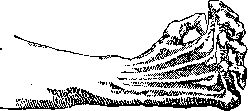
bone, during injury to the medial epicondyle of the humerus. The nerve can be involved in suffering not only during a fracture or contusion but also years after the trauma due to compression by a bone callus. In subjects with a flat sulcus n. ulnaris, dislocation or subluxation of the nerve may occur during trauma; painful phenomena in such a case are caused by subsequent neuritis. Relatively light but prolonged pressure of the elbow on a hard support (during sleep, under anesthesia) can cause mild paralysis due to compression of the nerve. Occupational paralysis of the ulnar nerve has been described in glassblowers, telephone operators, etc., due to habitual leaning on the elbow, as well as compression of the branches of the ulnar nerve in the hypothenar in cyclists. Mild lesions of the ulnar nerve are expressed in pain syndromes proceeding by the type of pure neuralgia or accompanied by motor disturbances; trophic disorders characteristic of neuritis are not observed in this case. Isolated primary neuritis of the ulnar nerve is observed rarely as a complication of certain infectious diseases, for example, typhus, sepsis, dysentery, syphilis, appendicitis, and poisoning by chemical warfare agents. The ulnar nerve often suffers during polyneuritis. Neuritis of the ulnar nerve, in addition to pain symptoms, is expressed in motor disorders (atrophic paralysis, paresis), sensory disturbances (Fig. 3), and trophic disturbances of the skin; Dupuytren's contractures have also been described in connection with neuritis of the ulnar nerve. In paralysis of the ulnar nerve, flexion of the hand is weakened
and does not occur in the ulnar direction, movements of the little finger are impossible, when spreading the fingers the little finger remains abducted, flexion of the IV and V fingers and partly the III is limited; adduction of the thumb is weakened. Due to atrophic paralysis of the interosseous and lumbrical muscles, hyperextension of the proximal and flexion of the middle and distal phalanges occurs when the fingers are spread; a "claw hand" results—the Aran-Duchenne hand (Fig. 5). In paralysis of the ulnar
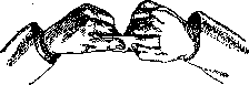
nerve, the patient has disturbed fine movements in the fingers; he cannot bend the distal phalanges of the last three fingers, he cannot scratch with the nail of the little finger. Froment's test consists of the patient having to hold a sheet of paper tightly between the index finger and thumb of both hands, pulling it to the sides. Instead of adduction of the thumb, flexion of its distal phalanx occurs (Fig. 6). Treatment and prevention—see Neuritis, Neuralgia, Polyneuritis. In cases of dislocation of the ulnar nerve, osteoplastic operations may take place, aiming to secure the ulnar nerve in the sulcus n. ulnaris.
S. Bychkov.
Related articles
Mentioned in
Cite this page
“Ulnar Nerve.” Soviet Medical Encyclopedia. English translation of Bolshaya Meditsinskaya Entsiklopediya, 1st ed. (Moscow, 1928–1936), ed. N. A. Semashko. https://sovietmedicalencyclopedia.com/article/ulnar-nerve/