Forearm
Historical document, translated for reference. It reflects medical knowledge of the 1920s–30s and is not medical advice.
Summary
This article describes the anatomy and development of the human forearm, including its skeletal structure, joints, and rotational mechanics. It details the comparative anatomy of the radius and ulna across different mammals and explains the processes of ossification and movement.
Encyclopedia article (1928–1936)
FOREARM, antibrachium, s. antebrachium, the anterior part of the human upper limb and the anterior limb of animals, homodynamic to the lower leg. The upper boundary of the forearm is determined by the transverse groove (plica cubiti) and a line passing at the level of the epicondyles of the humerus. The lower boundary corresponds to the well-defined transverse volar fold, which separates the hand from the forearm, and by a circular line connecting the bases of the styloid processes. The shape of the forearm corresponds to an elongated cone, compressed in the anterior-posterior direction and gradually tapering distally, which depends on the arrangement of the muscle bodies of the forearm musculature in the upper section and their relatively thin tendons in the lower third of the forearm, as well as on the fact that some muscles belong only to its proximal section. The average length of the forearm in a man is 24.6 cm, in a woman — 22.8 cm. In the ontogenesis of human forearm bones, a relatively greater length of the forearm bones is observed in relation to the humerus, which gradually decreases with age. The increase in the entire length of the free upper limb must be attributed primarily to the increase in the length of the forearm bones; this is observed particularly clearly in anthropoid apes and in a whole series of anterior limbs of other mammals (chiroptera, etc.). The human forearm differentiates from the limb bud of the upper limb, by dividing it into the arm, forearm, and hand, approximately at the 6th week of embryonic development. The forearm bones of mammals — the ulna and radius — arise as separate primordia, in the form of a thickening of mesenchymal cells. If the anterior limb develops as an organ of grasping and climbing, then anatomically distinct bones are formed from the initial mesenchymal primordia of the bone-forming tissue of the forearm. The distal end of the radius thereby gains the possibility of rotational movement together with the hand around the ulna. With an increase in this rotational movement, the ulna is gradually excluded from direct connection with the hand, while the lower surface of the radius is supplemented with cartilage and fully enters the radiocarpal joint; if, however, the animal's anterior limb becomes an organ of pendulum-like movements on the ground (walking), then the forearm bones lose their mutual mobility, are fixed by immobile joints, or fuse entirely at the lower epiphyses (monotremes, edentates, and insectivores), or a complete and partial reduction of the ulna occurs due to a corresponding strengthening of the radius. An example of such a reduction of the ulna with its incomplete fusion with the radius can be the forearm of ruminants; in odd-toed ungulates (horse), there is a complete reduction of the body of the ulna. The supporting skeleton of the human forearm is represented by an osteo-ligamentous-fascial apparatus. The bony skeleton of the forearm consists of two long tubular bones located one next to the other — the ulna (ulna; syn.: cubitus, focile majus), homodynamic to the fibula, and the radius (radius; syn.: focile minus, additamentum ulnae, manubrium manus), homodynamic to the tibia. The proximal and distal ends of the forearm bones are connected to each other by relatively independent joints, which are functionally combined into one combined rotational joint; the axis of rotation of the latter passes through the heads of the ulna and radius. Around this axis, the rotation of the radius together with the hand occurs; the volume of this movement at full rotation reaches 133–140° and does not change from the position of the ulna in the elbow joint. With the arm freely lowered, the radius crosses the ulna and lies ventrally; the flexor surface is then turned dorsally, the thumb is located medially. Such a position of the forearm bones corresponds to its initial (ancient) position, which arose during the period of phylogenetic formation of the animal's anterior limb, in connection with the lifting of the torso above the ground and the ventral displacement of the limbs in relation to the animal's body, as well as the rotation of the hand with the distal part of the radius inward. Supinatio — the rotation of the hand and radius outward — must be considered a further complication of the functions of the forearm, belonging already to higher mammals and man. In the supinated limb, the forearm bones are located one next to the other so that the radius and the thumb lie along the lateral edge of the limb, and the ulna and the little finger occupy a medial position. It is most convenient to proceed from this conventional position when examining the parts of the forearm. Each forearm bone has a body (corpus) or diaphysis and two ends — epiphyses. The beginning of ossification of the forearm diaphyses refers to the 8th week of fetal life, the heads (epiphyses) of the ulna and radius — to the 5th–6th year, the olecranon of the ulna — to the 10th year, and the distal end of the radius — to the 2nd–3rd year. The growth of the forearm bones ends by 20–24 years, with the proximal epiphyses fusing to the diaphyses 2–3 years earlier than the distal ones. The forearm bones are curved along their length so that only their upper and lower ends are in contact with each other, forming joints (articulatio radio-ulnaris distalis et proximalis). Thus, a slit-like space remains between the bodies of the bones (spatium interosseum antibrachii), which is filled throughout its entire extent by a very strong fibrous membrane (membrana interossea antibrachii); the latter attaches to the interosseous crests facing each other (crista interossea ulnae et radii) and passes into the periosteum. The middle part of the membrana interossea is the strongest: in this section, a group of the strongest oblique fibers, running from the radius downward to the ulna, stands out from the ascending and descending fibers that cross each other; at the ends, the interosseous membrane is thinner; here, there are openings through which blood vessels and nerves pass. In the upper section of the forearm, a bundle of oblique fibers (chorda obliqua) separates from the interosseous ligament, which runs from the tuberosity of the ulna to the tuberosity of the radius — it acts as a brake during hypersupination. The most thickened section of the radius is its lower epiphysis; in the ulna, on the contrary, the most thickened is the upper epiphysis. The diaphyses of the forearm bones approach a triangular shape; accordingly, three edges and three surfaces are distinguished on them. The interosseous crest (crista interossea) separates the concave palmar surface (facies volaris) from the convex, dorsal surface (facies dorsalis). The outer surfaces (facies med. ulnae et facies lat. radii) pass with rounded edges into their palmar and dorsal surfaces. The largest nutrient foramina of the forearm bones (foramina nutritia) are located primarily on the anterior surface of their bodies; the nutrient canals are directed along the long axis of the forearm bones. The proximal epiphyses of the forearm bones are combined with the lower end of the humerus into one complex elbow joint (see). The distal end of the radius, namely its lower surface, is supplemented by a triangular cartilage located on its medial side, between the distal end of the ulna and the first (proximal) row of the corresponding carpal bones; the distal articular surface of the radius, together with this triangular cartilage, articulates with the proximal row of carpal bones, forming the radiocarpal joint (see). The proximal end of the ulna has a semilunar-shaped articular notch (incisura semilunaris, s. cavitas sigmoidea); this notch, together with the articular surface of the ulna, corresponds to the articular surface of the trochlea of the humerus. The semilunar notch ends with two processes, namely: behind and above — the olecranon curved forward (olecranon, s. processus anconaeus), the apex of which, in the extended position of the forearm, is located in the olecranon fossa of the humerus (at the level of lineae epicondylicae) and serves as a brake during hyperextension of the elbow; in front and below, the ulnar notch passes into another, ventral (coronoid) process (proc. coronoideus). On the lateral side of the latter, there is an articular notch into which the head of the radius enters (incisura radialis); from the posterior edge of this notch, a rough line (crista m. supinatoris) runs obliquely backward and downward along the lateral side of the bone — a reflection of the attachment of the corresponding muscle. On the volar surface, just below the coronoid process, there is a tuberosity — the area of attachment of the m. brachialis. The dorsal surface of the olecranon has rough traces of the attachment of the fibers of the terminal tendon of the m. triceps brachii. The distal end of the ulna ends in a conical head, which bears two articular surfaces: one of them (circumferentia articul. ulnae) faces the radius and articulates with it in the corresponding notch, occupying approximately a semicircle; the second occupies the lower surface of the head of the ulna and articulates with the triangular cartilage. Laterally and posteriorly, the head of the ulna ends in a styloid process (processus styloideus ulnae). The proximal end of the radius represents the head (capitulum radii), which connects to the articular surface of the humerus with a special articular fossa (fovea capituli radii); the latter directly passes into the articular surface of a cylindrical shape, the so-called surface surrounding the head of the radius (circumferentia articul. radii), with which the radius articulates here with the ulna.
The head of the radius continues further into a narrowed part of the body—the neck (collum radii); the latter is encircled in the form of a loop by a particularly important ligament—lig. annulare radii, attached along the edges of the incisura radialis. On the volar surface of the radius, closer to its medial edge and somewhat below its neck, is located a tuberosity (tuberositas radii), to the medial edge of the base of which is attached the tendon of the m. biceps brachii; here, between this muscular tubercle and the tendon of the m. biceps brachii, is located a constant mucous bursa—Bursa m. bicipitis (bicipito-radialis). The distal end of the radius, in contrast to the proximal one, is significantly widened; its anterior surface is concave, the posterior is convex—and has a series of elevations and depressions; in the latter lie the tendons of the extensor muscles of the hand and fingers. The lower articular surface is concave (facies articularis carpea radii), articulating with two bones of the proximal row of the wrist; on the medial side, the articular notch—incisura ulnaris—directly adjoins it, in which the head of the ulna rotates, separated from the bone of the wrist by a triangular cartilage; the latter is fixed by its base to the incisura ulnaris and by its apex to the processus styloideus ulnae. The dorso-lateral distal end of the radius ends in a styloid process (processus styloideus radii). Through the thickness of the skin and a moderately developed layer of subcutaneous fatty tissue, the following are well palpable from the dorso-medial side of the forearm: the olecranon, the entire posterior edge of the body of the ulna, the head and the styloid process; from the dorso-lateral side, the head of the radius is palpable (in the posterior lateral fossa of the elbow joint), as well as the distal half of the body and the styloid process of the same bone (at the bottom of the fossa, the so-called anatomical snuffbox). Congenital defects of the development of the bones of the forearm in the majority of cases are associated with defects of the bones of the hand. Most often, of the two bones of the forearm, the radius is absent. Significantly less often, congenital defects of the ulna occur. According to statistical data, the ulna was absent or underdeveloped 21 times (Bereznikovsky). And finally, the simultaneous absence of both bones of the forearm is described in the literature as isolated cases. As for the etiology of congenital developmental defects, they apparently (Gruber, G. Ivanov, et al.) reduce to two different groups—one group of defects (acquired) has mechanical factors in its etiology (amniotic bands and other anomalies of the amnion); the other group of defects (congenital) must be attributed to hereditary developmental defects. Its explanation, besides taking into account ontogenetic moments of development, can also be substantiated by individual phylogenetic data. The musculature of the upper limb, and in particular the musculature of the forearm, is multi-segmented; it differentiates in an embryo 11 mm in length, with two groups of muscles being laid down initially—anterior and posterior, and then from the first group, in turn, flexors and pronators differentiate, and from the second—extensors and supinators. The greater part of the muscles of the forearm belongs to the long and multi-articular ones. By functional characteristic, the musculature of the forearm can be divided into the following groups: 1) muscles rotating the radius around the ulna (pronators and supinators), with attachment points on the radius; 2) muscles setting the hand into motion as a whole (flexors and extensors, adductors and abductors), with attachment points on the bases of the metacarpal bones; 3) muscles setting the II-III-IV-V fingers into motion (flexors and extensors), with attachment points on the ungual and middle phalanges; 4) muscles setting the first metacarpal bone and the phalanges of the thumb into motion (flexors, extensors, and abductors), with attachment points on the corresponding bones [see vol. XIX, art. 713-748 (fig. 23-30) and art. 729-734 (fig. 39-43)]. By topographic characteristic, the musculature of the forearm can be distributed into two groups: muscles of the ventral, or anterior, surface of the forearm and muscles of the dorsal, or posterior, surface; each of the named groups consists of two layers—superficial and deep. The muscles of the ventral group belong to the flexors and pronators; of them, the superficial layer takes its origin from the medial epicondyle of the humerus, the proximal ends of the bones of the forearm, and from the fascia of the forearm; the muscles of the deep layer originate predominantly from the volar surface of the bones of the forearm and the interosseous membrane. The dorsal musculature of the forearm consists of extensors and supinators; it is also divided into two layers: the muscles of the superficial layer take their origin from the lateral epicondyle of the humerus, the adjacent bones of the forearm, and from the fascia of the forearm; the muscles of the deep layer originate on the dorsal surface of the bones of the forearm and on the interosseous membrane; thus, the muscles of the deep layer are shorter than the superficial ones; their function does not extend to the elbow joint. Description of the musculature of the forearm by topographic characteristic. 1. Musculature of the ventral group: a) deep layer—m. pronator quadratus, m. flexor pollicis longus, and m. flexor digitorum profundus; b) superficial layer: this includes muscles which originate from the epicondylus med. humeri et fascia antibrachii by a common muscle belly. Thus, the muscles of this layer in the upper third of the forearm can be separated only by artificial means, following their fascial septa. To the musculature of the superficial layer belong: m. flexor digitorum sublimis, m. pronator teres, m. flexor carpi radialis, m. palmaris longus, and m. flexor carpi ulnaris. 2. Musculature of the dorsal side of the forearm, just like the volar one, is divided into two layers: a) deep and b) superficial. The musculature of the latter layer is in turn divided into radial and ulnar groups. To the muscles of the deep layer belong: m. supinator, m. abductor pollicis longus, mm. extensor pollicis brevis et longus, m. extensor indicis proprius. To the muscles of the superficial layer of the dorsal side belong (ulnar group): m. extensor carpi ulnaris, m. extensor digitorum communis, m. extensor digiti proprius, and the radial group—m. brachioradialis, m. extensor carpi radialis (longus et brevis). (Regarding individual muscles, their shape, position, origin, attachment, innervation, function, and variations—see Human Muscles.) Innervation of the musculature of the forearm occurs at the expense of the long branches of the brachial plexus, with the muscles genetically belonging to the dorsal surface of the forearm receiving nerve branches from the n. radialis [see vol. XXI, art. 753-754 (fig. 61); art. 757-758 (fig. 65); art. 759-760 (fig. 67-70)]. The superficial and deep branches of the n. radialis (r. superficialis et r. profundus) are located: the first in front under the m. brachioradialis and outward in relation to the a. radialis, the second—goes to the back of the forearm. The muscles of the volar surface of the forearm—m. flexor carpi ulnaris and the ulnar head of the m. flexor digitorum profundus—are innervated from the n. ulnaris; this nerve lies in the medial posterior groove of the elbow joint, then emerges onto the anterior surface of the forearm between the ulna and the ulnar flexor of the hand, and lies outward in relation to the a. ulnaris; in the middle third of the forearm, it divides into dorsal and palmar branches. All other muscles of the volar surface of the forearm are innervated by the n. medianus, which passes between the heads of the m. pronator teres and then between the superficial and deep layers of the flexors of the forearm; in the lower third of the forearm, the n. medianus goes in the sulcus medianus, between the tendon of the m. flexor carpi radialis and the m. flexor digitorum sublimis, covered by the m. palmaris longus. Arteries of the forearm—The radial and ulnar arteries (a. radialis et a. ulnaris) are the direct continuation of the a. brachialis, the bifurcation of which occurs in the region of the cubital fossa, under the lacertus fibrosus m. bicipitis brachii. The a. radialis lies in the sulcus radialis, namely in the upper third of the forearm between the m. pronator teres and the m. brachioradialis (covered by the edge of the latter), in the middle third of the forearm—in the groove between the tendons of the m. brachioradialis and the m. flexor carpi radialis; here it is covered only by fascia and skin; this section is usually used for feeling the pulse. In case of bleeding from the system of branches of the a. radialis, the latter can be ligated at any level of the forearm, in the radial groove, or pressed against the radius along almost its entire length. Immediately above the wrist joint, the radial artery gives off the r. volaris superficialis, and itself continues to the back of the hand. On its path, not far from the beginning, the a. radialis gives off a significant branch (a. recurrens radialis) to the articular network of the elbow joint; at its end, two branches depart (rr. carpeus volaris et dorsalis), entering the articular network of the wrist joint. In addition, numerous muscular branches depart from the a. radialis.—The a. ulnaris in the upper third of the forearm lies on the processus coronoideus ulnae and the m. flexor digitorum profundus, covered by the superficial layer of flexors.
In the middle third of the forearm, the ulnar artery becomes more superficial; distally, it appears in the ulnar sulcus, i.e., between the flexor carpi ulnaris muscle and the flexor digitorum superficialis muscle; here it is covered by the lateral edge of the latter throughout its further course in the region of the forearm; therefore, this site is the most accessible for ligation of the distal section of the ulnar artery; in the proximal section of the forearm, such a site for the artery is the cubital fossa—here the ulnar artery can be ligated without great difficulty, immediately below the bifurcation of the brachial artery. Branches of the ulnar artery: the ulnar recurrent artery—a significant branch to the elbow joint, and the common interosseous artery; the latter, in turn, gives off branches to all muscles of the dorsal surface of the forearm and to the deep layer of muscles of its volar surface; the diameter of this artery is often equal to the diameter of the ulnar artery or even exceeds it. The common interosseous artery arises at the level of the radial tuberosity and immediately divides into two branches: the volar and dorsal interosseous arteries. The latter, entering through an opening in the proximal part of the interosseous membrane, emerges on the dorsal side of the forearm, where it gives off a small branch (recurrent artery) to the arterial network of the elbow joint, while it itself nourishes the muscles of the superficial and deep layers of the back of the forearm. The volar interosseous artery is located directly on the volar surface of the interosseous membrane, under the deep layer of flexors, and distally under the pronator quadratus muscle, penetrating to the back of the forearm through a corresponding opening in the interosseous membrane. The volar interosseous artery participates in the formation of the arterial network of the wrist joint: along its course, it gives off a small branch (median artery), which accompanies and nourishes the median nerve, as well as numerous branches nourishing the bones of the forearm (nutrient arteries) and muscles (muscular branches). Veins of the forearm. The deep veins of the forearm, in the amount of two main trunks, often anastomosing with each other, accompany the corresponding arteries of the forearm, anastomosing in addition along their course with the superficial veins. The superficial veins lie on the proper fascia of the forearm; they originate on the back of the hand and pass to the flexor surface of the forearm in the form of three highly variable systems; of these systems, the basilic vein (main vein) is located along the medial surface of the forearm, the system of the cephalic vein—along the lateral surface, and the smallest system—the median antebrachial vein—along the flexor surface of the forearm. The median cubital vein in the region of the elbow bend is composed of two trunks, one of them anastomoses with the basilic vein, the other with the cephalic vein (see Vol. XVI, p. 343, Figure 15). Cutaneous nerves of the forearm. The medial antebrachial cutaneous nerve branches out in the skin of the anterior-medial and ulnar surface of the forearm, penetrating into the skin through the proper fascia of the arm at the border of its middle and lower third.
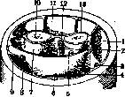
The lateral antebrachial cutaneous nerve (lateral nerve of the forearm) constitutes a continuation of the musculocutaneous nerve, innervates the skin of the lateral side of the forearm, and emerges through the fascia of the forearm in the region of the anterior-lateral groove of the elbow joint. The dorsal antebrachial cutaneous nerve (dorsal nerve of the forearm, a branch of the radial nerve) innervates the skin of the back of the forearm, passing to the surface through the fascia of the arm in its lower third. The sites where cutaneous nerves pass through the fascia are the most important points for anesthesia of the entire peripheral section of the nerve. Lymphatic vessels of the forearm—see Lymphatic system, human lymphatic system. The fasciae of the forearm (superficial fascia and proper fascia) are a continuation of the fascia of the arm; the proper fascia is most strongly developed in the proximal section, where the muscles of the forearm originate from it; on the anterior-medial side, the fascia of the forearm is reinforced by the tendinous expansion of the biceps muscle of the arm (bicipital aponeurosis). The fascia of the forearm constitutes a sheath and at the same time a soft, resilient, elastic skeleton for groups and individual muscles of the forearm (Figure 1); it fuses with the periosteum of the ulna along the entire posterior edge, which ensures its firm fixation. In the distal section, above the wrist joint, the fascia of the forearm is additionally significantly strengthened by transverse connective fibers, as a result of which the common carpal ligament is formed, the general ligament of the wrist, which in turn is divided into volar and dorsal (volar and dorsal carpal ligaments); the latter is expressed significantly better than the former, gives off inward septa running between the tendons of the extensors, and is fixed on the dorsal side of the bones of the forearm. (Figure 1: Fascial compartments of the forearm: 1—compartment of lateral extensors; 2—volar interosseous artery and nerve; 3—radial artery and nerve; 4—flexor carpi radialis muscle; 5—median nerve; 6—palmaris longus muscle; 7—compartment of deep flexors; 8—ulnar artery and nerve; 9—flexor carpi ulnaris muscle; 10—ulna; 11—compartment of extensors; 12—interosseous membrane; 13—radius. (According to Corning))
l. Shangina. Pathology of the forearm. In frequency of traumatic injuries, the forearm occupies one of the first places after the fingers and hand. Impact of the hand on moving parts of machines, gears, drums, transmissions, etc. gives severe injuries to the forearm, accompanied by scalped wounds of the skin, ruptures of muscles, tendons and fractures of bones. According to their nature, injuries can be: a) closed-contusions, fractures, b) open-wounds, open fractures, gunshot wounds, c) burns and frostbite. Closed injuries to the forearm can sometimes be accompanied by detachment of the skin over a large area, subcutaneous ruptures of large vessels with increasing hematoma, which requires surgical intervention-ligation of vessels and suturing of torn tissues. In open wounds, it is necessary no later than 12-24 hours to perform "primary treatment" of the wound, i.e., to excise all crushed, contaminated tissues within the limits of healthy tissues, followed by suturing of nerves, muscles, tendons. With large defects of individual muscles, their tendons are sutured to the bellies or tendons of the remaining functionally similar muscle groups. To avoid necrosis, the skin should be sutured without any tension; in cases with large defects of the skin, the latter are closed by means of primary plastic surgery according to Tirsch or a flap on a pedicle, taken from the abdomen. In the postoperative period, early physio-mechano-therapeutic treatment is applied. Small punctured and incised wounds with a knife, glass, often a broken neck when opening a bottle by drunkards can be accompanied by injury to vessels (more often a. radialis), nerves and tendons. With each injury to the forearm, it is always necessary to carefully examine the skin sensitivity and active movements of the fingers and hand, in order not to miss injuries to nerves or tendons. There are frequent cases when a patient, after suturing an ambulatory skin wound, comes with complaints of disturbance of sensitivity, absence of movement of the fingers and hand due to overlooked and not timely sutured injury of a nerve or tendon. When the n. radialis (deep branch) is injured, paralysis of the extensors occurs-the hand and fingers hang down, movements of dorsal extension are impossible. The thumb is adducted to the palm (m. adductor pollicis-innervation by n. ulnaris), cannot be abducted. When the superficial branch is injured, sensory disturbances occur, less constant due to anastomoses with the branches of ulnaris and medianus. The area of spread of sensory disturbances-the radial half of the dorsum of the hand.-Paralysis of the n. ulnaris manifests as weakening of the movements of flexion and abduction of the hand to the ulnar side. Flexion of the little finger is impossible; flexion of III and IV fingers is incomplete due to partial paralysis of m. flexoris digit, profundi. The thumb cannot approach the wrist bone of the index finger (paralysis of m. adductoris pollicis). The most characteristic feature of paralysis of the n. ulnaris is hyperextension of the main phalanges of the fingers with simultaneous flexion of the middle and nail ones, as a result of which the fingers take the form of claws. With prolonged paralysis of the n. ulnaris, atrophy of the interosseous muscles occurs, and the hand acquires the typical form of "claw hand". Disturbance of skin sensitivity occurs* on the ulnar half of the dorsum of the hand.-In paralysis of the n. mediani, flexion of the hand to the radial side is impaired, movements of flexion of the middle and nail phalanges of I, II, III fingers are impossible, the main phalanges flex due to mm. interossei. The thumb cannot flex and oppose; under the action of the extensors and m. adductoris pollicis, the thumb is applied to the index finger, as a result of which the hand has the appearance of "monkey hand". Skin disorders are noted on the radial side of the palmar surface of the hand (from I to the radial side of IV finger) and on the dorsum of the terminal phalanges of I, II, III fingers. Prolonged paralysis of the n. mediani leads to atrophy of the muscles of the thumb.* If a diagnosis of injury to a nerve or tendon is made, then to find the severed ends, the wound is expanded, having previously excised its edges. The severed ends of the tendons, especially the central one, "recede" due to the contraction of the muscles, for which* it is necessary to make additional incisions along the length. After finding the peripheral and central end of the nerve or tendon, they are refreshed (conveniently with a safety razor blade), after which they are sutured with observance of the corresponding technique of the suture. The nerve is sutured through the perineurium, without capturing its fibers, while the fibers should not be bent, but should be applied end to end. When suturing a tendon, it is necessary to ensure that its ends do not fray and no folds and nodes are formed that hinder free sliding. Convenient is the suture with two needles, in the form of a plait, with punctures on the cut surface; after tying the knots, they do not protrude on the surface of the tendon. The operation should be performed with minimal trauma to the tendon, with strict observance of asepsis, since even a weak inflammatory process causes adhesions of the tendon with surrounding tissues, hindering its movement. With a smooth course, early movements are started (after 7 days, Iselin), mainly active movements, baths, massage. With simultaneous injury to the n. mediani and tendons, erroneous suturing of the tendon to the nerve is possible (see Radiocarpal joint).-Gunshot wounds can be caused by jacket bullets, soft lead bullets and fragments of artillery shells. Wounds from jacket bullets, more often through with small entrance and exit openings and a direct bullet track, usually proceed aseptically. Treatment in the absence of damage to large vessels and nerves is conservative. Injury to vessels can be complicated by the formation of an aneurysm. If the injured nerves are not sutured immediately and the wound is complicated by suppuration, then suturing of the nerve can be started no earlier than 6 months after the cessation of suppuration, if the wound healed per primam, then the best time for suturing is after 2-2th/ months. Incomparably more severe are wounds from shrapnel bullets and especially artillery shells. Wounds are often blind, with the formation of cavities, with crushing of deep tissues; on-, the bottom are fragments of clothing, earth and fragments of the projectile. Such wounds require primary surgical treatment, with large defects: in
tissues the wound is left open. When the wound is contaminated with earth (gunshot wounds, street injuries, agricultural, especially garden), the introduction of anti-tetanus serum is mandatory.
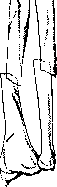
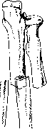
Fractures of the P. constitute, according to Bruns, up to 20% of all fractures. The cause of diaphyseal fractures of both bones, as well as isolated fractures of the radius and ulna diaphysis, is direct force, less often falling on the hand. - Isolated fractures of the ulna occur from direct impact of the poorly protected bone surface against a hard object - falling, protecting with hands from stick blows during a fight or attack. In the latter case, bilateral fracture of the elbow may occur. As a complication of isolated fracture of the ulna, dislocation of the radial head is observed. In fresh cases, dislocation reduction is sometimes possible, in neglected cases bloody reduction surgery is indicated, and even better resection of the radial head, because due to the subsequent scar tissue changes in the ruptured joint capsule and lig. annularis, the radial head cannot always be reduced and held after reduction, and when reduction is successful, recurrences often occur. In fractures of both bones from direct force (direct fractures, impact, lateral compression), the bones break at the same level, more often transversely (fig. 2). Indirect fractures - usually oblique or rotational depending on the mechanism. The radius breaks in the middle, the ulna at the border of the lower and middle thirds (the weakest places). In 3/4 of cases of fractures of both bones, the radius breaks above the ulna (Matti). The fragments are displaced in the direction of the force, secondary displacement - along the length. The radial muscle group is stronger than the ulnar, which is why shortening of the radius is greater. The pronator group is stronger than the supinator group, due to which the peripheral fragment (hand) is positioned in pronation (Bonier). The position of the central radial fragment depends on the height of the fracture: in fracture below the attachment of m. pronatoris teres, the central fragment is in the median position, above the attachment of m. pronatoris teres - the central fragment is in supination position (cross displacement).-With large displacement (fig. 3), especially cross displacement, it is difficult to reduce, and when reduction is successful, it is difficult to hold the fragments in the correct position. The difficulty of reduction is explained by the large number of muscles with different functions and the presence of membranae interosseae. Incorrect position of fragments can lead to healing in cross position, healing of both bones to each other; healing at an angle leads to shortening of the interosseous
^S5?!
area at a typical location.
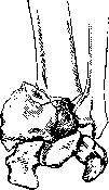
and limitation of rotational movement. Reposition of fragments is performed under local anesthesia (10-20 cm3 of 1-2% novocaine between fragments), after reposition the forearm is fixed in a 'semi-physiological' position with a dorsal plaster splint. In this semi-pronated position, the bones are at the greatest distance from each other, the interosseous ligament is stretched, and the danger of the bones fusing together is reduced. The hand is set in a position of dorsal flexion. Further treatment consists of early physio-mechano-therapy. When it is impossible to achieve the correct position of the fragments, surgery to suture the bones is indicated. During the operation, the ulna is first exposed and fixed in the correct position with Lambotte forceps, then the radius is exposed and sutured, after which the suturing of the ulna is completed. Oblique fractures with a large surface area of fracture can be sutured with wire, while transverse fractures are fixed with a metal or preferably a bone plate. The prognosis for fractures of the diaphysis of both forearm bones worsens due to the relative frequency of pseudarthroses (fourth place, according to Matti), explained by the great mobility of the fragments, as well as the ease with which muscles are trapped between them, which are compressed between the diaphyses in extreme pronation (mm. flexor pollicis longus, flexor digitorum profundus) and in extreme supination (mm. extensor carpi ulnaris, extensor digiti minimi) (Wegner). In cases of established pseudarthrosis, resection of the altered ends to normal bone and medullary canal is indicated, followed by bone grafting taken from the tibia. Improperly united at an angle and united together forearm bones also require surgical intervention; in the latter case, after separating the bones, muscles, fascia, or fat are placed between them. Open fractures require 'primary treatment' followed by treatment after suturing, like closed fractures. Particularly unfavorable are open infected fractures: fractures of the forearm from horse bites (coachmen) with crushing of soft tissues and comminution of bone. Treatment of infected fractures consists of immobilization of the forearm as much as possible in the correct position, with main attention paid to combating infection. Purulent extensions are widely opened, counter-incisions are applied for better drainage, etc. In cases of severe infection, amputation may be required. Incorrect position of fragments is corrected secondarily after the inflammatory process has ceased. Fracture of the epiphysis of the radius (fractura radii in loco classico, typico) occurs when falling on an outstretched hand; in this case, the epiphysis is as if knocked out by the carpal bones. The line of fracture when falling on the hand in dorsal flexion is located on the dorsal surface proximally and on the palmar surface distally; in the rarer fracture in palmar flexion, the position of the fracture plane is the opposite. The peripheral fragment is displaced dorsally, due to which the hand acquires the characteristic shape of a bayonet; the deformation is more pronounced on the radial side due to simultaneous supination of the epiphysis. Less constant is the displacement of the fragment to the radial side (Fig. 4). With a longitudinal impact, the dorso-lateral edge of the central fragment is wedged into the spongy part of the epiphysis. The wedging can lead to splitting of the peripheral fragment into pieces and result in T- and V-shaped fractures. The extension form of fracture of the radius is also observed in drivers and tractorists when the starter handle kicks back. In 50-80% of cases, the styloid process of the ulna is simultaneously torn off. The fracture may be accompanied by ligament and joint capsule ruptures and contusions of the metacarpal bones, especially their cartilage covering; in these cases, even after good anatomical union of the fragments, persistent functional disorders remain. Displacement of fragments causes disturbance of finger and hand movement, the protruding central fragment on the palmar surface presses against the flexor tendons, injuring them. Clenching the hand into a fist is impossible. Simple transverse fractures and epiphyseolyses without displacement that occur in children do not require immobilization; treatment with massage, active and passive movements from the first day of fracture is sufficient. When fragments are displaced, complete reposition is necessary, without which satisfactory functional results cannot be achieved. Under local anesthesia, gradually increasing traction is applied to the hand; the arm is bent at the elbow joint at a right angle to relax the two-joint finger and hand flexors; initially, to mobilize the fragments, the angle of displacement is increased dorsally and laterally, after which strong palmar flexion, pronation, and abduction to the ulnar side, with simultaneous pressure on the fragments with the fingers, places them in position. After reposition, a dorsal plaster or wooden splint is applied from the elbow joint to the metacarpophalangeal joint with palmar flexion and abduction of the hand to the ulnar side. The dorsal position of the hand prevents the development of flexor shortening. The splint is kept for 2-2½ weeks until the mobility of the fragments disappears. Immediately after removing the splint, massage, movements, and heat treatment are begun. Movements of the fingers are performed from the first day of fracture. Intra-articular fractures of the upper epiphysis of the radius and elbow joint—see Elbow joint. Burns of the forearm are more common in chemical production (acids), as well as in the dyeing workshop when the hand gets between hot rollers. Treatment of burns by the open method, which eliminates painful dressings, gives faster healing with less coarse scars in third and second degree burns. Deep burns with sloughing of the necrotic area may later cause bleeding from the devitalized vessel wall. Large skin defects are closed by plastic surgery (usually according to Thiersch), which prevents scar contractures. Plastic surgery is undertaken when the wound has cleaned and is covered with healthy granulations. Acute inflammatory diseases of the forearm. Of skin diseases, furuncles are more common (see Furuncle, Carbuncle). Phlegmon of the forearm. Infection penetrates primarily through the skin (furuncles, pyoderma) or as a complication of traumatic injuries, or by extension of purulent processes from the hand, especially in phlegmons of the tendon sheaths of the I and V fingers, which pass under the transverse volar carpal ligament to the forearm. In phlegmons of tendon sheaths, the affected finger is flexed, and light attempts to straighten it are sharply painful; pain is felt along the entire length of the tendon sheath. After destruction of the tendon sheath by pus, the purulent extension spreads upward between the pronator quadratus and flexors, then between both layers of flexors to the shoulder. Depending on location, phlegmon can be superficial, subcutaneous, or deep—subfascial, intermuscular. Subcutaneous phlegmon manifests as rapidly spreading redness, swelling, elevated temperature, and early gives foci of fluctuation. In deep forms, general septic phenomena prevail, and fluctuation may be absent, as the pus is deep under the fasciae and muscles. Treatment consists of early incisions widely opening the purulent extensions; in deep phlegmons, the fascial spaces involved in the process are opened strictly anatomically. The most favorable for pus drainage are incisions in phlegmons of tendon sheaths—along the lateral sides of the palmar surface of the forearm above the wrist joint, giving access to the most sloping parts of the tendon sheath. The incisions are drained with thin rubber strips (cut from old rubber gloves) (Iselin). The prognosis in early detected and properly treated cases is favorable; neglected, severe cases give high disability due to disfiguring scars and subsequent contractures. The prognosis worsens with the addition of putrefactive and especially anaerobic infection (gas gangrene). Osteomyelitic process relatively rarely affects the bones of the forearm (according to Haag's statistics, radius 5.1%, ulna 3.4%). For the clinic of osteomyelitis—see Osteomyelitis. Tuberculous lesion of the diaphyses of the forearm bones is more common in childhood. Often the process spreads from the epiphyseal part to the diaphyseal part through the metaphysis. Characteristic for tuberculosis is the prolonged course with insidious onset: the patient cannot specify the exact time of onset of the disease. The limb quite soon acquires a spindle-shaped form, similar to spina ventosa of the fingers, due to muscle atrophy and simultaneously developing circular ossifying periostitis around the focus of lesion (Oehlecker). The outcome of the process is more often the formation of small sequestra and sinuses with characteristic liquid, crumbly pus. Diagnosis is usually easy due to existing tuberculous lesions of other organs. It is mainly necessary to differentiate from chronic osteomyelitis and syphilitic lesions. Besides the history, the decisive factor is the X-ray picture. In tuberculosis, destructive processes prevail over reparative ones: 1) marked bone atrophy (osteoporosis), 2) absence of reactive bone process around, 3) sequestrum, if present at all, is small, delicate, and has a rounded shape. Conversely, in osteomyelitis: 1) bone atrophy is absent, 2) reactive sclerosing processes prevail, the sequestrum capsule is well expressed, 3) the sequestrum is more massive, elongated in shape, with large toothed edges.
Treatment consists of general strengthening treatment and local treatment of the focus, which consists of immobilization of the extremity, treatment with sunlight, quartz. Sometimes good results are obtained from the application of congestive hyperemia according to Viru. In cases with the formation of sequestra, as well as in long-unhealing fistulas, opening of the cavity with careful curettage of all affected tissue is indicated, otherwise recurrences occur. The resulting cavity is plugged with iodoform paste. The wound is sewn up tightly (Oehlecker). Syphilis of the bones P. is a rather rare disease. More often it is a matter of tertiary period lesions - osteo-periostitis gummosa. Gummatous lesions can be limited or diffuse. Superficial, periosteal gummas represent painful dense nodules with unchanged skin over them. With specific treatment, they can disappear without a trace; when they progress to a necrotizing form, necrotic masses are discharged, the ulcer has a dirty, greasy appearance with a sequestrum at the bottom. After healing, star-shaped scars remain, fused with the bone. Central gummas initially give little external change, later when the process reaches the surface of the bone, a firm diffuse swelling of it forms. Night pains are characteristic when the extremity is warmed in bed; in people working at night, pains appear during the day. The X-ray picture with periosteal proliferations and the presence of parallel processes of bone sclerosis and destruction is quite typical. The greatest difficulty is represented by the differential diagnosis with bone sarcoma. The sclerotic and periosteal changes in syphilis help to exclude it. The Wassermann reaction is also necessary. Treatment is specific - salvarsan, mercury. Tumors of P. In sarcoma of the bones P., the radius is affected three times more often than the elbow. Periosteal and central forms are encountered. Periosteal forms are distinguished by their malignant growth, quickly penetrate into surrounding tissues, which is why treatment usually consists of amputation of the extremity. Central sarcomas with an intact capsule can be resected with subsequent bone grafting. Cancer has the appearance of a cutaneous keratotic cancer; as a professional disease, it occurs in workers of paraffin factories and radiologists. The course is slow with late metastases. Treatment consists in the surgical removal of the tumor with subsequent skin plastic surgery. Among vascular tumors, lymphangiomas and hemangiomas are encountered. Unlike angiectasia, angiomas of embryonic origin develop as a result of abnormal excessive vascular laying. According to structure, simple angiomas (angioma simplex, capillare) and angiomas with cavity formation (angioma cavernosum) are distinguished. With their infiltrating growth and penetration of surrounding tissues, angiomas resemble malignant tumors, differing from them by a much slower growth and absence of metastases. A special type is the so-called racemose angioma (angioma racemosum), representing a tangle of tortuous vessels with arterial blood due to numerous communications between arteries and veins without the intervention of capillaries. These angiomas pulsate, on auscultation a blowing noise is heard, as in an aneurysm. Treatment of small angiomas - excision within healthy tissue; in well-developed inoperable cavernous angiomas, infiltration, galvanocautery can be tried. In angioma racemosum, its excision with ligation of all afferent branches is necessary. In severe cases, with penetration of all tissues, amputation may be required. Besides the described tumors, fibromas, osteomas can be encountered; treatment - surgical. A unique disease of the wrist joint is Madelung's deformation, manus valga. The disease arises in childhood and ends by the period of puberty. Both P. are symmetrically affected, in women more often than in men. The history shows no indication of trauma and inflammatory etiology. The basis of the disease lies in changes in the metaphysis and epiphysis of the radius (meta-epiphysitis radialis inf.) with disruption of the normal ossification process. Under the influence of muscle traction, the carpal bones slip from the articular surface of the radius toward the missing, not yet ossified palmar-ulnar part. Os lunatum as if becomes wedged between the bones of P., due to which the first row of carpal bones forms a triangle instead of the normally present arch. The ulna is dislocated at the same time to the back and outward. P. has the appearance of a bayonet or fork (Gabelhand), the anteroposterior diameter of the joint is sometimes doubled. The function of the joint usually suffers little. Treatment during the period of deformation development consists in wearing a special cuff that to a certain degree delays the development of deformation. In completed cases, surgical assistance may be required with exclusively cosmetic purposes in the form of excision of a wedge from the epiphysis of the radius. Unlike the described 'true' Madelung deformation, a so-called 'pseudomadelung' deformation is distinguished, similar in external joint changes but developing after trauma or inflammatory diseases. Operations on P. Ligation of vessels in bleeding, aneurysm (fig. 5). Ligation of a. radialis does not present difficulties along the entire course of the vessel. In the proximal half, the artery is exposed by a small incision of 5-6 cm between m. brachioradialis and m. flexor carpi ulnaris. The muscular edge of m. brachioradialis is pulled to the radial side, the common belly of the superficial flexors is moved toward the elbow; the artery lies under a fascial sheet through which it shines. It is necessary to remember about the nerve branch running alongside in the middle third of P. - r. superf. n. radialis. When ligating in the lower third of a. radialis, it is exposed in the middle between the radial bone and the tendon of m. flexor carpi radialis, at the pulse counting site. Ligation of a. ulnaris in the upper half is more difficult than ligation of a. radialis, due to its deeper location and abundance of muscular branches. A. ulnaris lies between the superficial and deep flexors (on m. flexor digitorum profundus). A skin incision 6-7 cm long along the line connecting the medial epicondyle of the humerus with the pisiform bone, along the radial edge of m. flexor carpi radialis. After incision of the fascia, the superficial flexor of the fingers is pulled to the radial side, the artery lies under the fascia accompanied by two veins and n. ulnaris coming from the ulnar side. The higher the artery is exposed, the more the superficial flexors have to be pulled to the ulnar side. The incision for exposing a. ulnaris in the distal third is made along the radial edge of m. flexor carpi ulnaris, the tendon of which is closest to the ulna. A. ulnaris lies in the fatty tissue under the fascia between the tendons of m. flexor digitorum sublimis and m. flexor carpi ulnaris. Exposure of the bones of P. is performed in incorrectly united fractures, pseudarthroses, osteomyelitis, tumors. Access to the ulna, due to its superficial location, is simple along its entire course from processus olecranon to processus styloideus. The radius is most easily accessible from the dorsal-lateral side, with P. in a middle position between pronation and supination, with the elbow joint bent at an obtuse angle. The incision is made along the line connecting the posterior-radial edge of the head with its styloid process. After incision of the fascia in the upper half, the bone is exposed between m. extensor digitorum communis and m. extensor carpi radialis, for which it is necessary to cut some fibers of m. supinator, which covers the radius (it is necessary to remember about the deep branch of n. radialis passing through the muscle). In the distal half, it passes between the tendons of mm. abductor pollicis longi et brevis on one side, extensor carpi radialis brevis on the other. In the lower third, the tendons of mm. abductor pollicis longi and extensor pollicis brevis need to be pulled to the back. Amputations of the forearm are performed under an exsanguinating tourniquet. Technically the operation is simple and can be performed by any method (formation of a skin cuff with the formation of dorsal and palmar flaps, method of oblique incision). The periosteum is incised at the same level as the interosseous ligament; tears of the periosteum should be avoided due to the danger of osteophyte formation. The bones are sawed off at the same level. To avoid painful neuromas and fusion with the scar, the nerves are cut off as high as possible.
The aa. radialis, ulnaris, interossea volaris et dorsalis are ligated, and the muscle flaps are sutured over the ends of the bones. The technique of the operation is somewhat complicated in the distal third due to the large number of tendons. It is recommended to prevent complete atrophy of the muscles by suturing antagonists in front of the stump. In amputation of the P., it is necessary to strive to preserve the stump as long as possible (see Prostheses).
Related articles
Mentioned in
Cite this page
“Forearm.” Soviet Medical Encyclopedia. English translation of Bolshaya Meditsinskaya Entsiklopediya, 1st ed. (Moscow, 1928–1936), ed. N. A. Semashko. https://sovietmedicalencyclopedia.com/article/forearm/