Mohrenheim's Fossa and Moria
Historical document, translated for reference. It reflects medical knowledge of the 1920s–30s and is not medical advice.
Summary
This article from the 1928-1936 Great Medical Encyclopedia describes Mohrenheim's fossa, an anatomical triangle below the clavicle significant for surgical approaches to axillary vessels, and moria, a psychopathological syndrome characterized by euphoric mood and compulsive jesting often associated with frontal lobe lesions.
Encyclopedia article (1928–1936)
MOHRENHEIM'S FOSSA (Mohrenheim), a depression named after the surgeon and gynecologist Mohrenheim, who first drew attention to it at the end of the 18th century; it is located below the clavicle at the boundary of its middle and outer thirds and is also known as the infraclavicular fossa or the deltopectoral triangle (fossa infra-clavicularis, s. trigonum deltoideo-pectorale).

Being a continuation of the cleft (sulcus deltoideo-pectoralis) between the deltoid and pectoral muscles diverging at the clavicle,
Mohrenheim's fossa has the shape of a triangle, the sides of which are formed by the named muscles, and the base by the clavicle (Fig. 2). This fossa is more clearly expressed in slender subjects, especially at the moment of pulling the arms forward. Within its limits, one can palpate (easier in individuals with poorly developed musculature) the coracoid process and the coracoclavicular ligament (Batuev, Brosike, and others), as well as the pulsation of the axillary artery. In some cases, the latter can be compressed against the second rib (Testut and Jacob), for which Mohrenheim proposed a special pad that no one uses to this day. The skin of the described region is very mobile, devoid of hair. The subcutaneous tissue consists of two layers: a superficial one, more or less rich in fat, and a deep, layered one, representing the true fascia superficialis. Between the two leaves of the latter are located the fibers of the lower part of the platysma, which contributes to the gaping of transverse wounds in this place. In this same layer, sensory nerves of the region, the supraclavicular nerves, descend from the cervical plexus. Beneath the superficial pectoral fascia and the adipose tissue filling Mohrenheim's fossa lies the deep pectoral fascia
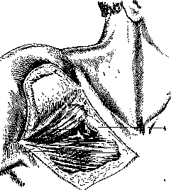
Figure 2. Mohrenheim's fossa and its relation to the cephalic vein: 1 - deltoid muscle; 2 - cephalic vein; 3 - pectoralis major muscle; 4 - clavicle. (fascia pectoralis profunda, s. coraco-clavi-axillaris Richet), in the splitting of which beneath the pectoralis major muscle the pectoralis minor muscle is enclosed. Directly behind the upper section of this deep fascia (clavipectoral fascia of Gruber), lying above the latter muscle, is located the axillary vein and, lateral to it, the axillary artery with the bundle of the brachial nerve plexus (Fig. 3). In the depth of the deltopectoral
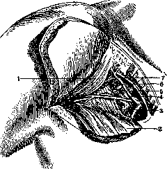
Figure 3. Relation of the deep axillary vessels to Mohrenheim's fossa: 1 - deltoid muscle; 2 - severed pectoralis major muscle; 3 - pectoralis minor muscle; 4 - brachial plexus; 5 - axillary vein; 6 - axillary artery; 7 - cephalic vein.
groove runs the cephalic vein. Having reached Mohrenheim's triangle, it goes together with lymphatic vessels from the pectoral muscles into the depths, pierces the clavipectoral
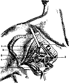
Figure 4. Contents of Mohrenheim's fossa: 1 - deltoid muscle; 2 - cephalic vein; 3 - thoracoacromial vein; 4 - thoracoacromial artery; 5 - severed pectoralis major muscle; 6 - clavicle; 7 - anterior thoracic nerve.
fascia and flows into the axillary vein. Sometimes the cephalic vein divides into two branches, one of which is thrown over the clavicle and flows into the subclavian vein above it, while the other preserves the usual position of the cephalic vein. In other instances, the cephalic vein does not empty into the axillary vein at all, but, passing over the clavicle, dumps blood into the external jugular vein. Cases have been noted where it forms a ring around the clavicle. Within the limits of Mohrenheim's fossa, the cephalic vein meets branches of the thoracoacromial artery, which, together with the anterior thoracic nerves, penetrates the clavipectoral fascia and supplies the pectoral muscles (Fig. 4). Along the course of the cephalic vein and in the region of Mohrenheim's fossa, lymph nodes (infraclavicular lymph nodes) are frequently located. Pathological smoothing of Mohrenheim's fossa is one of the earliest signs of subpectoral and especially interpectoral abscesses. A bulge in the region of Mohrenheim's fossa, in the presence of other symptoms, makes it possible to recognize a subcoracoid dislocation of the shoulder. Fractures of the clavicle significantly alter the relief of this region. Enlarged infraclavicular lymph nodes can be palpated in Mohrenheim's fossa (breast cancer). Gunshot and stab wounds of Mohrenheim's fossa present great danger due to the location of large blood vessels in the depth of this region. Mohrenheim's fossa is of great importance for the approach to the axillary and subclavian vessels below the clavicle, during their temporary or permanent ligation or other operations on them. Along the deltopectoral groove and triangle, the cephalic vein leads directly to the axillary vein. This significantly facilitates finding the artery located lateral to it. D. Vaza. MORIA, moria, a psychopathological syndrome characterized by an elevated, good-naturedly cheerful (euphoric) mood and a tendency toward foolish pranks. This term was previously abused to a significant extent, used to designate states observed in various mental diseases (amentia of Meynert, dementia praecox); at present, on the initiative of Jastrowitz, it is used exclusively to designate those peculiar states that sometimes develop in brain tumors and other organic lesions of the brain and which are most characterized by the patient's desire to joke and tell witticisms at all costs, a desire that disharmonizes with their severe objective state (hence the name Witzelsucht given by Oppenheim). In different cases, this syndrome presents fairly significant differences, and while in some patients with pronounced euphoria, jocularity is expressed to a comparatively insignificant degree, in others, on the contrary, the urge to jest is not accompanied by euphoria at all, but bears a peculiar tense-obsessive character. The very jokes of such patients usually do not differ in wit at all; on the contrary, they are flat and even downright absurd. The proposition put forward by Jastrowitz concerning the connection of moria with the localization of the pathological process in the frontal lobes remains controversial: while some assert that such localization is confirmed in no more than a third of cases coming to autopsy, others continue to insist on the connection between moria and the frontal lobes, pointing to the possibility of secondary damage to the latter by processes localized in other parts of the brain as well. Thus, the pathogenesis of this syndrome remains unclear at present.
I. Vaza
Related articles
Mentioned in
Cite this page
“Mohrenheim's Fossa and Moria.” Soviet Medical Encyclopedia. English translation of Bolshaya Meditsinskaya Entsiklopediya, 1st ed. (Moscow, 1928–1936), ed. N. A. Semashko. https://sovietmedicalencyclopedia.com/article/mohrenheims-fossa/