Colporrhaphy
Historical document, translated for reference. It reflects medical knowledge of the 1920s–30s and is not medical advice.
Summary
This article from the 1928–1936 Soviet medical encyclopedia describes the historical techniques and surgical procedures for colporrhaphy, an operation to repair vaginal wall prolapse. It details various methods of vaginal wall freshening and suturing, including the techniques of Hegar, Simon, and others, as well as post-operative care.
Encyclopedia article (1928–1936)
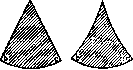
Colporrhaphy, colporrhaphia (from Greek kolpos—vagina and rhaphe—suture), is an operation on the vaginal walls for their prolapse. The first attempts in this direction date back to the first half of the 19th century. Initially, elytrorrhaphia was proposed, which consisted of excising pieces from the vaginal walls; then episiorrhaphia, which consisted of narrowing the genital slit by freshening and suturing the edges of the labia majora. The basis of elytrorrhaphia was the idea not only to narrow the vagina but also, through subsequent scarring, to achieve greater strength from the loosened paravaginal tissue. The latter idea found vivid reflection in the 'pincement du vagin' operation proposed by Desgranges, which consisted of clamping areas of the vaginal walls with small forceps and bringing them to necrosis; the forceps remained for 5-10 days, i.e., for as long as it took for them to fall off along with the necrotic tissue areas. Paulis inserted two uterine rings into the vagina, one above the other, of such sizes that significant pressure was exerted on the walls, resulting in inflammation and the formation of scars. Simon was the first to point out the irrationality of these methods and proposed the operation colpoperineorrhaphia, which aimed to significantly narrow the vagina and lengthen the perineum anteriorly. Subsequent improvements were made by Hegar, who proposed combining anterior colporrhaphy with colpoperineorrhaphy, which, in Simon and Hegar's methods, differed in the figure of freshening. Simon cut out a pentagonal figure, Hegar—a triangular one. Other various figures of freshening were also proposed; thus, some authors bypassed the columna rugarum posterior, while others removed it (Figs. 6 and 7). Hegar proposed for anterior colporrhaphy to excise an oval from the anterior vaginal wall. Colporrhaphy operations without additional operations on the uterus are performed for prolapse or descent of the vaginal walls alone without uterine prolapse and yield good results only in cases where the gap between the contracted crura of the levator ani in the middle of their extent is no wider than 1.5–2 cm; in all other cases, colporrhaphy must be combined with operations on the m. transversus perinei profundus and the crura of the levator ani and the uterus. Anterior colporrhaphy is performed as follows: in the lithotomy position, a short and wide Simon speculum is inserted into the vagina, the anterior lip of the vaginal portion is grasped with bullet forceps, and the uterus is pulled strongly downward and backward, so that the anterior vaginal wall is stretched. Depending on the degree of prolapse, a larger or smaller flap is excised from the anterior wall, the edges of the wound are sutured, and the vaginal lumen is thus narrowed. The figure of freshening varies among different authors: Simon proposed a horseshoe shape, the open side of which is directed toward the vaginal portion. Emmet excised three separate areas. Hegar proposed a figure of freshening that is used most frequently. It represents a longitudinal oval, the upper point of which is located 1–1.5 cm below the orificium ext. urethrae, and the lower one 1.5–2.5 cm above the orificium ext. uteri; the diameter in the widest part is 3–5 cm. The flap is outlined, and some excise it entirely, while others make a longitudinal incision between both ends and separate each half of the oval separately (Figs. 1 and 2). Fehling proposed preserving a longitudinal section 1–1.5 cm in diameter in the middle of the anterior vaginal wall and excising two ovals on both sides of it. In this method, he sees the advantage that with extensive freshening, the tension of the walls is reduced,
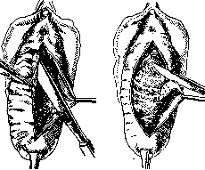
Figure 1.
Figure 2. and the influence of intra-abdominal pressure and the filled bladder on the sutured vaginal wall is also reduced, which promotes healing and facilitates it. When separating the flap, which can be either thickened or thinned, it must be remembered that one can injure the urethra, bladder, or ureters, and therefore the separation must be performed in the layer of connective tissue; it is recommended to direct the tip of the knife toward the tissue being removed, i.e., toward the vaginal wall. If the connective tissue is loose, separation can be achieved by blunt dissection. After removing the flap, the posterior surface of the bladder is exposed. Before
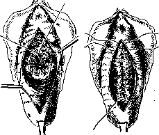
Figure 3.
Figure 4. the operation, it is better to empty the bladder to avoid injuring it. Bleeding vessels are ligated. If the freshened surface turns out to be uneven, it should be leveled with curved scissors. The edges of the wound are sutured; to avoid the formation of pockets, buried sutures are applied to the connective tissue. The material for the suture for most authors is catgut, iodine-catgut; the suture can be interrupted, or continuous, or interrupted-continuous (Vydrin). The latter consists of the thread being passed through the loop twice, which ensures that the continuous suture is as if it consists of tied interrupted sutures; with this suture, each individual section of it is completely independent in terms of integrity from the other section, and there is not that mass of knots that would be present if only interrupted sutures were applied. If after removing the flap the bladder bulges strongly, i.e., there is a cystocele, then it is necessary to apply either one or concentrically several purse-string sutures according to Schroeder, Gershuni (Fig. 3) on the bladder wall or a sagittal suture on the anterior retinaculum uteri according to Bumm and Martin (Figs. 4 and 5). Posterior colporrhaphy is performed in the same way, but since with prolapse of the posterior vaginal wall there is usually a strongly gaping genital slit and an old perineal tear, in these cases the colpoperineorrhaphy proposed by Simon-Hegar is performed. The technique is as follows: having determined the necessary degree of perineal elevation according to the case, bullet forceps or two threads are applied to the extreme points, which are tied at the ends, and thus loops are obtained, with which assistants stretch the perineum and the posterior vaginal wall. The resulting almost straight line at the border of the vaginal mucosa and the perineal skin
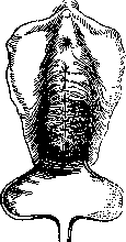
Figure 5.
is the base of the triangle, the apex of which is located on the posterior vaginal wall, more or less high depending on the degree of prolapse of the latter; a Kocher forceps is applied to the apex of the triangle. Assistants, pulling on the threads applied to the extreme points of the base of the triangle and on the Kocher forceps applied to the apex of the triangle, straighten the area of the posterior vaginal wall to be removed. A transverse incision is made at the border of the mucosa of the posterior vaginal wall and the perineal skin between the threads, the posterior vaginal wall is separated by sharp (Fig. 8) and blunt dissection from the rectum to the upper point of the triangle; the entire separated flap is removed. Lawson Tait proposed not to remove the flap, but to suture its edges in the sagittal direction and leave it (flap plasty). Then, to restore the septum recto-vaginale, the muscles and fasciae that are part of the centrum tendineum of the perineum in front of the rectum are sutured. To do this
rectum with the index finger of the left hand, gloved in a rubber glove. After applying three or four interrupted a
sutures of iodine-catgut
on the separated or torn muscles and fasciae of the septum recto-vaginale, an interrupted-continuous suture of iodine-catgut is applied from top to bottom on the edges of the triangle of the posterior vaginal wall in the sagittal direction until the extreme points of the base of the triangle, on which the loops are applied, are connected. This concludes the actual posterior colporrhaphy. Then, either interrupted silk sutures or Michel clips are applied to the perineal skin (Figs. 9-10). After the operation, the patient is put to bed and remains in the supine position until
Figure 7. Left—Hegar's figure of freshening. Right—corrected Hegar's figure of freshening. the morning of the next day; then she turns on her side, and she is allowed to change position independently, but not to sit up. A light diet is prescribed. Giving opium is not recommended. On the fourth day, the patient receives a laxative, usually Magnesia sulfurica. Care
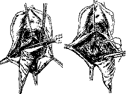
Figure 8.
Figure 9.
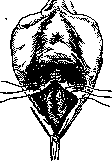
Figure 10.
for the wound consists of irrigating it several times a day (mandatory after each urination) with a lysoform solution. On the 5th or 6th day, the sutures or Michel clips are removed from the perineum. On the 12th day, the patient gets up, and before this, sitting in bed is not allowed. On the 14th
day, the patient is discharged. It is also necessary to mention the operation first proposed by Neugebauer in Warsaw, and some time later by Le Fort in Paris, for large uterine prolapses. The operation consists of excising flaps of the same shape from the anterior and posterior vaginal walls that face each other, and the freshened surfaces thus obtained are sutured together; as a result of this operation, a longitudinal septum is obtained in the vagina. This operation did not receive wide distribution. Colporrhaphy can be performed under general or local anesthesia. Anterior colporrhaphy is performed without any anesthesia (Vydrin), colpoperineoplasty—under local anesthesia: 1/2% solution of novocaine and three drops of Sol. Adrenalini hydrochlorici (1:1,000) for each gram of solution.
M. Vydrin
Related articles
Mentioned in
Cite this page
“Colporrhaphy.” Soviet Medical Encyclopedia. English translation of Bolshaya Meditsinskaya Entsiklopediya, 1st ed. (Moscow, 1928–1936), ed. N. A. Semashko. https://sovietmedicalencyclopedia.com/article/colporrhaphy/