Hysterosalpingography
Historical document, translated for reference. It reflects medical knowledge of the 1920s–30s and is not medical advice.
Summary
This historical article from the 1930s Soviet Medical Encyclopedia describes hysterosalpingography (metrosalpingography), a method of X-ray imaging of the uterine cavity and Fallopian tubes using contrast media. It outlines the indications, contraindications, technique, and diagnostic interpretation of the resulting radiographs, particularly for assessing tubal patency in cases of infertility.
Encyclopedia article (1928–1936)
METROSALPINGOGRAPHY (metrosalpingographia), a method of radiography of the uterine cavity and Fallopian tubes filled with a contrast medium. The idea of introducing a contrast medium into the uterine cavity belongs to Rubin (1915), and the practical use of metrosalpingography as a method belongs to Kennedy (W. J. Kennedy; 1923). The method is used to determine the patency of the tubes and the site of their possible obliteration (in cases of infertility), to diagnose tumors by changes in the normal pattern of the uterine cavity (metrography), and to study the physiology of the tube and uterus (tubal peristalsis and uterine contractions). Contraindications for performing metrosalpingography should include: 1) the slightest suspicion of pregnancy; 2) a febrile or subfebrile state of the patient; 3) signs of local and peritoneal irritation, even if minor; 4) the presence of even minor uterine bleeding and the menstrual period. The technique of metrosalpingography is not complicated, but requires hospitalization of the patient for at least 24 hours. After a thorough examination of the patient and the exclusion of the above-mentioned contraindications, the bowel (enema) and bladder are emptied. The patient is placed on a chair or table in the lithotomy position (with legs raised and abducted). After thorough disinfection of the vagina and cervix, the latter is grasped by the anterior and posterior lips with tenaculum forceps, after which the contrast medium is introduced into the uterine cavity. At present, iodipin or lipiodol are used almost exclusively as contrast media. The advantage of the latter is its high contrast, low viscosity, antiseptic properties, and at the same time, the almost complete absence of any local irritation. One of the most convenient devices for introducing the contrast medium is the hysteromanometer of C. Béclère. It consists of a syringe enclosed in a metal frame (Figure b). A manometer is attached to the syringe, allowing monitoring of the pressure under which the contrast medium is introduced. At the end of the syringe, there is a metal tip onto which a soft rubber probe (Figure d) with an oval thickening at the end is placed, which is inserted beyond the internal os of the uterus. An indispensable condition for correct technique is the tight closure of the cervix, which is achieved with the help of the probe and two tenaculum forceps (Figure c) grasping the cervix, or with the help of special forceps (Figure a) containing a hollow obturator probe inside. In the absence of Béclère's special hysteromanometer, an ordinary 10-gram syringe can be used in extreme cases. The contrast medium should be introduced under a pressure not exceeding 30 cm of mercury, in an amount of 15–20 cm3. The most favorable time for metrosalpingography is the middle of the intermenstrual period. By the time the contrast medium is introduced, the cassette with the plate must already be under the patient's pelvis, and the X-ray tube must be ready for the exposure. For a more accurate diagnosis, it is always recommended to take an en face image, a profile image, and a repeat image after 24 hours. The best images are obtained using a Bucky-Potter diaphragm, with two

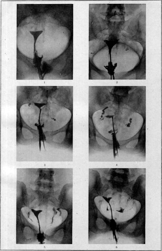
Figure 1. Bilateral tubal occlusion.
Obliteration of the tubes at the uterine horns. The contrast medium has filled only the uterine cavity.

Figure 2. Bilateral tubal occlusion. Obliteration of the tubes in the ampullary portion. The contrast medium has filled the tubes but has not penetrated into the peritoneal cavity.


Figures 3 and 4. Bilateral tubal occlusion. Obliteration of the tubes in the ampullary portion.
The contrast medium has filled the tubes and accumulated in the dilated, obliterated ampullary portion of the tubes. The contrast medium has not penetrated into the peritoneal cavity.

Figure 5. Patency of the tubes. The contrast medium has penetrated into the tubes and spilled through the ampullary portion into the peritoneal cavity (left tube).

Figure 6. The same case, imaged a second time after some time. Due to the peristaltic movements of the tube, the contrast medium has flowed from the tube into the peritoneal cavity and remained there in the form of accumulations. (Radiographs from the State X-ray Institute of the People's Commissariat of Health of the RSFSR.) Diagram for the illustrated article Metrosalpingographia.
For the article Metrosalpingographia
intensifying screens, 40–50 mA, a spark gap of 13–15 cm, and an exposure of 3–4 seconds for en face images and 6–8 seconds for profile images. In some cases, observing the entry of the contrast medium into the uterine cavity and tubes on the screen (metrosalpingoscopy) is extremely valuable. The results of metrosalpingography can be extremely diverse. The most characteristic patterns are as follows. 1) Both tubes are occluded, with the site of obliteration in both tubes located at the uterine end of the tubes. In this case, the image will show a triangular

a—forceps with a hollow probe inside; b—hysteromanometer;
d—soft probe with tenaculum forceps (c). shadow of the uterine cavity filled with the contrast medium. The tubes are not visible, and (what is most important) no contrast medium is visible in the peritoneal cavity (see separate plate, Figure 1). 2) Both tubes are obliterated, but the site of obliteration in both tubes is in the ampullary portion. In this case, the triangular uterine cavity and both tubes are visible, well-filled with the contrast medium, which accumulates particularly abundantly in the ampullary portion of the tube, which is dilated and obliterated at this site. The most characteristic sign of actual tubal obliteration is the absence of contrast medium in the free peritoneal cavity, distinct filling of the tube along its entire length (due to the impossibility of free outflow of the contrast fluid into the peritoneal cavity and the consequent emptying of the tube), and sharp delineation of the ampullary end of the tube filled with the contrast medium (see separate plate, Figures 2, 3, and 4). 3) Both tubes are patent along their entire length. In this case, the decisive sign is the presence of the contrast medium in the free peritoneal cavity, predominantly at the bottom of the rectouterine pouch (pouch of Douglas), which gives a pattern of poorly contoured, shapeless spots on the radiograph. Supporting signs of patency are the absence of accumulations of contrast medium in the ampullary portions of the tube and a weak, in places even disappearing, contour of the tubes (due to the outflow of the contrast medium into the peritoneal cavity) (see separate plate, Figures 5 and 6). In cases where the pattern of contrast medium entering the peritoneal cavity is not sufficiently convincing, it is recommended to take a repeat image after 24 hours. If the tube is patent, the contrast medium flows out of the tube during this period and gives a pattern of indistinct, diffuse spots in the peritoneal cavity on the repeat image. Conversely, if the tube is obliterated, the contrast medium that entered the dilated ampullary portion remains unchanged, giving a pattern of sharply contoured spots identical to those obtained the day before. The method of metrosalpingography for the diagnosis of tumors is used as an auxiliary method in determining the presence of polyps, submucosal fibroids, etc., in the uterine cavity. However, in this respect, the method has not acquired great practical significance. The method of metrosalpingography is of great scientific interest for studying tubal peristalsis and uterine contractions, which can be well observed by taking serial images.
Related articles
Cite this page
“Hysterosalpingography.” Soviet Medical Encyclopedia. English translation of Bolshaya Meditsinskaya Entsiklopediya, 1st ed. (Moscow, 1928–1936), ed. N. A. Semashko. https://sovietmedicalencyclopedia.com/article/hysterosalpingography/