Vesiculography
Historical document, translated for reference. It reflects medical knowledge of the 1920s–30s and is not medical advice.
Summary
Vesiculography is a radiological technique used to visualize the seminal vesicles by filling them with a contrast medium. The procedure, which can be performed via the ejaculatory ducts or the vas deferens, is used to diagnose obstructions, morphological changes, and chronic pain in the prostate and seminal vesicle region.
Encyclopedia article (1928–1936)
VESICULOGRAPHY, the obtaining of an X-ray image of the seminal vesicle. In view of the fact that seminal vesicles do not cast a shadow during standard radiography, it is necessary to fill their cavity with a contrast medium in order to obtain an image.
Figure 1. Cannula for filling the seminal vesicles through a urethroscope.
This is done with a 5% solution of collargol, a 10–15% solution of sodium iodide, iodipin, etc.
The contrast medium can be introduced either through the ejaculatory ducts, into which a special cannula (see Figure 1) connected to a Record syringe is inserted during posterior urethroscopy, or through the vas deferens by means of vasopuncture (see). Locating the ejaculatory ducts and filling them through a urethroscope is technically difficult and requires special instrumentation. Vesiculography via the vas deferens is technically simple. With the latter method, the radiograph shows an image not only of the seminal vesicle but also of the vas deferens (see Figure 2).
Vesiculography has found relatively wide application in America (Lowsley and Delzell, Young, and others); in Europe (Luys, François), vesiculography is used significantly less often.
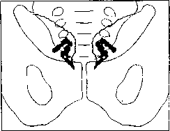
According to Young, vesiculography is indicated: 1) in cases of suspected narrowing of the vas deferens or ejaculatory duct to determine their patency in cases of infertility, if an orchio-vasoanastomosis is planned; 2) to determine morphological changes in the seminal vesicle, ampulla, and vas deferens in cases of spermatocystitis;
Figure 2. Bilateral vesiculography. Both seminal vesicles and the vasa deferentia are filled with contrast medium (from a radiograph).
and 3) to determine the condition of the vesicles in cases of undefined and difficult-to-explain pain in the region of the prostate, bladder, and seminal vesicles. Ya. Gotlib.
Related articles
Cite this page
“Vesiculography.” Soviet Medical Encyclopedia. English translation of Bolshaya Meditsinskaya Entsiklopediya, 1st ed. (Moscow, 1928–1936), ed. N. A. Semashko. https://sovietmedicalencyclopedia.com/article/vesiculography/