Spectroscopy
Historical document, translated for reference. It reflects medical knowledge of the 1920s–30s and is not medical advice.
Summary
This article defines spectroscopy as the observation of spectra, explaining the physical principles of light dispersion through prisms and diffraction gratings. It details the types of spectra (continuous, line, and band), the function of various instruments like spectroscopes and microspectroscopes, and their applications in chemical and medical analysis.
Encyclopedia article (1928–1936)
SPECTROSCOPY, SPECTROSCOPE. Spectroscopy is the observation of a spectrum. A spectrum is the colored strip (rainbow) obtained on a screen when light passes through a prism or a dense grating, a diffraction optical grid. The reason for the appearance of such a strip (rainbow) lies in the physical properties of a white solar ray to split into constituent rays when passing through a prism. A white ray, as Newton showed as early as 1666, is a mixed ray. It consists of rays colored in the various colors of the rainbow and possessing different angles of refraction when passing through a glass prism in connection with their different wavelengths. Rays with the shortest wavelength—violet—possess the strongest refraction. Therefore, they will be located in the spectrum toward the base of the prism, whereas rays with the longest wavelength—red—possessing less strong refraction, will fall toward the side facing the apex of the prism. In the interval between the red and violet, rays colored in other colors will be located, according to their refraction, and a full continuous spectrum will thus represent a series of colored strips gradually transitioning into one another in the following sequence: violet, indigo, blue, green, yellow, orange, and red. But this is only the visible part of the spectrum, covering rays with a wavelength in the range between 400 mμ and 760 mμ. On one side of the visible spectrum are located infrared rays, possessing a longer wavelength—thermal rays; on the other—ultraviolet rays, possessing a shorter wavelength—chemical rays. Prisms made of ordinary glass to a significant degree block ultraviolet rays, whereas prisms made of rock crystal or Iceland spar transmit them. Spectra obtained as a result of the decomposition of a light ray by a prism differ among themselves. Thus, there are spectra: continuous, when the colored strip represents a gradual transition from one color to another (spectra of solid bodies heated to white heat); line, or discontinuous, when instead of a strip we have a whole series of lines colored in various colors (spectra of luminous vapors or gases, e.g., He, H, Hg); and band spectra, consisting of a mass of indistinct lines (observed during the luminescence of vapors and chemical compounds). Their general name is emission spectra. If, in the path of light rays producing a continuous spectrum, one places a solution of substances capable of absorbing any rays, then against the background of the continuous spectrum, dark bands or individual lines appear corresponding to those wavelengths which are absorbed by the given intermediate body. The spectrum observed in this case is called an absorption spectrum. Examples of the latter include the solar spectrum, the spectra of blood pigments—Hb and its derivatives, etc. For the observation of spectra, there are several instruments, which are called spectroscopes. Spectroscopes are of different systems. Thus, the spectroscope of the Kirchhoff and Bunsen system consists of three tubes, which is why it is called a three-tube spectroscope. The 1st tube is a collimator, the 2nd is a viewing telescope, and the 3rd is a tube with a scale for measuring the relative distance of spectral lines. The collimator, in turn, consists of a narrow rectilinear slit located at the focus of an achromatic collecting lens, so that the light ray that has passed through the slit and fallen onto the collecting lens, after passing through it, is transformed into a bundle of parallel rays and in this form will fall onto the prism of the spectroscope. The prism is the main component of any prismatic spectroscope: thanks to it, the white ray is separated into its constituent parts by virtue of the different angles of refraction of these rays. In diffraction spectroscopes, the decomposition of a white ray into a colored strip is carried out with the help of a diffraction grating, located in the same place as the prismatic prism. Spectra obtained from a prism and from a grating differ from each other. While the spectrum from a prism is compressed in the red and yellow regions and strongly dispersed in the regions of blue, indigo, and violet rays, the diffraction spectrum presents a picture of more uniform distribution of rays along the length of the entire spectrum. The viewing telescope or eyepiece contains a biconvex lens and has the purpose of increasing the size of the spectrum. The 3rd tube contains a translucent ruler, on which divisions are marked, expressing the lengths of light waves in millimicrons (mμ). This ruler can be moved with the help of a micrometric screw. Moving the ruler is necessary before observations of spectra are carried out, because it is necessary to correctly orient this ruler in relation to the spectrum, i.e., so that the division with the number 589 falls or, more correctly, coincides with the Fraunhofer line D in yellow light (see Spectral analysis). A direct-vision spectroscope (a vision directe, Amici prism), released by Zeiss or Reichert (see figure), is so called because the spectrum is observed in a direct direction. This is achieved by the fact that one prism of an ordinary spectroscope is replaced by a combination of 5 prisms, folded together in such a way that their bases face in opposite directions. In front of the prisms, the pocket spectroscope also has a micrometric ruler, which is set in motion with the help of a screw.
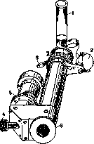
Reichert's spectroscope: 1—test tube with the liquid being tested; 2—small mirror at a 45° angle for obtaining the upper spectrum (solar); 3—eyepiece; 4—small screw for setting the micrometric ruler; 5—screw for adjusting the ruler to the eyes; 6—small shutter for dividing the spectrum into two parts (above—solar spectrum, below—spectrum of the liquid being tested); 7—screw for setting the proper width of the slit (so that the Fraunhofer lines are visible). The Zeiss firm in Germany has released a direct-vision spectroscope adapted to a microscope—a microspectroscope. This attachment can be of importance for the study of small colored spots. For photographing spectra, spectrographs are used, the peculiarity of which, besides various attachments of a photographic nature, or rather attachments connected with the setting of the negative, is that all the optics in the spectrograph are made of quartz glass in view of the circumstance that ordinary glass absorbs ultraviolet rays. For quantitative analysis, for determining the concentration of certain colored liquids, a special instrument is used—a spectrophotometer. Several systems of spectrophotometers are known. The spectroscopes of the English firm Hilger and the spectroscopes of the German Konig-Martens system enjoy particularly great fame. Spectrophotometers are instruments representing a combination of a spectroscope and a polarization apparatus. The determination of the quantity of the pigment being investigated is based on comparing the color of one half of the field of view, obtained from the pigment being tested, with the color of the other half, obtained from a specific quantity of pigment. Furthermore, the spectrophotometer makes it possible, when mixing two pigments, e.g., oxyhemoglobin and methemoglobin or other pigments, to determine the quantities of one and the other pigments found in the mixture by the value of the quotient, using tables provided by Hüfner.
Related articles
Mentioned in
Cite this page
“Spectroscopy.” Soviet Medical Encyclopedia. English translation of Bolshaya Meditsinskaya Entsiklopediya, 1st ed. (Moscow, 1928–1936), ed. N. A. Semashko. https://sovietmedicalencyclopedia.com/article/spectroscopy/