Eyepiece
Historical document, translated for reference. It reflects medical knowledge of the 1920s–30s and is not medical advice.
Summary
This article from the 1928–1936 Great Medical Encyclopedia describes the optical system of lenses known as the eyepiece, used in instruments like microscopes and telescopes to magnify the image produced by the objective lens. It details the physical principles, types such as the Huygens, Ramsden, and Kellner eyepieces, and their applications including micrometers and projection.
Encyclopedia article (1928–1936)
EYEPIECE, a system of lenses in optical instruments (microscope, telescope, etc.) facing the observer's eye and playing the role of a magnifying glass. The purpose of the eyepiece is to magnify the real image produced by the objective (see). The image from the objective must fall at a distance less than the main focal length of the eyepiece, and in order to obtain the greatest magnification, this image must lie as close as possible to the main focus of the eyepiece. Examining the image through the eyepiece as through a magnifying glass (see), we obtain a virtual image located from the eye at the distance of distinct vision. A good eyepiece must give a colorless image, and for this it is necessary that the red and blue rays passing through the eyepiece produce virtual images of the same angular magnitude. This requires that the focal length of the eyepiece be the same for both kinds of rays. This problem is solved in the complex achromatic eyepiece, consisting of two lenses (of the same material) separated from each other by a distance equal to half the sum of their focal lengths. This condition is satisfied by the main types of Huygens and Ramsden eyepieces used in the microscope (for a detailed description of them see Microscope) and in telescopes. Usually, the eyepiece is calculated and corrected for major optical aberrations separately from the objective. Therefore, the eyepiece can be changed in optical instruments, selecting the focal length according to the required magnification. However, eyepieces with a focal length of less than 5 mm are not used, because in such short-focus eyepieces the brightness of the image becomes too small, and its clarity is affected by diffraction. In a terrestrial telescope, the eyepiece consists of two parts—one, as in all eyepieces, being a magnifier, and the other, located between the first part of the eyepiece and the objective, usually consisting of two collecting lenses and serving to turn the inverted image produced by the objective into an upright one. The eyepiece of a terrestrial telescope can be viewed as a microscope through which the real image ab produced by the objective of the telescope is observed. The diagram of the terrestrial telescope eyepiece is given in Fig. 1, where lenses A and B are the Huygens eyepiece, and C and D are the objective (erecting system of the telescope). In the telescopes of spectroscopes, cathetometers, etc., the Ramsden eyepiece is usually used. The problem of the eyepiece in Galileo's telescope (binocular) is solved very originally: here the eyepiece consists of a diverging lens (single or a system of lenses) placed behind the objective at a distance less than the focal length of the latter, and equal to the difference between the focal lengths of the objective and the eyepiece. Rays from the object MN, refracted by the objective B, enter the eyepiece before they meet at the focus, and are transformed into a diverging beam. As a result, a virtual, magnified, and upright image of the object mn is obtained for the eye.
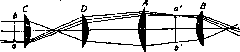
FIG. 1. -(Fig. 2). In eyepieces used for measurements, either a crosshair A (spider silk) or a transparent scale is placed at the location of the image of the object produced by the objective. The observer's eye in this case sees the image of the object superimposed on the scale. Such a device is called an ocular micrometer. To better illuminate the scale or crosshairs, an aperture B (Gauss eyepiece) (Fig. 3) is made in the eyepiece mount, through which light from a source L, reflected either from a mirror plate P or from a prism with total internal reflection, falls on the crosshairs or scale. - If

Figure 2.
in a microscope, the eyepiece is moved further than the focal length of the image produced by the objective, the system will produce a real magnified image of the object, which can be projected onto a photographic plate (see Photomicrography) or onto a screen. In the latter case, we turn the eyepiece into a projection one. Zeiss released a number of special eyepieces («Homal» and others) for microprojection. (For special eyepieces used in microscopes, see Microscope.) Among simple eyepieces, the orthoscopic Kellner eyepiece is also encountered. It consists of two identical plano-convex

Figure 3.
lenses B and C, facing with their convex side toward the objective D, having identical focal lengths and separated from each other by a distance also equal to their focal length. The eyepiece is installed in such a way >(Fig. 4) that the image produced by the objective coincides with the first lens of the eyepiece C. The first lens of the eyepiece will not produce magnification, but will deflect the rays toward the main optical axis. The second lens B gives an image that is virtual and magnified.
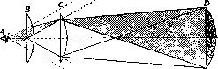
Figure 4.
magnified. To avoid the effect of chromatic aberration, the second lens is made achromatic. The advantage of the orthoscopic eyepiece is a very large field of view. It is used in simple microscopes, e.g., for examining wood sections, coarse tissue structures, etc. The main disadvantages of this eyepiece are strong spherical aberration (marginal parts of the image are disproportionately magnified) and image distortion due to the fact that the image of the surface of the first eyepiece lens with all the dust and dirt covering it is superimposed upon it.
a. Irisov.
Related articles
Mentioned in
Cite this page
“Eyepiece.” Soviet Medical Encyclopedia. English translation of Bolshaya Meditsinskaya Entsiklopediya, 1st ed. (Moscow, 1928–1936), ed. N. A. Semashko. https://sovietmedicalencyclopedia.com/article/ocular/