Ophthalmometer
Historical document, translated for reference. It reflects medical knowledge of the 1920s–30s and is not medical advice.
Summary
This article from the 1928–1936 Great Medical Encyclopedia describes the ophthalmometer, an optical instrument used to measure the radii of curvature of the eye's refracting media, primarily the cornea and lens. It details the historical Helmholtz ophthalmometer, the widely used clinical Javal-Schiötz ophthalmometer, and their role in determining corneal astigmatism.
Encyclopedia article (1928–1936)
OPHTHALMOMETER (from Greek ophthalmos - eye and metron - measure), an optical instrument designed to measure the radii of curvature of the surfaces of the refracting media of the eye (cornea and lens). The basic principle of ophthalmometry is as follows: the surfaces of the refracting media of the eye are mirror-like and, like all spherical mirrors, under certain conditions produce reduced images of nearby objects. The size of the image obtained in a mirror depends on three factors: the size of the object, the distance of the object from the mirror, and the radius of curvature of the mirror. Therefore, if the size of the object, the distance of the object from the mirror, and the size of the image are known, the radius of curvature of the mirror r can be easily calculated using the formula: r = (A / a) B, where A and a are the sizes of the object and its image, and B is the distance of the object from the mirror. A and B are easily accessible to measurement, and therefore ophthalmometry ultimately reduces to measuring the size of the image obtained from a specific object upon reflection from various surfaces of the refracting media of the eye. This measurement is usually performed using a telescope in which a special doubling device serving measurement purposes is inserted between the objective and the eyepiece.
The prototype of all ophthalmometers is the Helmholtz ophthalmometer. It represents a very precise instrument,
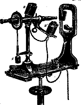
Figure 1. Javal-Schiötz apparatus.
which played a huge role at the time, because it was used to first obtain accurate information about the radii of curvature of the refracting surfaces of the cornea and lens, on the basis of which all our knowledge about the refracting apparatus of the eye is built. In view of the complexity of the study and the calculations associated with it, the Helmholtz ophthalmometer, however, did not acquire clinical significance. For clinical purposes, the Javal-Schiötz ophthalmometer is of great importance. (The general view of the apparatus is shown in Fig. 1, and the path of the rays in it in Fig. 2.) This ophthalmometer consists of a telescope, inside of which, between the objective and the eyepiece, two prisms are inserted as a doubling device, folded along their hypotenuses so that they form
a parallelepiped. Attached to the middle of the telescope
is a graduated
rotating arc
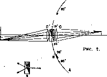
on which there is one sliding figure on each side, closed in front by milk or colored glass. These glasses are illuminated from behind by an electric bulb. The measurement is performed in a dark room. The patient sits at the apparatus from the objective side, and their head is immovably fixed on a special stand placed precisely in the center of the arc of the ophthalmometer. When the subject looks straight into the tube, images of both figures appear on their cornea (one of them has the shape of a ladder, and the other of a rectangle). Thanks to the doubling device, the physician sees two ladders and two rectangles through the telescope, and only the two middle figures (the ladder and the rectangle) are important for measurement purposes. During the measurement, using a special screw, both figures are moved together or apart along the arc of the ophthalmometer until both middle figures come into contact in the corneal image. At this moment, the radius of curvature of the anterior surface of the cornea in millimeters can be read off the arc.
The clinical significance of the Javal-Schiötz ophthalmometer lies in the fact that it allows one to determine the degree of corneal astigmatism extremely easily and quickly, for which the following procedure is used: having set the middle figures to touch with the horizontal position of the arc of the ophthalmometer, the arc is then turned by 90°. If there is no corneal astigmatism, the figures do not change their mutual position and remain in a state of contact. If corneal astigmatism is present, the mutual position of the figures changes, and they either diverge or overlap. From the degree of overlapping, it is easy to determine the degree of corneal astigmatism, since the apparatus is constructed in such a way that each step of the ladder corresponds to one diopter. The different mutual arrangements of the middle figures of the corneal image at individual moments of measurement are shown in Fig. 3. The keratometer of Sutcliffe is built on a slightly different principle and also serves to determine corneal astigmatism. It has the advantage that it can determine the degree of astigmatism immediately in a single setup, without any additional rotations. The radii of curvature of the lens
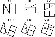
Figure 3.
surfaces can be determined relatively quickly and simply using Tscherning's ophthalmophotometer. However, this apparatus still has no clinical application.
E. Tron.
Related articles
Mentioned in
Cite this page
“Ophthalmometer.” Soviet Medical Encyclopedia. English translation of Bolshaya Meditsinskaya Entsiklopediya, 1st ed. (Moscow, 1928–1936), ed. N. A. Semashko. https://sovietmedicalencyclopedia.com/article/ophthalmometer/