Ophthalmoplegia
Historical document, translated for reference. It reflects medical knowledge of the 1920s–30s and is not medical advice.
Summary
This article from the 1928–1936 Soviet Great Medical Encyclopedia defines ophthalmoplegia as the simultaneous paralysis of several eye muscles innervated by different nerves. It covers complete and partial forms, internal and external types, acute and chronic manifestations, and various underlying etiologies such as infections, intoxications, and neurological disorders.
Encyclopedia article (1928–1936)
OPHTHALMOPLEGIA (from Greek ophthalmos - eye and plesso - I strike), simultaneous paralysis of several eye muscles innervated by different nerves (nn. oculomotorius, trochlearis, abducens). Most commonly, ophthalmoplegia refers to a condition of the aforementioned character in both eyes; however, the combined paralysis of two or three nerves of a single eye also falls under this designation. Ophthalmoplegia is divided into complete and partial. In c o m p l e t e ophthalmoplegia, both the external muscles of the eye (straight, oblique, and the levator palpebrae superioris) and the internal muscles (sphincter of the iris and ciliary muscle) are affected. Partial ophthalmoplegia is of two kinds: e x t e r -
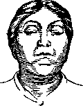
Figure 1. Complete ophthalmoplegia. Hutchinson's face. Bilateral ptosis.
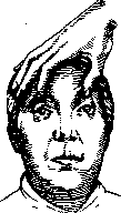
Figure 2. Complete ophthalmoplegia. Hutchinson's face. When both eyelids are artificially raised, immobility of the eyeballs is revealed. n a l ophthalmoplegia, when only the external eye muscles are affected, and i n t e r n a l ophthalmoplegia, when exclusively the internal musculature of the eye is paralyzed. In the vast majority of cases, ophthalmoplegia depends on an organic lesion of the peripheral neurons of the aforementioned three nerves (more often their nuclei, less often the roots and the nerves themselves). - The symptomatology of pronounced ophthalmoplegia reduces to drooping of both eyelids and immobility of the eyeballs—the so-called "Hutchinson's face" (Figures 1 and 2); in internal ophthalmoplegia, the pupils are dilated and react neither to light nor to convergence; accommodation often suffers due to paralysis of the m. ciliaris. In less sharp cases, paralysis is observed in some muscles and only paresis in others. - D i a g n o s i s of complete external or internal ophthalmoplegia presents no difficulties—complete absence of mobility of the eyes, ptosis in external ophthalmoplegia, and absence of pupillary movements and accommodation in internal ophthalmoplegia are easily determined. In incomplete ophthalmoplegies with paralysis or paresis of individual muscles, diagnosis requires the study of diplopia, the field of gaze, etc., as is done in determining paralytic strabismus (see Diplopia, Strabismus). By course, acute and c h r o n i c ophthalmoplegia are distinguished. The latter usually has a progressive character. Acute unilateral ophthalmoplegia is often the result of acutely developing necroses of vascular origin, being then combined with a number of other symptoms indicating lesions of neighboring systems (see Alternating syndromes). More often, acute ophthalmoplegia is bilateral. It occurs under the influence of various intoxications and infections. Among chemical substances, alcohol is in first place, followed by lead, ergotin, and sulfur derivatives. Methyl alcohol causes isolated acute internal ophthalmoplegia. Not infrequently, ophthalmoplegia owes its onset to poisoning by meat, fish, or sausages; in this case, it is sometimes difficult to decide whether it is a matter of poisoning by chemical substances contained in these products, or
Figure 3. Ptosis of the left eyelid, outward deviation of both eyes, dilation of the right pupil,
si. in basilar specific meningitis. ite- infection entering the organism along with them. Acute ophthalmoplegia occupies a prominent place in the symptomatology of a number of infectious diseases of the brain and its membranes (epidemic encephalitis, tuberculous meningitis, acute hemorrhagic polioencephalitis, etc.). Sometimes acute ophthalmoplegia occurs in fractures of the base of the skull with subsequent hemorrhage. - S u b a c u t e ophthalmoplegia sometimes develops in tumors of the brain stem, base of the brain, and cerebellum; it is significantly more often observed in syphilitic diseases of the brain (Figure 3). C h r o n i c ophthalmoplegia very rarely represents an independent disease; in the vast majority of cases, it develops against the background of an already existing disease of the nervous system or precedes it (sometimes long before). Among these diseases, tabes dorsalis and progressive paralysis are in first place, followed by myasthenia gravis, funicular myelosis, multiple sclerosis, chronic poliomyelitis, and even more rarely Basedow's disease, cysticercosis, and sarcomatosis of the leptomeninges. Isolated internal ophthalmoplegia can be for many years the only manifestation of neurosyphilis. External chronic ophthalmoplegia usually has a progressive character, and as a rule, in its further development it does not pass into complete, i.e., it does not spread to the internal musculature of the eye, which speaks in favor of its nuclear localization. The pathomorphological essence of chronic ophthalmoplegia usually reduces to an inflammatory process at the floor of the third ventricle and the cerebral aqueduct, which ultimately leads to the destruction of the nuclei of the third, fourth, and sixth nerves. - A peculiar place is occupied by ophthalmoplegia in migraine (see Hemicrania). In rare cases of "migraine ophthalmoplégique," ophthalmoplegia occurs on the side of the pain. It either disappears after the migraine attack or leaves residual paresis that intensifies with a new migraine attack and turns into chronic ophthalmoplegia. - S t a n d i n g a p a r t is congenital ophthalmoplegia, depending on a deficiency in the development of the nuclei of the third, fourth, and sixth nerves. This ophthalmoplegia as a rule is external, and the elevators of the eyelid are affected with particular readiness, while the capacity for convergence is usually preserved. Double images do not occur with this ophthalmoplegia. Sometimes this form of ophthalmoplegia is familial; essentially it relates to aplastic or abiotrophic processes and can be considered as a partial atavism: in a number of low-organized animals, the eyes sit immobly in the orbits. - In individual cases of pseudobulbar palsy, external ophthalmoplegia has been observed, depending on the interruption of central pathways from the cortex to the nuclei of the third, fourth, and sixth nerves. This form of ophthalmoplegia is characterized by the absence or limitation of voluntary eye movements while reflex movements are preserved. This form was named by Wernicke pseudoophthalmoplegia. - To false ophthalmoplegias can be attributed those cases where it is caused by a primary lesion not of the nervous apparatus, but of the muscular one. Such is ophthalmoplegia in scleroderma. - F i n a l l y , hysterical ophthalmoplegia also occurs. In it, the internal musculature of the eye does not suffer, automatic eye movements are preserved, and the ophthalmoplegia itself sometimes disappears when the patient's attention is distracted.

Related articles
Mentioned in
Cite this page
“Ophthalmoplegia.” Soviet Medical Encyclopedia. English translation of Bolshaya Meditsinskaya Entsiklopediya, 1st ed. (Moscow, 1928–1936), ed. N. A. Semashko. https://sovietmedicalencyclopedia.com/article/ophthalmoplegia/