Phakoscope
Historical document, translated for reference. It reflects medical knowledge of the 1920s–30s and is not medical advice.
Summary
The phakoscope is an instrument proposed by Helmholtz to observe Purkinje-Sanson images during accommodation. This article details its construction, optical setup, and how it demonstrates changes in the curvature of the crystalline lens.
Encyclopedia article (1928–1936)
PHAKOSCOPE, an instrument proposed by Helmholtz for observing the Purkinje-Sanson images during accommodation; it is a box with blackened interior walls of triangular shape, whose corners are cut off by three planes. In one of the planes (Figure 1) there is a circular opening where the observed eye is positioned. Directly opposite it in the opposing wall, a quadrangular window with a thin needle-point is made. The test subject, through the opening, either fixes their gaze on the needle point or, relaxing accommodation, looks into the distance in the same direction at some sufficiently distant point. Such an arrangement achieves the alignment of the test subject's eye and two fixation points along a single straight line. If a light source is now placed opposite the wall containing two glass prisms embedded in it with their bases turned toward each other, three pairs of reflexes (glints) will appear on the test subject's eye: a pair of the brightest ones (Figure 2) (a) from the cornea, a second pair somewhat dimmer and larger in size from the anterior surface of the crystalline lens (b), and finally the dimmest one, barely distinguishable, from its posterior surface (c). The first two reflexes will present
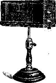
Figure 1.
a direct image, as reflections from convex surfaces, and the third an inverted image, as from a concave surface. If the observer now, placing their eye at opening B, looks at the test subject's eye during the period when the latter is fixing a distant point, they will see three A
pairs of points diff- 'a b c'
erent points and at this Figure 2.
time the observer notes that the reflex from the cornea in the form of the brightest points has not changed (a), the pair following it (b) has shifted forward closer to each other and the images themselves have become smaller—the anterior surface of the crystalline lens has become more convex and has approached the cornea; finally, the third pair (c), although very barely noticeable, has nevertheless drawn somewhat closer together and has become brighter (Figure 2, B). All this indicates an increase in the convexity of the crystalline lens, and primarily of its anterior surface. D. Romashov.
Related articles
Cite this page
“Phakoscope.” Soviet Medical Encyclopedia. English translation of Bolshaya Meditsinskaya Entsiklopediya, 1st ed. (Moscow, 1928–1936), ed. N. A. Semashko. https://sovietmedicalencyclopedia.com/article/phacoscope/