Spectral Analysis
Historical document, translated for reference. It reflects medical knowledge of the 1920s–30s and is not medical advice.
Summary
An overview of spectral analysis methods and their application in clinical laboratories during the 1930s, primarily for examining blood and urine pigments for diagnostic purposes.
Encyclopedia article (1928–1936)
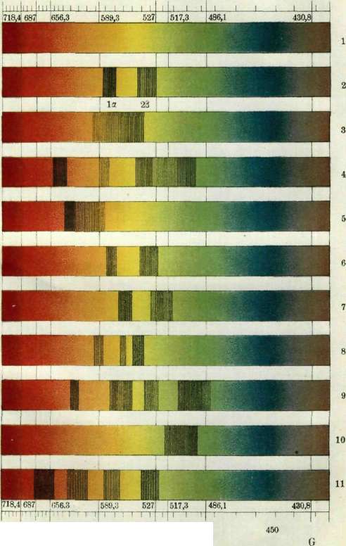
SPECTRAL ANALYSIS, one of the methods of analysis in which spectra (see Spectroscopy, spectroscope) are used, produced by various bodies when they are heated or when rays that produce a continuous spectrum are passed through solutions. For the study of solutions and obtaining absorption spectra of any pigments, a sunbeam can also be successfully used as a light source, which itself produces an absorption spectrum, as evidenced by the presence of Fraunhofer lines in it. Fraunhofer lines are narrow strips intersecting the solar spectrum, while the bands of the spectra of pigment solutions have a rather large width and can be easily distinguished from Fraunhofer lines, although essentially both represent traces of rays retained by pigment solutions or heated vapors or gases. Fraunhofer lines were discovered by the German scientist Fraunhofer in 1802. They play a major role for orientation purposes. Thanks to them, the entire spectrum is divided into specific sections, and this helps to determine the location of the investigated bands or lines. The determination of the wavelengths corresponding to different colored rays was made by Fraunhofer using a diffraction grating, and therefore, by observing the solar spectrum along the Fraunhofer lines, it is possible to determine not only the location of the band, but also the wavelengths themselves, expressed in mμ. Spectral analysis was discovered in 1859 by Kirchhoff, and since then the scope of its application has been constantly expanding. Recently, it has begun to find application in clinical laboratories for the study, mainly, of blood and urine for diagnostic purposes. Solutions of the blood pigment hemoglobin (Hb) are capable of absorbing certain rays included in the composition of the white sunbeam, and therefore when viewing such a sunbeam passing through an Hb solution in a spectroscope, we will observe the so-called adsorption spectrum, or Hb absorption spectrum, i.e., along with narrow Fraunhofer lines, we will see rather wide dark strips in various sections of the spectrum with a width of 10-30 mμ. For each pigment, there are its own absorption bands, and from these bands, by their location, one can judge the presence of one or another pigment. Thus, for example, oxyhemoglobin found in the normal blood of a healthy person gives a spectrum with 2 absorption bands I λ = 589-577 mμ and II λ = 556-536 mμ. To obtain this spectrum, it is necessary to dilute the blood at least 200 times. At a lower dilution, both bands can merge and give one continuous absorption band. Undoubtedly, the width of the vessel in which the blood solution is located also plays a role here. When 1-2 drops of ammonium sulfide [(NH4)2S] are added to this solution, the picture changes: the spectrum of reduced Hb with one wide band λ = 596-543 mμ is obtained. In case of carbon monoxide poisoning, the pigment carboxyhemoglobin appears in the blood, characterized by a spectrum with 2 absorption bands shifted somewhat towards the violet end of the spectrum, if compared with the oxyhemoglobin spectrum. The wavelength of the absorbed rays is I λ = 579-564 and II λ = 548-530 mμ. Upon reduction with ammonium sulfide of this solution, the picture should not change if the amount of carboxyhemoglobin is 15 - 20% according to Ziemke and 10% according to Schumm. In case of aniline poisoning, the pigment methemoglobin appears in the blood, again giving a characteristic spectrum. But to detect it, it is necessary to make observations on a denser blood solution, approximately 0.1 cm3 of blood in 0.5 cm3 of H2O. At such a dilution, an absorption band characteristic of methemoglobin will appear in the red region of the spectrum with λ = 630-620 mμ. When diluted with water in the yellow-green and green regions of the spectrum, 2 more bands will appear, coinciding with the bands of oxyhemoglobin, and finally the 4th band, characteristic of methemoglobin, lies between 518-486 mμ. This band is barely noticeable, and therefore the presence of methemoglobin in the blood can be established, as already mentioned, by the presence of a band in the red region of the spectrum. In case of poisoning with arsenic hydride, as well as in other diseases, hemolysis of blood corpuscles can occur in the blood and, in connection with hemolysis, the breakdown of the pigment into globin and hemochromogen; but since there is a large excess of O2 in the blood plasma, hemochromogen is in a state of oxidation—in the form of hematin, and when viewing this pigment in a spectroscope, we will detect a band in the red light with λ = 640-630 mμ. In the region of yellow-green and green colors, there will also be absorption bands, but the characteristic one is the band in red light. When (NH4)2S is added, hematin is reduced and turns into hemochromogen with two absorption bands I λ = 565-554 and II λ = 536-523 mμ. In case the band in red light is due to the presence of methemoglobin, then upon reduction with (NH4)2S, the Hb spectrum with 1 wide band λ = 596-543 mμ will be obtained, and not that of hemochromogen with 2 bands. Among the pigments easily recognizable in the spectroscope, mention should also be made of urobilin and porphyrin. The latter is found in insignificant quantities in the urine of healthy people, but in certain diseases, the amount of this pigment can be significantly increased. Porphyrin is characterized by a spectrum with bands I λ = 597-587 mμ, II λ = shade around 576-565 and III λ = 557 - 541 mμ. It can be obtained from urine in lead poisoning according to Garrod's method, by precipitating 40 cm3 of 10% NaOH from 200 cm3 of urine (porphyrin is carried down by the precipitating phosphates). The washed precipitate is then dissolved in 0.5-1.0 cm3 of 25% HCl, and the absorption spectrum is viewed in a spectroscope. In case of sulfonal poisoning, in congenital porphyria, porphyrin appears in the urine, very similar in spectrum, but different in chemical properties from porphyrin—the so-called uroporphyrin. Urobilin, found in urine in certain diseases, is characterized by a spectrum with a wide absorption band λ = 510 - 490 mμ. A pocket spectroscope is very convenient for such an analysis (see Spectroscopy, spectroscope).
K. Lavrovsky. Biological spectral analysis is an extremely interesting and important new method for studying the fine metabolism of tissues and cells without disrupting their structure. The application of biological spectral analysis became possible thanks to the discovery of mitogenetic rays (see). It was established that various chemical processes underlying the emergence of these rays qualitatively differ from one another by the set of wavelengths (lines) inherent to them. Thus, for each chemical radiation source there is a characteristic spectrum. When studying a source of mitogenetic radiation, it is currently necessary to know its spectrum. By comparing the obtained picture with already studied templates of the main radiation sources (glycolysis, proteolysis, splitting of phosphoric acid, etc.), it is possible in each specific case to clarify the nature of the processes underlying the given radiation. Complex physiological radiation sources turned out to contain essentially a whole set of simple chemical processes—glycolytic, proteolytic, etc. Subsequently, it turned out that the fluctuations of spectral patterns are extremely subtle and allow conclusions to be drawn whose significance goes far beyond the problem of mitogenesis, being of essential importance for physiology, chemistry, and other disciplines. Thus, by means of spectral analysis, subtle differences were established in the chemistry of nerve excitation caused by various stimuli—thermal, mechanical, etc.; a completely peculiar spectrum is obtained in the case of physiological excitation. The character of metabolism at the site of stimulation differs spectrally from the site of conduction, and so on. Certain data of spectral analysis are of interest in solving a number of problems of biological and general chemistry. Of particular interest is the fact that the so-called secondary radiation (see Mitogenetic rays) is resonant, i.e., responds spectrally to the spectrum of primary irradiation; it is very important to note that under monochromatic action, a single line of a given spectrum is sufficient to secondary evoke the entire spectrum as a whole. This phenomenon, the study of which is only now beginning, is of high theoretical interest. The experimental technique itself is extremely simple and boils down to the fact that the studied radiation source is located in front of the entrance slit of a quartz spectrograph, in the exit plane of which, at places corresponding to various wavelengths and marked by a special scale, a radiation detector—a liquid or solid yeast culture—is located. By the presence or absence of an effect in one or another detector, one can judge the presence of various lines of the spectrum. By comparing with chemically simplest sources, the content of this spectrum is clarified. This method, which allows investigating the spectral content of a source with an accuracy of up to 10 Å, can be significantly refined when working with a monochromator movable slit equipped with a scale and allowing to cut out sections several (1–3) Å wide along the entire extent of the spectrum. This same method leads to a B C
500 450 ' Oxyhemoglobin Hemoglobin Methemoglobin (neutr. sol.) Hematine acid sol. Hematine alk. sol. Carboxyhemoglobin Hemochromogen Hematoporphyrin ACID SOL. Hematoporphyrin (alk. sol.) Urobilin (ammon. sol.+ZnCl2) Chlorophyll
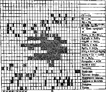
revising the initial coarse spectrum. It turns out that in many broad lines, the mitogenetically active component is a strip a few angstroms wide, while the remaining sections (bands) are mitogenetically empty. To obtain general preliminary results, templates are used—the arrangement of the detector only at a few points of the spectrum corresponding to the main chemical processes. The main spectrally investigated radiation sources (see figure): 1) Glycolysis—its best-studied sources are: a) lactic acid fermentation, b) hemolyzed blood with the addition of glucose, c) alcoholic fermentation, etc. The coincidence of the spectra of these chemically very different processes suggests that the glycolytic radiation is associated with the first stage of the process—the breakdown of the glucose molecule into its two constituent trioses; only in this initial stage does the chemistry of such processes as, for example, lactic acid and alcoholic (yeast) fermentation coincide; the further course of glycolysis in various cases is different. The most characteristic lines for glycolysis are the following: 1,900–20 Å, 1,940–50 Å, 1,960–70 Å, 2,170–80 Å. 2) Proteolytic spectrum—an example is the digestion of fibrin or serum albumin by gastric juice and dipeptides (glycyl-glycine) by erepsin. The coincidence in the spectra of these two processes forces us to associate the radiation with the moment common to them of the cleavage of the NH2 group. The most characteristic lines are 1,980–90 Å, 2,030–50 Å, 2,110–30 Å, 2,300–10 Å, 2,340–50 Å, 2,390–2,400 Å, 2,410–20 Å. 3) Phosphatase spectrum—the action of phosphatase on lecithin and nucleic acid was studied as an object. The most characteristic lines studied on the phosphatase of a cancer cell are 2,150–60 Å, 2,240–50 Å, 2,280–90 Å, 2,350–60 Å, 2,460–80 Å, 2,480–2,500 Å—the longest of the lines of mitogenetic radiation known to us so far. The action of liver phosphatase shows new lines—1,980–90 Å, 1,990–2,000 Å. 4) Spectrum of di- and polysaccharide breakdown—maltose and sucrose were used as objects; in accordance with the difference in their chemical structure, differences in the spectral pattern were also obtained. These differences allow us to approach the question of the structure of polysaccharide (starch); the coincidence of its spectrum with that of maltose allows us to assert that it is a polymer of the latter. Lines characteristic of maltose are 1,970–80 Å, 1,980–90 Å, 2,020–30 Å, 2,230–40 Å, 2,320–30 Å, 2,370–80 Å, 2,400–10 Å, 2,410–20 Å, 2,430–40 Å; sucrose is characterized by the absence of the first two lines. 5) The spectrum of creatine phosphoric acid breakdown is found in a number of physiological radiation sources—in muscles, nerve, flowing blood, etc., and is characterized by lines 2,000–20 Å, 2,030–60 Å, 2,090–2,110 Å, etc. 6) The spectrum of the enzyme urease (which causes the breakdown of urea) coincides with the absorption and destruction spectra of this substance; the most characteristic lines are 1,940–50 Å, 1,950–60 Å, 2,040–50 Å, 2,050–60 Å, 2,080–90 Å, 2,290–2,300 Å.
2,000
2,500 7) The spectrum of oxidative processes was studied on the oxidation of pyrogallol in an alkaline medium, the oxidation of glucose by permanganate, and blood serum by hydrogen peroxide, and especially on inorganic oxidation models, e.g., K2Cr2O7+FeSO4, HgCl2+SnCl2, etc. (Braunshtein and Pototskaya). In all these cases, oxidative processes are understood in the broadest sense as processes of electron exchange between two chemical systems (oxidation-reduction processes); special experiments show that the moment of radiation is associated with the process of attachment of electrons to the system, i.e., with reduction. The spectra of various oxidation reactions are very similar, but not identical; lines in the middle part of the spectrum are characteristic; for pyrogallol oxidation, for example, lines 2,250–70 Å and 2,280–2,300 Å are typical. A number of lines found in certain physiological radiation sources cannot yet be chemically identified at present. S. Zalkind.
Related articles
Mentioned in
Cite this page
“Spectral Analysis.” Soviet Medical Encyclopedia. English translation of Bolshaya Meditsinskaya Entsiklopediya, 1st ed. (Moscow, 1928–1936), ed. N. A. Semashko. https://sovietmedicalencyclopedia.com/article/spectral-analysis/