Laryngofissure
Historical document, translated for reference. It reflects medical knowledge of the 1920s–30s and is not medical advice.
Summary
A historical overview of the surgical procedure known as laryngofissure (or laryngotomy), detailing the techniques for complete and partial dissection of the larynx. The article covers indications, anesthesia methods, surgical steps, and postoperative care, including variations like transverse laryngotomy and combined pharyngolaryngotomy.
Encyclopedia article (1928–1936)
LARYNGOFISSURE (from Greek larynx-larynx and Latin fissura-cleft), dissection of the larynx; it is more correct to call this operation laryngotomia, since laryngofissure—a cleft of the larynx—is already the result of the operation. A distinction is made between complete and partial laryngotomy; in the former, the thyroid and cricoid cartilages, the cricothyroid ligament, and the upper rings of the trachea are dissected along the midline; in the latter, the cricothyroid ligament, the cricoid cartilage, and the upper rings of the trachea are dissected. (It is preferable to perform a partial laryngotomy, as the vocal cords are not injured during it, and the voice does not suffer.) A transverse laryngotomy, according to Gersuny, is also used, in which a transverse incision of the larynx is made above the region of the vocal cords, and a longitudinal incision along the midline is added above it upwards. For small lesions of the larynx, it is sometimes sufficient to dissect only the thyroid cartilage. Indications for laryngotomy include foreign bodies, neoplasms, cicatricial stenoses, injuries of the larynx, and tuberculosis. The operation is performed under local anesthesia with the head hanging down or in the Trendelenburg position, and the patient is placed in this position only at the moment of opening the trachea to avoid increasing suffocation due to venous congestion. Technique of the operation. In a complete laryngotomy, the skin incision is made along the midline from the hyoid bone down to the jugular notch; following the skin, the fascia is dissected, and it is necessary to ligate and cut several small arteries running along the cricothyroid membrane and at the isthmus of the thyroid gland. Having pushed the muscles to the side, the hyothyroid ligament, the thyroid cartilage, the cricothyroid ligament, and the cricothyroid cartilage are exposed; to expose the trachea, it is necessary to bluntly separate the isthmus
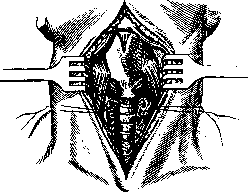
Figure 1. First moment of the operation.
of the thyroid gland, ligate it in two places, and cut it (Fig. 1); it is even better to perform the exposure of the trachea according to Bose: the deep fascia of the gland, attached to the cricoid cartilage, is separated from the cartilage by a transverse incision and bluntly pushed downwards together with the gland. Then the trachea is opened, the edges of the incision are spread apart with blunt hooks, and through the resulting opening, the inner surface of the trachea and larynx is swabbed with 20-percent cocaine-adrenaline. A tracheostomy tube is inserted, and one waits 5–10 minutes for the onset of complete anes
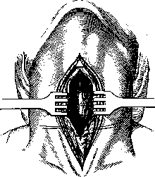
Figure 2. Second moment of the operation.
thesia, after which, with strong scissors (or, in case of ossification of the cartilage, Liston's scissors), the cricoid and then the thyroid cartilages are dissected, trying to keep strictly to the midline so as not to damage the vocal cords. Having spread the halves of the thyroid cartilage with hooks (Fig. 2), the interior of the larynx is examined, the changes found there are determined, and one proceeds according to what is found. Upon completion of the intralaryngeal operation, the wound is sutured: the perichondrium and muscles with catgut, the skin with silk; if there is no bleeding and breathing is free, the trachea is also sutured, placing a strip of gauze in the lower corner of the wound; if there is a suspicion of the possibility of stenosis, it is better to insert a cannula for a few days. If the intralaryngeal intervention was quite extensive and there is a danger of bleeding, it is safer to tampon the laryngeal cavity and bring the tampon out above the tracheostomy tube; after the removal of the tampon and cannula, the opening in the trachea closes with granulations. Where the laryngeal lesions are deeper and where the postoperative wound requires prolonged observation and treatment, the split larynx is not sutured, and treatment of the open larynx is conducted; laryngotomy is thus transformed into laryngostomy (see). In partial laryngotomy, the skin incision begins slightly above the lower edge of the thyroid cartilage, and the cricothyroid membrane, cricoid cartilage, and upper rings of the trachea are dissected. Instead of an ordinary tracheal cannula, various laryngotracheal cannulas constructed on the principle of T-shaped tubes (Dupuis, Schimmelbusch, Killian) have been proposed for laryngotomy. When the tumor is located closer to the laryngeal inlet, it is necessary to combine laryngotomy with pharyngotomy (pharyngo-laryngotomia subhyoidea mediana, according to Kocher); in this case, the larynx is split in the middle, the sternohyoid and thyrohyoid muscles are cut at the hyoid bone, then the thyrohyoid membrane is cut, the hyoid bone is strongly pulled upwards with a hook, and the mucous membrane of the pharynx is opened; thus, a wide access to the pharyngeal space and the laryngeal inlet is obtained.
Related articles
Mentioned in
Cite this page
“Laryngofissure.” Soviet Medical Encyclopedia. English translation of Bolshaya Meditsinskaya Entsiklopediya, 1st ed. (Moscow, 1928–1936), ed. N. A. Semashko. https://sovietmedicalencyclopedia.com/article/laryngotomy/