Human Nerves
Historical document, translated for reference. It reflects medical knowledge of the 1920s–30s and is not medical advice.
Summary
This article provides a systematic anatomical and physiological overview of human peripheral nerves, detailing their origins, segmental relationships, branching, and clinical significance. It includes a detailed table covering specific cranial nerves, such as the abducens, accessory, and auditory nerves, describing their pathways and functions.
Encyclopedia article (1928–1936)
HUMAN NERVES. [For the anatomy, physiology, and pathology of the nerve, see the article Nerves in volume XX; in the same place (pp. 667–782) are drawings of Human Nerves]. Below is a table of nerves, illuminating in systematic order the most important points of the anatomy and physiology of each peripheral nerve (its origin, relation to segments, course, branching, connections with other nerves, point of termination, general topography, function, variations). * When examining a patient, familiarity with all these data is necessary for making a topical diagnosis of a disease of a given nerve and especially for surgical intervention upon it. * * Bilateral symmetry, observed in spinal nerves, and the presence of a spinal ganglion at the beginning of each nerve allow us to consider the nerve as a segmental formation that has preserved quite clearly traces of metamerism, which has almost disappeared in the central nervous system, where it can be traced only with the help of comparative anatomical and embryological studies. A nerve segment consists of a central organ from which roots emerge—an anterior (motor) and a posterior (sensory); the latter has a thickening on its path—the intervertebral ganglion, outward from which both roots join, forming a mixed spinal nerve—nervus spinalis, which divides into 4 branches (see Nerves). Each nerve segment innervates the corresponding segment of the body. This innervation is symmetrical on both sides, but on both the anterior and posterior surfaces, there is a transition of branches to the other side for a short distance (into the skin and muscles). The nerve segment in the indicated form is preserved only in the thoracic region, while in other sections (in the cervical, lumbar-sacral) there is a displacement of nerves within significant limits (formation of plexuses—see Nerve Plexuses), and only through careful study is it possible to establish the connection of any peripheral nerve with the corresponding segments of the spinal cord. The second important point noted in the table is the anastomoses between nerves. True, the term "anastomoses" in relation to nerves can be used only very conditionally (for types of anastomoses—see Nerves, vol. XX). The presence of these anastomoses explains how a nerve that is motor in its origin becomes mixed, and also how vegetative fibers enter cerebrospinal nerves. At their origin, nerves lie deep and close to the skeleton, then, with few exceptions, become more superficial. Fibers in a nerve are arranged in a known order: short fibers for collaterals occupy a peripheral position, while terminal long fibers are located in the center; therefore, in a mixed nerve, sensory fibers occupy a central position in most nerves, while motor fibers are located more peripherally (see also Nerve Fibers). Such a deep position of sensory fibers in a nerve explains the preservation to a greater or lesser degree of sensitivity during contusion or compression of the corresponding nerve and the disturbance of sensitivity during the transection of a nerve. As for the anatomy of the nerve, here it is necessary to note a very important fact—the presence of variations and anomalies that have great significance in the clinic. Anomalies in the formation of a nerve are expressed in the fact that the nerve originates not from the roots as usual. As for anastomoses, the latter may either be absent or, conversely, be in greater quantity or in other places along the length of the nerve. Variations in relations to surrounding formations, in particular to vessels, must be taken into account during operations on nerves, when it is easy to injure vessels located in an unusual place. Anomalies in distribution are more often observed for sensory nerves, less often for motor ones. Nerves: Latin Name, Russian Name, Synonyms, Origin, To which segment it corresponds, With which nerves it anastomoses, Which branches depart from it, What they innervate and where the nerve ends, Topography of the nerve and its branches, Character of the nerve and its function, Variations. 1. Abducens (abducting). Syn.: oculo-muscularis externus, par sextum nn. encephali. Medulla oblongata—rhomboid fossa, colliculus facialis. Exits along the posterior edge of the pons between it and the pyramid. Sympathetic fibers from the sinus cavernosus and sensory fibers from the n. ophthalmicus. Peripheral. M. rectus lateralis oculi. Exiting from the medulla oblongata along the posterior edge of the pons, the nerve is directed along its ventral surface forward and laterally, pierces the dura mater, lies in the sinus cavernosus medially from the trigeminal nerve and laterally from the oculomotor nerve and laterally from the internal carotid artery, passes through the superior orbital fissure into the orbit, where it lies under the oculomotor nerve, and then between both origins of the m. rectus lateralis oculi and penetrates into this muscle from its medial side. Motor, but sympathetic fibers from the plexus caroticus internus and sensory fibers from the oculomotor nerve also pass along its path. Abducts the eye outward. Accessorius: r. internus and r. externus (accessory nerve, Willis, XI cranial N.). Syn.: Willisi, recurrens, respiratorius externus, par undecimum. The nucleus of the accessory nerve lies: a) in the medulla oblongata: in the mass of the floor of the IV ventricle, laterally from the nucleus n. hypoglossi and the sensory nucleus of the nn. vagi and glosso-pharyngei, behind their motor nucleus—nucleus ventralis, s. ambiguus, being a continuation of the latter backward; the nucleus reaches anteriorly to the posterior third of the olivae inferioris; this nucleus gives the r. internus; b) in the spinal cord: in the anterior horns of the spinal cord dorsolaterally from the medial cell group in the form of a continuous cell mass up to the level of the exit of the V (VI or even VII) cervical nerves; this nucleus gives the r. inferior, s. externus. With the posterior roots of the three upper cervical nerves, receives motor roots from the III (IV) cervical nerve and sensory fibers from the vagus and III and IV cervical. 1) R. internus (accessorius vagi), 2) r. externus (accessorius spinalis). 1) Follow the path of the n. vagi, 2) m. sterno-cleido-mastoideus and m. trapezius. Exiting from the medulla oblongata by two or three bundles from the sulcus lateralis posterior dorsally from the olive and behind the roots of the vagus nerve (ramus internus) and by several bundles from the spinal cord between the posterior and anterior roots of the upper cervical nerves (ramus externus) and having merged into one trunk, the nerve in one common sheath of the dura mater together with the vagus nerve passes through the jugular foramen and divides outside the cranial cavity again into both its branches, which diverge: the internal branch passes into the nervus vagus and follows together with it into the rami pharyngei, laryngei and cardiaci n. vagi; the external branch, having received sensory fibers from the vagus nerve and its jugular nucleus, is directed outward, backward, and downward, lies laterally from the vagus nerve and behind the internal jugular vein, reaches the medial surface of the musculi sterno-cleidomastoidei, pierces the muscle, or, having given it part of its branches, passes with its other branches through the regio lateralis colli to the musculus levator scapulae, reaches the musculus trapezius, lies on its medial surface and enters its mass, on the border of the middle and lower thirds. Motor, but sympathetic fibers from the plexus caroticus internus and sensory fibers from the oculomotor nerve pass along its path. Acusticus: radix vestibularis et radix cochlearis (auditory nerve: vestibular root, or vestibular nerve, or static nerve, or nerve of equilibrium, and cochlear root, or nerve of the cochlea or auditory nerve proper). Radix vestibularis begins in the nuclei of the vestibule from their ganglion cells; the peripheral processes of these cells constitute five nerves and end in the following five places: 1. Ampulla membranacea superior of the superior semicircular canal—its neuroepithelium (crista ampullaris)—this is nervus ampullaris superior. 2. Ampulla membranacea lateralis of the lateral semicircular canal—its neuroepithelium (crista ampullaris)—this is n. ampullaris lateralis. 3. Ampulla membranacea posterior of the posterior semicircular canal—its neuroepithelium (crista ampullaris)—this is n. ampullaris posterior. 4. Utriculus membranaceus—its neuroepithelium (macula acustica utriculi)—this is nervus utricularis. 5. Sacculus membranaceus—its neuroepithelium (macula acustica sacculi)—this is n. saccularis. The central fibers of these ganglion cells in the form of radix, s. n. vestibularis go as part of the auditory nerve into the medulla oblongata and end in three nuclei. 1) R. communicans (Arnold!) externus and 2) internus, both—with nervus facialis. I. Radix, s. n. vestibularis: 1) n. ampullaris superior, 2) n. ampullaris lateralis, 3) n. ampullaris posterior, 4) n. utricularis, 5) n. saccularis. II. Radix, s. n. cochlearis.
The nerve may divide into two, may give a branch to the ganglion ciliare, or may be absent. It is purely motor, but sensory fibers from the vagus nerve and from the cervical nerves also pass along its path. The r. internus, traveling along the path of the vagus nerve, takes part in the innervation of the pharynx, larynx, and heart. The r. externus innervates the m. sterno-cleido-mastoideus and the musculus trapezius. The nerve may end in the m. sterno-cleido-mastoideus or the ramus externus, may divide into two independent branches for each muscle separately, and may anastomose with the ramus descendens nervi hypoglossi, with the n. phrenicus, and with the ganglion cervicale superius. Emerging from the medulla oblongata caudally from the pons and laterally from the facial nerve, the nerve is covered together with the facial nerve by one common process of the dura mater, enters the porus and meatus acusticus internus, connects here with the facial nerve by means of fila anastomotica, and terminates with both its roots in the aforementioned places. In the depth of the meatus acusticus internus, it is divided by the crista transversa into two levels; in the upper one, which in turn also divides into two fossae, the n. facialis passes in the anterior fossa—area nervi facialis, and in the posterior—nervus vestibularis—area nervi vestibularis superior; in the lower one—the radix cochlearis: in the anterior fossa—area cochleae, which has the shape of a spiral row of openings—tractus spiralis foraminosus, and part of the fibers of the r. vestibularis, in the posterior fossa—area vestibularis inferior. It is sensory. The radix vestibularis serves in the statics of the organism as a connection between the peripheral static apparatus (vestibule and semicircular canals) on one side and the brain on the other. The radix cochlearis—auditory nerve. No. Latin and Russian name—Synonyms. Origin. Which segment it corresponds to. With which nerves it anastomoses. Which branches depart from it. Rhomboid fossa: nucleus medialis (Schwalbe), lateralis (Deiters), et superior (Bechterew). The radix cochlearis begins in the spiral nucleus of Corti, which is located in the mass of the bony spiral lamina of the cochlea—lamina spiralis ossea—in its canalis spiralis modioli; the peripheral processes of these cells go in the mass of the spiral lamina of the cochlea in its fissura spiralis to the cells of the organ of Corti; the central fibers of these ganglionic cells are directed in the form of the radix, s. n. cochlearis into the medulla oblongata as part of the auditory nerve and terminate in two nuclei of the rhomboid fossa: nucleus ventralis et nucleus dorsalis, s. tuberculum acusticum. Alveolares inferiores anteriores (lower anterior alveolar nerves) 5| Alveolares inferiores posteriores (lower posterior alveolar nerves) See n. alveolaris inferior. See n. alveolaris inferior. 6| Alveolares superiores: posteriores, medius et anterior (upper alveolar nerves: posterior, middle, and anterior). Syn.: nn. dentales superiores: anterior, medius et posteriores. Nervus infraorbitalis throughout the extent from the pterygopalatine fossa to the infraorbital foramen. 1) Rr. alveolares superiores posteriores, 2) r. alveolaris superior medius, 3) r. alveolaris superior anterior. 7| Alveolares superiores posteriores (posterior upper alveolar branches). See nn. alveolares superiores. Alveolaris inferior (lower alveolar nerve). Syn.: dentalis inferior, s. mandibularis. N. lingualis. N. mandibularis. 1) N. mylo-hyoideus (see), 2) nn. alveolares inferiores posteriores (lower posterior alveolar nerves), 3) nn. alveolares inferiores anteriores (lower anterior alveolar nerves).
1) What they innervate and where the nerve ends. Topography of the nerve and its branches. Character of the nerve and its function. Variations. See n. alveolaris inferior. See n. alveolaris inferior. They take part in the formation of the upper dental plexus and give branches: a) r. dentales superiores and b) r. gingivales superiores. 1. Rami alveolares superiores posteriores lie on the facies posterior of the upper jaw—on its tuber maxillare, pierce the bone, enter the maxillary sinus, go along its posterior lateral wall, form the upper dental plexus, which lies partly under the mucous membrane of the sinus and partly in the mass of the bone, and give branches to the mucous membrane of the maxillary sinus, the posterior part of the cheek, to the alveoli of the molars, and to the gums around them. 2. Ramus alveolaris superior medius begins in the infraorbital canal, enters the maxillary sinus, participates in the formation of the upper dental plexus, and terminates in the alveoli of the premolars and in the corresponding parts of the gum. 3. Ramus alveolaris superior anterior begins in the infraorbital canal near its exit foramen, enters the maxillary sinus, goes along its anterior wall, participates in the formation of the upper dental plexus, and terminates in the alveoli of the incisors and canines, in them themselves, and in the corresponding parts of the gum and in the mucous membrane of the nasal cavity—ramus nasalis. Sensory. Innervates the mucous membrane of the cheek in its posterior part, the mucous membrane of the maxillary sinus, the gums of the upper jaw, the teeth of the upper jaw, and also partly the mucous membrane of the nasal cavity. See nn. alveolares superiores. 1) M. mylo-hyoideus et venter anterior m. digastrici and the skin of the chin, 2) molars and premolars and the gum around them, 3) canine and incisors and the gum around them. Having started from the mandibular nerve almost immediately after the latter's exit from the foramen ovale, the nerve is located in the infratemporal fossa between the mm. pterygoideus externus et internus, then lies on the musculus pterygoideus internus—on its lateral surface, between it and the ramus of the lower jaw, next to the nervus lingualis, crosses the chorda tympani, lying deeper than it, and the arteria maxillaris interna, lying more superficially than it; having given off 1) nervus mylo-hyoideus, it enters further through the foramen mandibulae into the canalis mandibulae, here it gives 2) nn. alveolares inferiores posteriores et 3) anteriores, and then exits. Mixed, but after the departure of the n. mylo-hyoideus, into which all its motor fibers enter, it becomes exclusively sensory. Transmits sensory irritations from the skin of the chin, lower lip, gums, and teeth of the lower jaw. The nerve may begin with two roots, divide early into three branches (even before entering the canalis mandibularis), or divide into two trunks, between which the a. maxillaris interna passes.
No. Latin and Russian name. Synonyms. Origin. Which segment it corresponds to. With which nerves it anastomoses. Which branches depart from it. 4) n. mentalis (mental nerve), which breaks up into a) rr. mentales, b) rr. labiales inferiores. Alveolaris superior anterior (anterior upper alveolar nerve). See nn. alveolares superiores. 10| Alveolaris superior medius (middle upper alveolar nerve). See nn. alveolares superiores. 11| Ampullaris lateralis (lateral ampullary nerve). 12| Ampullaris posterior (posterior ampullary nerve). 13| Ampullaris superior (superior ampullary nerve). Syn.: r. minor n. vestibuli lateralis et posterior, s. inferior, s. r. inferior n. vestibuli. See n. acusticus. See n. acusticus. See n. acusticus. 14| Anastomotici nervi hypoglossi cum plexu cervicali (rr.) (anastomoses of the hypoglossal nerve with the cervical plexus).
Use the English headword: Human Nerves.
(cervical plexus) 151 Anastomotici rami dorsalis manus (n. ulnaris) cum ramo superficiali nervi radialis (branches) [anastomoses of the dorsal branch of the ulnar nerve (digital nerves) with the superficial branch of the radial nerve]. See hypoglossal nerve and cervical nerves. See dorsal digital nerves. 16 Anastomoticus cum plexu tympanico nervi facialis (branch) (anastomosis of the facial nerve with the tympanic plexus). 17 Anastomoticus ganglii otici cum chorda tympani (branch) (anastomosis of the otic ganglion with the chorda tympani). Anastomoticus ganglii otici cum nervo auriculo-temporali (branch) (anastomosis of the otic ganglion with the auriculo-temporal nerve). See facial nerve. See Nerve ganglia (otic ganglion) and chorda tympani nerve. See Nerve ganglia (otic ganglion). Anastomoticus ganglii otici cum n. spinoso (branch) (anastomosis of the otic ganglion with the spinous nerve). See Nerve ganglia (otic ganglion). Anastomoticus nervi auricularis posterioris (branch) (anastomosis of the posterior auricular nerve with the lesser occipital). See posterior auricular nerve and lesser occipital nerve. Anastomoticus nervi digastrici (branch) (anastomosis of the digastric nerve). See digastric nerve and glosso-pharyngeal nerve. What they innervate and where the nerve ends. Topography of the nerve and its branches. Character of the nerve and its function. Variations 4) the skin of the chin and the lower lip from the mandibular canal through the mental foramen in the form of a terminal nerve, which is called 4) mental nerve; the latter immediately divides into 2 groups of branchlets: a) mental branches, which pierce the triangular muscle and the quadratus muscle of the lower lip and branch in the skin of the chin, and b) inferior labial branches, which branch in the skin of the lower lip. See superior alveolar nerves. See superior alveolar nerves. See acoustic nerve. See acoustic nerve. See acoustic nerve. See hypoglossal nerve and cervical nerves. See dorsal digital nerves. See facial nerve. See Nerve ganglia (otic ganglion) and chorda tympani nerve. See Nerve ganglia (otic ganglion). See Nerve ganglia (otic ganglion). See posterior auricular nerve and lesser occipital nerve. See digastric nerve and glosso-pharyngeal nerve. ^ Latin name and No. |
Russian. Synonyms 22 Anastomoticus nervi facialis cum nervo auriculari vagi (branch) (anastomosis of the facial nerve with the auricular branch of the vagus). 23 Anastomoticus nervi hypoglossi cum ganglio cervicale superiore (branch) (anastomosis of the hypoglossal nerve with the superior cervical ganglion). Origin. To which segment it corresponds. With which nerves it anastomoses. Which branches depart from it. See facial nerve and vagus nerve. See hypoglossal nerve. Anastomoticus nervi hypoglossi cum ganglio nodoso nervi vagi (branch) (anastomosis of the hypoglossal nerve with the nodose ganglion of the vagus nerve). See hypoglossal nerve. 28! Anastomoticus nervi lacrimalis (branch) (anastomosis of the lacrimal nerve with the zygomatic). 26 Anastomoticus nervi laryngei superioris cum ganglio cervicali superiore (branch) (anastomosis of the superior laryngeal nerve with the superior cervical ganglion). Anastomoticus nervi laryngei superioris cum nervo laryngeo inferiore (branch) (anastomosis of the superior laryngeal nerve with the inferior laryngeal). Anastomoticus nervi laryngei superioris cum plexu carotico commune (branch) (anastomosis of the superior laryngeal nerve with the common carotid plexus). See superior laryngeal nerve. See superior laryngeal nerve. See superior laryngeal nerve. 29; Anastomoticus nervi laryngei superioris cum plexu pharyngeo (branch) (anastomosis of the superior laryngeal nerve with the pharyngeal plexus).
[plexus]) See superior laryngeal nerve. Anastomoticus nervi lingualis cum chorda tympani (branch) (anastomosis of the lingual nerve with the chorda tympani). See lingual nerve and chorda tympani. 311 Anastomoticus nervi lingualis cum ganglio submaxillare (branch) (anastomosis of the lingual nerve with the submaxillary ganglion). See lingual nerve. Anastomoticus nervi lingualis cum nervo alveolari inferiore (branch) (anastomosis of the lingual nerve with the inferior alveolar nerve). Anastomoticus nervi lingualis cum nervo hypoglosso (branch) (anastomosis of the lingual nerve with the hypoglossal). See lingual nerve. See lingual nerve. 34 Anastomoticus nervi mediani cum nervo musculo-cutaneo (branch) (anastomosis of the median and musculo-cutaneous nerve). 351 Anastomoticus nervi oculomotorii cum nervo ophthalmico (branch) (anastomosis of the oculomotor nerve with the ophthalmic).
nerve with the ophthalmic). See oculomotor nerve and ophthalmic nerve. What they innervate and where the nerve ends. Topography of the nerve and its branches. Character of the nerve and its function. Variations. See facial nerve and vagus nerve. See hypoglossal nerve. See hypoglossal nerve. See lacrimal nerve and zygomatic nerve. See superior laryngeal nerve. See superior laryngeal nerve. See superior laryngeal nerve. See superior laryngeal nerve. See lingual nerve and chorda tympani. See lingual nerve. See lingual nerve. See lingual nerve. See median nerve. See oculomotor nerve and ophthalmic nerve. 1 i No. i Latin and Russian name. Synonyms. Origin. To which segment it corresponds. With which nerves it anastomoses. Which branches depart from it. Anastomoticus nervi oculomotorii cum plexu cavernoso (branch) (anastomosis of the oculomotor nerve with the cavernous plexus). See oculomotor nerve. Anastomoticus nervi ophthalmici cum nervo abducente (branch) (anastomosis of the ophthalmic nerve with the abducens). See ophthalmic nerve and abducens nerve. Anastomoticus nervi ophthalmici cum nervo trochleari (branch) (anastomosis of the ophthalmic nerve with the trochlear). See ophthalmic nerve and trochlear nerve. 39! Anastomoticus nervi peronaei profundi (branch) (anastomosis of the deep peroneal nerve with the superficial peroneal nerve). See deep peroneal nerve. 40 Anastomoticus nervi recurrentis (vagi) cum ganglio cervicale medio et inferiore (branch) (anastomosis of the recurrent nerve with the middle and inferior cervical ganglia). See vagus nerve and recurrent. Anastomoticus nervi vagi cum nervo glosso-pharyngeo (branch) (anastomosis of the vagus nerve with the glosso-pharyngeal). See vagus nerve and glosso-pharyngeal nerve. Anastomoticus nervi vagi cum nervo hypoglosso (branch) (anastomosis of the vagus nerve with the hypoglossal). See vagus nerve and hypoglossal nerve. Anastomoticus peronaeus nervi ischiadici (branch) (peroneal anastomosis). See sciatic nerve, medial sural cutaneous and sural nerves. Anastomoticus peronaeus (branch) (anastomosis of the peroneal nerve to the lateral cutaneous nerve of the calf). See lateral and medial sural cutaneous nerves. Anastomoticus rami volaris manus (n. ulnaris) cum n. mediano- r. superficialis (n. digitalis volaris communis III) (branch) (anastomosis of the palmar branch of the ulnar nerve to the superficial branch of the median nerve). See ulnar nerve. Anastomoticus ulnaris (nervi radialis) (branch) (anastomosis of the ulnar and radial nerves). See radial and ulnar nerves. Anococcygeus (anococcygeal nerve). Ansa hypoglossi (loop of the hypoglossal nerve). Syn.: descending cervical nerve, or descending hypoglossal, or descending cervical, or descending ninth. Coccygeal plexus-sacrococcygeal loop. Sympathetic nerves from sacral ganglion and coccygeal ganglion. 1) Muscular branches, 2) cutaneous branches. See hypoglossal nerve. What they innervate and where the nerve ends. Topography of the nerve and its branches. Character of the nerve and its function. Variations. See oculomotor nerve.
d. I Latin name and No. Russian. Synonyms. Origin. To which segment it corresponds. With which nerves it anastomoses. Aortales (aortic nerves). 50 Arteriae femoralis proprius (proper nerve of the femoral artery). 51 Articulares nervi auriculo-temporalis (branches) (branches of the auriculo-temporal nerve to the temporomandibular joint). Articulares nervi cutanei dorsalis (pedis) lateralis (branches) (branches of the dorsal lateral cutaneous nerve to the ankle joint). 53 Articulares nervi glutaei inferioris (branches) (branches of the inferior gluteal nerve to the hip joint). Articulares nervi ischiadici (portionis peronaeae) (branch) [branches of the sciatic nerve (peroneal portion) to the knee joint]. Which branches depart from it. See sympathetic nerve. See femoral nerve. See auriculo-temporal nerve. See dorsal lateral cutaneous nerve (of the foot). See inferior gluteal nerve. See sciatic nerve. Articulares nervi masseterici (branches) (branches of the external masticatory nerve to the temporomandibular joint). See masseteric nerve. 56 Articulares nervi mediani (branches) (branches of the median nerve to the elbow [joint]).
(to the elbow joint) See n. medianus. Articular branches of the obturator nerve (branches of the obturator nerve to the hip joint) 58i Articular branches of the common peroneal nerve (branches of the common peroneal nerve to the talocalcaneal joint) See n. obturatorius See n. ischiadicus and n. peronaeus communis 50; Articular branches of the superficial peroneal nerve (branches of the superficial peroneal nerve to the talocalcaneal joint) See n. peronaeus superficialis 60 Articular branches of the ulnar nerve (branches of the ulnar nerve to the elbow joint) See n. ulnaris Articular branch of the axillary nerve (branch of the axillary nerve to the shoulder joint) . 62j Articular branch of the sciatic nerve (branch of the sciatic nerve to the hip joint) See n. axillaris See n. ischiadicus Articular branch of the femoral nerve (articular branch) See n. femoralis 64 Ascending (branch) (ascending branch) See n. cervicalis secundus 8
HUMAN NERVES
34 What they innervate
and where the nerve ends
Topography of the nerve and its branches See n. sympathicus See n. femoralis See n. auriculo-temporalis See n. cutaneus dorsalis (pedis) lateralis See n. glutaeus inferior See n. ischiadicus See n. massetericus See n. medianus See n. obturatorius See n. ischiadicus and n. peronaeus communis See n. peronaeus superficialis See n. ulnaris See n. axillaris See n. ischiadicus See n. femoralis See n. cervicalis secundus Character of the nerve and its function. Variations № Latin and Russian name. Synonyms Origin. To which segment it corresponds With which nerves it anastomoses Which branches depart from it Auriculares anteriores (anterior auricular nerves) See n. auriculo-temporalis Auricularis magnus (great auricular nerve) Plexus cervicalis—CII-CIII 1) Ramus anastomoticus cum n. accessorio, 2) r. posterior cum n. auriculari posteriore n. facialis et n. occipitali minore.
I 1) R. anterior, posterior, s. mastoideus Auricularis (branch of the posterior auricular nerve of the facial nerve) (branch) (auricular branch of the posterior auricular nerve) See n. auricularis posterior Auricularis nervi vagi (branch) (auricular branch of the vagus nerve) See n. vagus Auricularis posterior (posterior auricular nerve). Syn.: auricularis profundus, s. inferior, s. auriculo-occipitalis Nervus facialis in the space between foramen stylo-mastoideum and glandula parotis 1) R. auricularis nervi vagi, 1) Ramus anterior,
auricularis, 2) n. auricularis magnus (plexus cervicalis), 3) n. occipitalis minor (plexus cervicalis) 2) r. occipitalis, 3) the above-mentioned anastomoses Auriculo-temporalis (auriculotemporal). Syn.: nervus temporalis superficialis, s. cutaneus N. mandibularis 1) Ganglion oticum and further through the mediation of n. petrosus superficialis minor n. glosso-pharyngei (secretory fibers for the parotid gland), 2) n. facialis, 3) peripheral branches with peripheral branches from nn. frontalis, facialis et occipitalis 1) Rr. articulares, 2) rr. parotidei, 3) nn. meatus auditorii externi: superior et inferior et r. membranae tympani, 4) nn. auriculares anteriores, 5) rr. temporales superficiales Axillaris (axillary). Syn.: circumflexus humeri (brachii) Plexus brachialis—its fasciculus posterior. CV, CVI, CVII 1) R. muscularis, then divides into a) r. posterior, which gives: 2) r. muscularis, 3) n. cutaneus brachii (humeri) lateralis, 4) r. muscularis; and b) r. anterior, which gives: 5) rr. musculares, 6) rr. musculares, 7) r. articularis humeri, 8) r. intertubercularis (Rauber), 9) r. muscularis, 10) rr. cutanei perforantes
What they innervate and where the nerve ends Topography of the nerve and its branches Character of the nerve and its function. Variations See n. auriculo-temporalis 1) Skin over the regio parotideo-masseterica, the earlobe and the concave surface of the auricle, 2) skin on the nape behind the ear and the convex surface of the auricle. Emerging from under the m. sterno-cleido-mastoideus at the middle of the length of its posterior border, somewhat lower than the nervus occipitalis minor, the nerve lies on this muscle, on its lateral surface, crosses the muscle, lying under the superficial fascia of the neck and m. platysma behind the v. jugularis externa, and at the level of the lower border of the parotid gland divides into its: 1) r. anterior and 2) r. posterior, which with their peripheral branches spread in the skin over the region of the parotid gland and in the skin of the auricle. Sensory. Transmits sensory irritations from the region of the parotid gland and the auricle. Anastomosis with n. facialis can be at different levels; N. can start from the loop between the anterior branches of CII and CIII. See n. auricularis posterior. See n. vagus. 1) Muscles of the auricle: a) m. auricularis posterior, b) m. auricularis superior, c) m. transversus auriculae, d) m. obliquus auriculae, e) m. antitragicus, 2) m. occipitalis. Having started with two roots from the facial nerve immediately upon its exit from the foramen stylo-mastoideum, the nerve passes along the posterior surface of the processus mastoidei, then in the slit between the m. sterno-cleido-mastoideus behind and the muscles starting from the styloid process in front (Riolan's fan), emerges at the level of the base of the earlobe under the superficial fascia and divides into both its branches, heading toward the muscles. Motor. Innervates the muscles that move the auricle upward and backward, and the occipital muscle. Variations of the nerve reduce to the number of roots by which it departs from the n. mandibularis. 1) Joint of the lower jaw, 2) parotid gland, 3) wall of the external auditory canal, 4) skin of the anterior surface of the auricle, 5) skin of the temporal region. Having started from the posterior end of the trunk of the n. mandibularis with two roots, between which the a. meningea media passes, the nerve goes in the lateral direction around the neck of the articular process of the lower jaw immediately under the external auditory canal, gives here 1) rr. articulares for the joint of the lower jaw, 2) rr. parotidei for the parotid gland and 3) rr. meatus auditorii externi (superior et inferior) et ramus membranae tympani for the skin of the external auditory canal, enters the parotid gland, rises along the anterior border of the base of the ear upward, lying behind the a. temporalis superficialis immediately under the superficial fascia, gives 4) rr. auriculares anteriores for the skin of the anterior surface of the auricle and finally with its peripheral branches 5) rr. temporales superficiales branches in the skin of the temporal region. Sensory, but also carries secretory fibers for the parotid gland. 1) Transmits sensory irritations from the joint of the lower jaw, from the skin of the anterior surface of the auricle, the external auditory canal, the skin in the region of the temporal fossa and from the tympanic membrane. 2) Carries secretory fibers to the parotid gland. 1) M. subscapularis, its most lateral portion, 2) m. teres minor, 3) skin of the arm in the region of the deltoid muscle, 4) m. deltoideus (portio spinata), 5) m. deltoideus (portio acromialis), 6) m. deltoideus (portio clavicularis), 7) shoulder joint, 8) shoulder joint, 9) m. pectoralis (portio clavicularis), 10) skin over the deltoid muscle. Having started from the posterior trunk of the brachial plexus in the very dome of the axillary fossa laterally from the a. axillaris, the nerve bends from below the entire nerve bundle and, lying on the m. subscapularis next to and laterally from the n. subscapularis, heads together with the a. circumflexa humeri posterior around the surgical neck of the humerus into the lateral axillary slit, gives here 1) r. muscularis and divides into: a) r. posterior, which sends branches: 2) r. muscularis for m. teres minor, 3) n. cutaneus humeri lateralis for the skin over the deltoid muscle and 4) r. muscularis for m. deltoideus (portio spinata) and b) r. anterior, which gives 5) rr. musculares for the acromial portion of the deltoid muscle, 6) rr. musculares for its clavicular portion, 7) ramus articularis humeri and 8) r. intertubercularis for the shoulder joint, 9) inconstant branch for the clavicular portion of the pectoralis major muscle and 10) inconstant rr. cutanei perforantes for the skin over the deltoid muscle. Mixed. 1) Innervates the muscles that lift the arm to a horizontal position (abduction), move it in a horizontal direction forward, transferring it to a sagittal position, and backward—m. deltoideus; besides that, they participate in the rotation of the arm in the shoulder joint—musculus teres. 2) Transmits sensory irritations: a) from the skin in the region of the deltoid muscle and b) from the shoulder joint. # N. can pierce the m. subscapularis, can give a small branch for the m. teres major; fibers from different cervical nerves, starting from CV to CVIII and even to DI, can enter the composition of the nerve, most often from CV-VII
No. Latin and Russian name. Synonyms. Origin. Which segment it corresponds to. With which nerves it anastomoses. Which branches depart from it. Bronchiales (bronchial nerves). See n. sympathicus. Bronchiales: anteriores, posteriores (rr.) (bronchial branches). Syn.: nn. pulmonales, s. rr. bronchiales n. vagi. See n. vagus. Buccales (rr.) (buccal branches). Syn.: rr. infraorbitales, s. nn. faciales medii, s. nn. buccales. See n. facialis. Buccalis nervi dentalis superioris post. (r.) (buccal branch of the posterior superior alveolar nerve). See Nerve plexuses (plexus dentalis superior). Buccalis (plexus) (nerve plexus of the parotid gland). Syn.: plexus parotideus. Rr. buccales nervi facialis. With peripheral nerves of other branches of the facial nerve. Rami musculares. Buccinatorius (buccal). Syn.: buccalis, s. buccinatorio-labialis, s. bucco-labialis. (See n. masticatorius). N. masticatorius or independently from n. mandibularis. With peripheral branches of the facial nerve. 1) Branches to the mucous membrane of the cheek, 2) branches to the skin of the cheek and corner of the mouth. Calcanei laterales (rr.) (lateral calcaneal nerves). Syn.: calcanei externi nervi suralis. See n. suralis (cutaneus dorsalis pedis lateralis). N. suralis. 3-5 peripheral branches. Calcanei mediales (rr.) (medial calcaneal nerves). Syn.: calcanei interni n. suralis, s. cutanei plantares mediales nervi tibialis, s. nervi cutanei calcis, s. cutanei plantares mediales, s. interni. See n. tibialis. N. tibialis. 2-3 peripheral branches. Canalis musculo-peronaei (nerve of the musculoperoneal canal). See n. tibialis. Canalis pterygoidei (Vidii), (N. of the pterygoid canal, or Vidian nerve). Syn.: pterygoideus, s. plexus nervosus Vidianus. By this name is understood the n. petrosus superficialis major and n. petrosus profundus major, while they pass together in the form of one nerve in the canalis pterygoidei (Vidii) of the pterygoid process of the sphenoid bone. Cardiaci inferiores nervi recurrentis (rr.) (inferior cardiac branches of the recurrent nerve). See rami cardiaci inferiores nervi vagi. Cardiaci inferiores nervi vagi (rr.) (inferior cardiac branches of the vagus nerve). N. vagus in its lower cervical part slightly above the site. With all other cardiac nerves. Plexus cardiacus profundus. What they innervate and where the nerve ends. Topography of the nerve and its branches. Character of the nerve and its function. Variations. See n. sympathicus. See n. vagus. See n. facialis. See Nerve plexuses (plexus dentalis superior). 1) M. quadratus labii superioris, 2) m. zygomaticus, 3) m. risorius, 4) m. caninus, 5) m. buccinator, 6) m. orbicularis oris, 7) mm. incisivi superiores et inferiores, 8) m. triangularis, 9) m. quadratus labii inferioris. The buccal nerves of the facial nerve depart mainly from its upper branch and partly from the lower one in the mass of the parotid gland, where they form a plexus, which also lies on the lateral surface of the m. buccinatoris in the mass of the adipose tissue of the cheek, and sends its peripheral muscular branches to all the mimic muscles of the mouth. Motor. Moves the upper and lower lip, the muscles of the cheek, and the muscle of smiling. Having started from the n. masticatorius or independently from the trunk of the n. mandibularis still in the depth of the temporal fossa, the N. passes into the slit between the upper and lower portions of the m. pterygoidei externi, lies on the lateral surface of this muscle, crosses the arteria maxillaris externa, goes in an arc downward and continues along the lateral surface of the m. buccinatoris; on it, the nerve changes direction to horizontal, is directed toward the corner of the mouth, and gives off in this section its rather numerous branches, of which some pierce the musculus buccinator and, directed inside the oral cavity, branch in the mucous membrane of the cheek, and others pierce the mass of fat of the cheek and the m. risorius and branch in the skin of the cheek. Sensory. Carries sensory irritations: a) from the mucous membrane of the cheek and b) from the skin of the cheek and corner of the mouth. The N. can start differently: a) together with the n. temporalis profundus anterior, b) from the n. maxillaris, c) from the ganglion Gasseri, d) from the n. alveolaris inferior; the N. can pass between the mm. pterygoidei externus and internus. Skin over the lateral malleolus, skin of the lateral part of the heel and the lateral edge of the sole. Having departed from the n. suralis in the lower quarter of the lower leg on its posterior lateral surface with two branches that then ramify, the nerve lies immediately under the skin, spreading with its peripheral branches more or less parallel with the branches from the vena saphena parva around the lateral malleolus, on the heel, and on the lateral edge of the sole. Sensory. Carries sensory irritations from the skin around the lateral malleolus, the lateral half of the heel, and the lateral edge of the sole. Skin over the medial malleolus, skin of the heel, and the medial part of the sole. Having departed from the n. tibialis in the lower quarter of the lower leg on its medial surface with two or three branches, the nerve immediately spreads under the skin. Sensory. Carries sensory irritations from the skin around the medial malleolus, the medial half of the heel, and the medial edge of the sole. See n. tibialis. See rami cardiaci inferioris nervi vagi. Having started with several branches from the n. vagus in its lower cervical part, in the place where 48.
HUMAN NERVES
44 No. Latin and Russian Name, Synonyms. Origin. Which segment it corresponds to. With which nerves it anastomoses. Which branches originate from it. "I ° e origin of n. recurrens and from the last Cardiaci (nervi sympathici): superior, medius et inferior (cardiac sympathetic nerves). See n. cardiacus superior, medius et inferior. Cardiaci superiores nervi vagi (branches) (superior cardiac branches of the vagus nerve). Syn.: cardiaci supremi n. vagi, s. thoracici cardiaci. N. vagus in its cervical part, between n. laryngeus superior et n. recurrens. With the remaining cardiac nerves. Plexus cardiacus - its superficial part on the left and deep part on the right. Cardiacus ansae hypoglossi (cardiac nerve of the hypoglossal loop). A rarely encountered nerve which originates from the ansa hypoglossi and which apparently should be considered as fibers from the n. cardiacus nervi vagi or sympathici, running in the path of the hypoglossal nerve. Cardiacus crassus (thick cardiac nerve). A general name for n. cardiacus medius et inferior. Cardiacus imus (lowest cardiac nerve). Not recognized as an independent nerve by all anatomists. Ganglion thoracale primum. N. cardiacus inferior. Plexus cardiacus. Cardiacus inferior (inferior cardiac nerve). Syn.: cardiacus parvus. Ganglion cervicale inferius. With all cardiac nerves. Plexus cardiacus. Cardiacus medius (middle cardiac nerve). Syn.: cardiacus magnus, s. profundus. Ganglion cervicale medium or in case of its absence from the ramus interganglionaris. With all cardiac nerves. Plexus cardiacus. Cardiacus superior (superior cardiac nerve). Syn.: cardiacus superficialis, s. superficialis cordis. Ganglion cervicale superius. With all remaining cardiac nerves. Plexus cardiacus. Carotici (nn.) (sympathetic nerves of the common carotid artery). Ganglion cervicale medium. 1) Branches along the a. carotis communis, 2) branches along the a. thyreoidea inferior. What they innervate and where the nerve ends. Topography of the nerve and its branches. Character of the nerve and its function. Variations. It crosses the a. subclavia, and from the n. recurrens, the nerves descend, lying on the left on the anterior surface first of the a. subclaviae, then the arcus aortae, on the right—along the arteria anonyma, and then also along the anterior surface of the aortic arch, and penetrate into the heart. Parasympathetic. Take part in the innervation of the heart. Variations—see nn. cardiaci superiores. See n. cardiacus superior, medius et inferior. Having started with two or three small branches from the vagus nerve from its medial surface at the level of the laryngeal cartilages or somewhat below them, the nerve lies on the arteria carotis communis or between it and the trachea, descends along the a. carotis communis on the left to the lateral anterior surface of the arcus aortae, and on the right to the a. anonyma. The superior branch, which is called n. depressor cordis, can originate entirely or partially from the n. laryngeus superior; it often lies in a common sheath with the vagus nerve on its medial side. Parasympathetic. Take part in the innervation of the heart. Together with other cardiac branches, they are subject to very frequent variations in relation to number, place of origin, and interrelationships with other cardiac nerves. Having started from the ganglion thoracale primum in front of the head of the second rib or the first in the case when the ganglion thoracale primum fuses with the ganglion cervicale inferius, the nerve most often fuses with the n. cardiacus inferior into one common trunk or, running parallel to it, reaches the plexus cardiacus. Sympathetic. Takes part in the innervation of the heart. Having started from the ganglion cervicale inferius at the level of the last cervical vertebra above the head of the first rib, behind the a. subclavia, the nerve then goes under the a. subclavia, further on the right under the a. anonyma, and on the left under the a. carotis communis sinistra, then descends along the lateral surface of the trachea and reaches the heart. Sympathetic. Takes part in the innervation of the heart. Having started from the ganglion cervicale medium at the level of CVI, it lies on the m. longus colli, descends along the anterior medial surface of the a. thyreoideae infer., or the trunci thyreo-cervicalis, or the a. vertebralis, laterally from the nervus cardiacus superior, crosses the arteria subclavia, lying ventrally or sometimes dorsally to it, and reaches the heart. Sympathetic. Takes part in the innervation of the heart. Having started from the lower end of the ganglion cervicale superius approximately at the level of the transverse process of CIII on the fascia praevertebralis, it lies on the m. longus capitis and m. longus colli, descends down along the groove between the spine and the trachea, occupying the most medial position, reaches the apertura thoracis superior, crosses the a. subclavia, lying ventrally to it, further goes on the right along the a. anonyma along its lateral-ventral surface, and on the left along the a. carotis communis sinistra and finally, bypassing the arcus aortae from the front, reaches the heart. Sympathetic. Takes part in the innervation of the heart. 1) Enmesh the common carotid artery, 2) enmesh the inferior thyroid artery. Pass in the walls of the above-mentioned arteries. Sympathetic. Latin and Russian Name. Synonyms. Origin. Which segment it corresponds to. With which nerves it anastomoses. Which branches originate from it. Carotici externi (nn.) (external sympathetic nerves of the carotid artery). Syn.: carotici molles. Ganglion cervicale superius. Numerous small branches: a) ascending, b) descending. Carotico-tympanicus inferior et superior (inferior and superior caroticotympanic nerves). See n. caroticus internus et n. glosso-pharyngeus. Caroticus internus (internal carotid nerve). Syn.: caroticus adscendens, s. cerebralis (Arnold). Ganglion cervicale superius. 1) Plexus tympanicus, 2) n. facialis, 3) n. oculomotorius, 4) n. trochlearis, 5) n. abducens, 6) ganglion semilunare, 7) ganglion ciliare, 8) ganglion spheno-palatinum, 9) ganglion oticum. I. R. lateralis: 1) n. carotico-tympanicus inferior, 2) n. carotico-tympanicus superior, s. petrosus profundus minor, 3) n. petrosus profundus, 4) ramus anastomoticus, 5) ramus; II. R. medialis: 6) ramus, 7) ramus, 8) ramus, 9) ramus, 10) radix sympathica, 11) ramus, 12) ramus, 13) ramus, 14) ramus, 15) ramulus. Caroticus Vidiani. Syn.: n. petrosus profundus major. See n. petrosus profundus major. Cavernosi clitoridis minores (lesser cavernous nerves of the clitoris). Syn.: ramuli cavernosi. See Nerve plexuses (plexus cavernosus clitoridis). Cavernosi penis minores (lesser cavernous nerves of the male member). See Nerve plexuses (plexus cavernosus penis). 99 Cavernosus cerebri (plexus) (cavernous sympathetic plexus of the skull). See n. caroticus internus. 100 Cavernosus clitoridis (plexus) (cavernous plexus of the clitoris). Plexus vesico-vaginalis. 1) Nn. cavernosi clitoridis minores, 2) n. cavernosus clitoridis major. Cavernosus clitoridis major (greater cavernous nerve of the clitoris). See Nerve plexuses (plexus cavernosus clitoridis). What they innervate and where the nerve ends. Topography of the nerve and its branches. Character of the nerve and its function. Variations. 1) Glomus caroticum, 2) plexus thyreoideus superior et glandula thyreoidea, 3) plexus caroticus externus, 4) plexus lingualis, 5) plexus maxillaris externus et radix sympathicus ganglii submaxillaris, 6) plexus pharyngeus ascendens, 7) plexus occipitalis, 8) plexus auricularis posterior, 9) plexus temporalis superficialis, 10) plexus maxillaris internus, 11) plexus meningeus et ganglion oticum. Exactly corresponds to the topographical distribution of those branches of the external carotid artery along which the nerves pass. Sympathetic. Constrict vessels. See n. caroticus internus et n. glosso-pharyngeus. 1) Plexus tympanicus, 2) plexus tympanicus, 3) ganglion spheno-palatinum, 4) n. petrosus superficialis major to n. facialis, 5) n. abducens, 6) n. abducens, 7) n. oculomotorius, 8) n. trochlearis, 9) n. trigeminus et ganglion semilunare, 10) ganglion ciliare, 11) hypophysis cerebri, 12) a. cerebri anterior (plexus), 13) a. cerebri media (plexus), 14) a. chorioidea anterior (plexus), 15) a. ophthalmica (plexus). Having started from the ganglion cervicale superius, the nerve approaches the internal carotid artery even before its entry into the canalis caroticus, passes through this canal, divides here into its rami lateralis et medialis, which lie in the sinus cavernosus and form a plexus here and all the above-mentioned small branches. Sympathetic. See n. petrosus profundus. See Nerve plexuses (plexus cavernosus clitoridis). See Nerve plexuses (plexus cavernosus penis). See n. caroticus internus. Cavernous body of the clitoris. Sympathetic. See Nerve plexuses (plexus cavernosus clitoridis). L1 Latin and Russian Name. Synonyms. Origin. Which segment it corresponds to. With which nerves it anastomoses. Which branches originate from it. Cavernosus penis major (greater cavernous nerve of the male member). Syn.: profundus penis (Waldeyer). See Nerve plexuses (plexus cavernosus penis). Cavernosus penis (plexus) (cavernous plexus of the male member). Plexus hypogastricus (pars pelvina). 1) N. dorsalis penis, 2) n. pudendus. 1) Nn. cavernosi penis minores, 2) n. cavernosus penis major. Cervicalis descendens (superior) (superior cervical descending nerve).
Syn.: r. descendens n. hypoglossi. See n. hypoglossus. 105 Cervicalis descendens inferior (lower cervical descending nerve or loop of the hypoglossal nerve). Syn.: ansa hypoglossi. See n. hypoglossus et cervicalis secundus. Cervicalis octavus (eighth cervical nerve). Spinal cord in the cervical region. C viii 1) N. sympathicus, 2) Di I. R. communicans; II. R. meningeus; III. R. anterior with the following branches: a) short branches: 1) r. muscularis, 2) rr. muscularis, b) long branches together with cervical nerves: 3) n. subscapularis (with Cv-vn), 4) n. thoraco-dorsalis (with Cv-vn), 5) n. medianus (with Cv-vn), 6) n. ulnaris (with Di), 7) n. radialis (with Cv-vn), 8) n. cutaneus antibrachii medialis (with Di), 9) n. cutaneus brachii medialis (with Di); IV. R. posterior: a) r. lateralis, b) r. medialis. Cervicalis primus (first cervical nerve). Syn.: r. posterior, s. n. suboccipitalis, s. n. Aschianus. Spinal cord in the cervical region. Ci 1) N. cervicalis secundus (ansa cervicalis prima), 2) n. hypoglossus (ansa hypoglossi, s. ansa cervicalis profunda), 3) ganglion cervicale superius I. R. communicans; II. R. meningeus; III. R. anterior: 1) ramus, 2) r. muscularis, 3) r. muscularis; IV. R. posterior: 1) r. muscularis, 2) r. muscularis. What they innervate and where the nerve ends. Topography of the nerve and its branches. Character of the nerve and its function. Variations. See Nerve plexuses (plexus cavernosus penis) 1) Corpus cavernosum penis, 2) corpus cavernosum urethrae et corpora cavernosa penis. Having started from the plexus hypogastricus, the plexus enters the pars membranacea urethrae, penetrates through the m. transversus perinaei profundus and passes to the dorsal surface of the penis; from here the nervi cavernosi minores pass into the root of the cavernous bodies, and the n. cavernosus major penis continues along the dorsal surface of the penis, lying between the a. and v. dorsalis penis, anastomoses here with the n. dorsalis penis and penetrates into the corpus cavernosum urethrae et penis. Sympathetic. See n. hypoglossus. See n. hypoglossus et cervicalis secundus. 1) See n. sympathicus, 2) see Nerve plexuses (plexus meningeus anterior et posterior), a) 1) m. scalenus medius, 2) m. intertransversarius... b) see corresponding nerves, a) m. splenius, m. longissimus cervicis, m. ilio-costalis cervicis, b) deep muscles of the back and skin along the midline of the back. Having exited the vertebral canal through the seventh foramen intervertebrale, the nerve divides into its communicating, meningeal, anterior, and posterior branches. The anterior branch exits between both mm. intertransversarii, gives off its short branches to them, lies on the m. scalenus medius, gives off its branches to it, and exits from the spatium interscalenium, is placed above the a. subclavia and gives off its branches to the brachial plexus: nn. subscapularis, thoraco-dorsalis, radialis, medianus, ulnaris, cutaneus antibrachii medialis et cutaneus brachii medialis. The posterior branch gives short muscular branches for the deep back muscles and for the neck muscles, as well as for the skin along the midline of the body posteriorly. For the communicating and meningeal branches - see n. cervicalis secundus and n. sympathicus. Mixed. I. Carries sensory impulses from the medial part of the arm and forearm, from their palmar and dorsal surfaces, from the palmar edge of the hand, from the palmar surface of the little finger, and from the dorsal side of the little finger and the IV finger. II. Innervates the following muscle groups: 1) those tilting the head sideways, 2) respiratory muscles, 3) muscles producing all movements of the arm. III. Transmits sensitivity from the membranes of the spinal cord and from the spine of its segment. IV. Sends fibers along the course of the sympathetic nerve. I. N. sympathicus, II. Plexus meningeus; III. 1) ansa hypoglossi, m. genio-hyoideus, m. sterno-thyreoideus, m. omo-hyoideus, m. sterno-hyoideus, m. thyreo-hyoideus, 2) m. rectus capitis lateralis, 3) m. rectus capitis anterior; IV. 1) m. rectus capitis posterior major, 2) m. rectus capitis posterior minor, 3) m. obliquus capitis superior, 4) m. obliquus capitis inferior, 5) m. semispinalis capitis, 6) articulatio atlanto-occipitalis, 7) n. cervicalis secundus. Having exited the vertebral canal through the gap between the occipital bone and Ci, the nerve lies in the sulcus a. vertebralis under the a. vertebralis, divides into its communicating, meningeal, anterior, and posterior branches. The anterior branch exits to the anterior surface of the spine laterally, gives branches to the ansa hypoglossi, m. rectus capitis lateralis et m. rectus capitis anterior. The posterior branch gives numerous branches to the posterior muscles of the neck: m. rectus capitis posterior major et minor, m. obliquus capitis superior et inferior, m. semispinalis capitis, rami for the articulatio atlanto-occipitalis et r. anastomoticus cum n. cervicalis secundo. For the communicating and meningeal branches - see n. cervicalis secundus and n. sympathicus. Mixed. I. Carries sensory impulses from the skin of the occipital region. II. Innervates muscle groups: 1) which all together tilt the head backward, in particular: m. rectus capitis posterior major - backward and to its side, m. rectus capitis posterior minor - backward, m. obliquus capitis superior to the opposite side, m. obliquus capitis inferior - to its side, m. rectus capitis lateralis - sideways, 2) muscles connected with the ansa hypoglossi. III. Transmits sensitivity from the membranes of the spinal cord and from the spine of its segment. IV. Sends fibers along the course of the sympathetic nerve. Name (Latin and Russian). Synonyms. Origin. Which segment it corresponds to. With which nerves it anastomoses. Which branches depart from it. 3) r. muscularis, 4) r. muscularis, 5) ramus, 6) ramus articularis, 7) ramus anastomoticus. Cervicalis quartus (fourth cervical nerve). Spinal cord in the cervical region. Civ 1) Ciii, iv, 2) n. sympathicus. I. Ramus communicans; II. R. meningeus; III. R. anterior: 1) r. muscularis, 2) r. muscularis, 3) r. muscularis, 4) r. muscularis, 5) rami supraclaviculares (see) (with Ciii), 6) n. phrenicus (with Ciii), 7) r. trapezius; IV. R. posterior: r. lateralis: 8) r. muscularis, 9) r. muscularis, 10) r. muscularis; r. medialis: 11) r. muscularis, 12) r. muscularis, 13) r. sensitivus. Cervicalis quintus (fifth cervical nerve). Spinal cord in the cervical region. Cv 1) Civ, 2) Cvi, 3) n. sympathicus. I. R. communicans; II. R. meningeus; III. R. anterior: 1) r. muscularis, 2) r. muscularis, 3) r. muscularis, 4) r. muscularis, 5) n. dorsalis scapulae, 6) n. subclavius, 7) n. suprascapularis (with Cvi), 8) n. musculo-cutaneus (with Cvi-vii), 9) n. medianus (with Cvi-vii and Di), 10) nn. thoracales anteriores (with Cvi-vii), 11) n. radialis (with Cvi-viii and Di), 12) n. axillaris (with Cvi-viii and Di), 13) nn. subscapulares (with Cvi-viii), 14) n. thoracalis longus (with Cv-viii); IV. R. posterior: r. lateralis: 1) r. muscularis, 2) r. muscularis, 3) r. muscularis, 4) r. muscularis, r. medialis. What they innervate and where the nerve ends. Topography of the nerve and its branches. Character of the nerve and its function. I. N. sympathicus; II. Plexus meningeus; III. Plexus cervicalis: 1) m. longus colli, 2) mm. intertransversarii, 3) mm. scaleni, 4) m. levator scapulae, 5) skin in the shoulder region and upper part of the chest, 6) diaphragma, 7) m. trapezius, 8) m. splenius, 9) m. longissimus cervicis, 10) m. ilio-costalis cervicis, 11) m. multifidus, 12) m. interspinalis, 13) skin on the neck and on the back up to the shoulder blades. While still lying in the intervertebral canal, the nerve gives off its r. meningeus and exits from the intervertebral canal through the foramen intervertebrale tertium in the form of its branches: r. communicans anterior et posterior. R. anterior gives its short branches to the mm. intertransversarii, m. longus colli, mm. scaleni anterior et medius, levator scapulae, exits through the spatium interscalenium approximately in the middle of the neck, covered by the sternocleidomastoid muscle, gives off the n. phrenicus, which lies on the anterior surface of the m. scaleni anterioris, r. trapezius, which goes into the path of the n. accessorius, and finally exits from under the posterior edge of the m. sterno-cleido-mastoidei into the fascia superficialis colli in the form of three groups of peripheral branches: nn. supraclaviculares: anteriores, medii et posteriores (see). Ramus posterior exits along the lateral edge of the m. semispinalis cervicis and divides into rr. lateralis et medialis for the muscles and skin of the lower part of the neck. For the communicating and meningeal branches - see n. cervicalis secundus and n. sympathicus. Mixed. I. Carries sensory impulses from the skin of the back of the neck, its lower half, from the skin of the upper part of the chest in the region of the first ribs, and from the skin of the upper part of the shoulder above the deltoid muscle, the pericardium, pleura, and diaphragm. II. Innervates the following muscle groups: 1) those tilting the head forward, 2) respiratory, 3) muscles tilting the neck sideways. III. Transmits sensitivity from the membranes of the spinal cord and from the spine of its segment. I. N. sympathicus; II. Plexus meningeus; III.
Brachial plexus: 1) m. intertransversarius, 2) m. longus colli, 3) m. scalenus anterior, 4) m. scalenus medius, 5-14) see corresponding nerves. IV. 1) m. longissimus cervicis, 2) m. splenius, 3) m. semispinalis, 4) m. multifidus, skin and muscles along the midline of the back. While still lying in the intervertebral canal, the nerve gives off the r. meningeus and exits through the foramen intervertebrale IV in the form of its branches: r. communicans, r. anterior, and r. posterior. The r. anterior goes to the formation of the plexus brachialis, passes between the mm. intertransversarii, gives off short branches to the mm. intertransversarii dorsales et ventrales, mm. scaleni et longus colli, exits through the spatium interscalenium, and while still in this gap gives off the nn. dorsales scapulae, nn. suprascapulares, subclavius, thoracalis longus, thoracales anteriores (see), and then forms several nerves which enter the axillary fossa, where they take part in the formation of the trunks of the brachial plexus. The ramus posterior exits from under the lateral edge of the m. semispinalis and divides into its own: a) lateral branch, which gives twigs to the muscles: mm. longissimus cervicis, splenius, semispinalis, multifidus, and b) medial branch for the muscles along the midline of the back and for the skin. For the connecting and meningeal branches, see n. cervicalis secundus and n. sympathicus. Mixed. I. Carries sensory impulses from the skin of the back, shoulder, and forearm from their dorsal and palmar surfaces and from part of the palmar surface of the thenar eminence, as well as from the shoulder joint. II. Innervates the following muscle groups: 1) those that tilt the head forward (m. longus colli), 2) those that tilt the neck to the side (mm. scaleni anterior et medius, levator scapulae, mm. intertransversarii), 3) those that tilt the neck backward and to the side (mm. semispinalis, splenius, multifidus), 4) those that raise the ribs, inspiratory (mm. scaleni), 5) those that raise the scapula (mm. levator scapulae, rhomboidei), 6) all movements of the arm. III. Transmits sensitivity from the meninges of the spinal cord and from the spine of its segment. IV. Sends fibers along the course of the n. sympathicus. No. Latin and Russian name. Synonyms. Origin. Which segment it corresponds to. With which nerves it anastomoses. Which branches depart from it. Cervicalis secundus (second cervical nerve). Spinal cord in the cervical region. Cii. 1) Ci, 2) Ciii, 3) n. sympathicus, 4) n. hypoglossus, 5) nn. occipitales major et minor, n. auricularis magnus et n. auricularis posterior nervi facialis. I. R. communicans; II. R. meningeus; III. Ramus anterior: 1) n. occipitalis minor, 2) ramus cervicalis descendens inferior, 3) r. muscularis, 4) r. muscularis, 5) r. muscularis; IV. R. posterior: 1) r. ascendens, 2) r. descendens, 3) n. occipitalis major. Cervicalis septimus (seventh cervical nerve). Spinal cord in the cervical region. Cvii. 1) Cvi, 2) Cviii, 3) n. sympathicus. I. R. communicans; II. R. meningeus; III. Ramus anterior: 1) r. muscularis, 2) r. muscularis, 3) r. muscularis, 4) n. musculo-cutaneus (c Cvi-vii), 5) n. medianus (c Cvi, Cvii), 6) nn. thoracales anteriores (c Cvi-vii), 7) nn. subscapulares (c Cv, vi, vii, viii), 8) n. thoraco-dorsalis (c Cviii), 9) n. axillaris (c Cv, Cvi, vii, viii and Di), 10) n. radialis, 11) n. thoracalis longus (c Cv, vi); IV. Ramus posterior: 1) r. lateralis, 2) r. medialis. Cervicalis sextus (sixth cervical nerve). Spinal cord in the cervical region. Cvi. 1) Cv, 2) Cvii, 3) n. sympathicus. I. R. communicans; II. R. meningeus; III. Ramus anterior: 1) r. muscularis, 2) r. muscularis, 3) r. muscularis, 4) nervus musculo-cutaneus (c Cv-vii), 5) n. medianus (c Cv-viii and Di), 6) n. suprascapularis (c Cv-viii), 7) n. medianus (c Cv, Cviii and Di), 8) nn. thoracales anteriores (c Cv-vii), 9) nn. subscapulares (c Cv-viii), 10) n. axillaris (c Cvii, Cviii), 11) n. radialis (c Cv, Cvii and Di). What they innervate and where the nerve ends. Topography of the nerve and its branches. Character of the nerve and its function. I. N. sympathicus (see); II. Plexus meningeus anterior et posterior; III. Plexus cervicalis: 1) see n. cervicalis tertius, 2) ansa hypoglossi—muscles of the hyoid bone, 3) m. longus capitis, 4) mm. intertransversarii, 5) m. trapezius; IV. 1) mm. obliquus capitis inferior et superior, longissimus capitis, 2) mm. splenius capitis et cervicis, semispinalis capitis, 3) skin of the lateral occipital region. While still lying in the intervertebral canal, the nerve gives off its r. meningeus, which returns to the vertebral canal, anastomoses with similar twigs of adjacent segments, and takes part in the formation of the plexus meningei anterioris et posterioris, and upon exiting from the foramen intervertebrale primum gives the r. communicans to the n. sympathicus; the r. anterior passes between the mm. intertransversarii dorsales et ventrales, gives them and the m. longus capitis its twigs, then gives the n. occipitalis minor, the r. to the m. trapezius and the ansa hypoglossi. The r. posterior in the region of the m. obliquus inferior divides into its three main branches: r. ascendens, r. descendens, et n. occipitalis major. The first two branches give twigs to the muscles, and the first sometimes also to the skin; the n. occipitalis major (see) as a continuation of the nerve trunk itself rises in an arc upward, pierces together with the a. occipitalis the m. semispinalis capitis et m. trapezius near the protuberantia occipitalis externa, becomes a cutaneous nerve, and with its peripheral branches spreads throughout the entire occipital region up to the crown and the coronal suture, following the peripheral branches of the a. occipitalis. Mixed. I. Carries sensory impulses from the back of the head. II. Innervates the following muscle groups: 1) those that tilt the head to its side (m. obliquus capitis inferior), 2) those that tilt the head to the opposite side (m. obliquus capitis superior), 3) those that tilt the head forward (musculus longus capitis), 4) muscles connected with the hyoid bone through the mediation of the ansa hypoglossi. III. Transmits sensory impulses from the meninges of the spinal cord and from the spine of its segment. IV. Sends fibers along the course of the sympathetic nerve. I. N. sympathicus; II. Plexus meningeus anterior et posterior; III. Plexus brachialis: 1) m. scalenus anterior, 2) m. scalenus medius, 3) mm. intertransversarii, 4-11) see corresponding nerves; IV. 1) m. splenius, m. longissimus cervicis, 2) for the deep back muscles and for the skin of the middle part of the back. While still lying in the intervertebral canal, the nerve gives off its: r. meningeus, which returns to the vertebral canal and anastomoses with similar twigs of adjacent segments and takes part in the formation of the plexus meningei anter. et poster., and r. communicans, which passes into the n. sympathicus. The n. cervicalis VII exits from the intervertebral foramen through the foramen intervertebrale sextum in the form of the rr. anterior et posterior. The r. anterior passes between the mm. intertransversarii dorsales et ventrales, gives its short branches to these muscles, lies on the m. scalenus medius, gives it its twigs, exits through the septum interscalenium, gives off here its branches—nn. thoracales anteriores—and enters the cavum axillare, where it gives the nn. axillaris, subscapularis, thoraco-dorsalis, radialis, musculo-cutaneus, medianus et thoracalis longus. Mixed. I. Carries sensory impulses from the skin of a narrow strip on the chest, shoulder, forearm. II. Innervates the following muscle groups: 1) those that tilt the head to the side, 2) inspiratory muscles, 3) muscles of all groups of the upper limb. III. Transmits sensitivity from the meninges of the spinal cord and the spine of its segment. IV. Sends fibers along the course of the sympathetic nerve. I. N. sympathicus; II. Plexus meningeus anterior et posterior; III. Plexus brachialis: 1) m. longus colli, 2) m. scalenus anterior, 3) m. scalenus medius, 4-11) see corresponding nerves. While still lying in the intervertebral canal, the nerve gives off its: r. meningeus, which returns to the vertebral canal, anastomoses with similar twigs of adjacent segments and takes part in the formation of the plexus meningei anter. et poster., and r. communicans, which passes into the n. sympathicus. The n. cervicalis VI exits from the intervertebral foramen through the foramen intervertebrale quintum in the form of the rr. anterior et posterior. The r. anterior passes between the mm. intertransversarii dorsales et ventrales, gives its short branches to these muscles and to the m. longus colli, lies on the m. scalenus medius, gives twigs to it and to the m. scalenus posterior, exits through the spatium interscalenium and gives off here its nn. suprascapulares, thoracales anteriores, thoracalis longus, and then enters the cavum axillare, where it takes part in the formation of the trunks of the brachial plexus. The r. posterior exits from under the lateral edge of the m. semispinalis and divides into its rr. lateralis et medialis. Mixed. I. Carries sensory impulses from a narrow strip of skin on the chest, forearm, and shoulder, as well as from the palmar surface and partly the dorsal surface of the hand. II. Innervates the following muscle groups: 1) those that tilt the head forward, 2) those that tilt the neck to the side, 3) inspiratory muscles, 4) those that produce all movements of the upper limb. III. Transmits sensitivity from the meninges of the spinal cord and the spine of its segment. IV.
Sending fibers along the course of the sympathetic nerve. No. Name (Latin and Russian). Synonyms. Origin. To which segment it corresponds. With which nerves it anastomoses. Which branches depart from it. 112. a and b, K V o. 12) n. thoracalis longus (C5-C8); IV. Ramus posterior: 1) r. lateralis, 2) r. medialis. Cervicalis tertius (third cervical nerve). Spinal cord in the cervical region. C3. 1) C2, IV 2) n. hypoglossus, 3) n. sympathicus, 4) n. accessorius. I. Ramus communicans; II. R. meningeus; III. Ramus anterior: 1) r. muscularis, 2) r. muscularis, 3) r. muscularis, 4) r. muscularis, 5) r. muscularis, 6) n. occipitalis minor, 7) n. auricularis magnus, 8) n. cutaneus colli, 9) nn. supraclaviculares, 10) r. muscularis, 11) r. muscularis, 12) r. cervicalis descendens inferior, 13) n. phrenicus; IV. Ramus posterior: 1) r. lateralis, 2) r. medialis. Cervico-facialis (cervico-facial nerve). Name applied to the lower main branch of the facial nerve. Chorda tympani (tympanic cord). Ganglion geniculi. 1) N. lingualis, 2) ganglion oticum. Peripheral branches. Ciliares breves (short ciliary nerves). Syn.: ciliares profundi (Boucheron). Ganglion ciliare. 1) Radix motoria, s. brevis - n. oculomotorius, 2) radix sensitiva, s. longa - n. nasociliaris, 3) radix sympathica, s. media - plexus cavernosus. 3-6 peripheral branches, dividing into more numerous branchlets. Ciliares longi (long ciliary nerves). Syn.: ciliares interni. N. nasociliaris. Otherwise, like nervi ciliares breves. What they innervate and where the nerve ends. Topography of the nerve and its branches. Character of the nerve and its function. Variations. 1 see corresponding /
nerves, IV. 1) m. splenius, m. longissimus cervicis, 2) for the deep dorsal muscles and for the skin of the middle part of the back. I. N. sympathicus; II. Plexus meningeus: anterior et posterior; III. Plexus cervicalis: 1) m. longus colli, 2) m. longus capitis, 3) mm. intertransversarii, 4) m. scalenus medius et anterior, 5) m. levator scapulae, I
6-9)
! see corresponding i
nerves, 10) m. trapezius, 11) m. sterno-cleido-mastoideus, 12) ansa hypoglossi, 13) diaphragma; While still lying in the vertebral canal, the nerve gives off its r. meningeus, which returns to the vertebral canal, anastomoses with similar branchlets of neighboring segments and takes part in the formation of the plexus meningei anter. et poster.; N. cervicalis III exits from the intervertebral foramen through the foramen intervertebrale secundum in the form of the r. communicans, which passes into the n. sympathicus, rr. anterior et posterior. R. anterior passes between the mm. intertransversarii dorsales et ventrales, lies on the m. scalenus medius, gives its short branches for neighboring muscles: mm. intertransversarii, longus colli, longus capitis, scalenus medius et anterior, exits through the spatium interscalenium, gives the r. trapezius into the path of the n. accessorius, a branchlet to the r. cervicalis descendens inferior, a branchlet to the n. phrenicus and penetrates along the posterior edge of the m. sterno-cleido-mastoidei into the cervical superficial fascia, where it forms its peripheral cutaneous nerves: n. occipitalis minor, together with C2, auricularis magnus, cutaneus colli and branches into the nn. supraclaviculares (see). R. posterior exits along the lateral edge of the m. semispinalis cervicis and divides into a lateral branch - purely motor, and a medial branch, mainly sensory - n. occipitalis tertius (see). Mixed. I. Carries sensory impulses from the occipital region, from the skin over the parotid gland, from the anterior surface of the neck and partially from the upper part of the anterior surface of the chest, the pericardium, the pleura and the diaphragm. II Innervates the following muscle groups: 1) those tilting the head forward, 2) those tilting the neck to the side, 3) those tilting the head backward and to the side, 4) respiratory muscles. III Transmits sensitivity from the meninges of the spinal cord and from the spine of its segment. IV. Sends fibers into the course of the sympathetic nerve. IV. 1) Mm. splenius, longissimus cervicis et capitis, semispinalis, multifidus. In the two anterior thirds of the tongue. Having started from the ganglion geniculi, the nerve processes of these unipolar ganglionic cells go: a) as central collaterals in the medulla oblongata along the path of the n. intermedii Wrisbergi to the nucleus tractus solitarii - the sensory terminal nucleus of the glossopharyngeal nerve, and b) as peripheral collaterals in the form of the chorda tympani, first along the path of the facial nerve, from which the chorda tympani then separates in the canalis facialis, penetrates through the canaliculus chordae into the tympanic cavity, lies there under the mucous membrane between the crus incudis longum and the manubrium mallei, then through the fissura petrotympanica Glaseri exits from the mass of the bone to the base of the skull and is directed obliquely from the arteria meningea media to the nervus lingualis, having previously exchanged anastomotic fibers with the ganglion oticum; finally, along the path of the n. lingualis, the chorda tympani reaches the tongue and spreads in the mucous membrane of the two anterior thirds of the tongue. Mixed: of general and specific (taste) sensitivity. Carries taste impulses and general impulses from the two anterior thirds of the tongue. The nerve at a certain distance can consist of two independent branches; it may not merge with the nervus lingualis, but run along it independently; it may be directed into the n. alveolaris inferior or give anastomoses to it, give branchlets to the n. pterygoideus internus. 1) Cornea, 2) iris, 3) m. ciliaris, 4) choroid. Lie on the lateral side of the optic nerve and penetrate through the pole of the eyeball into the mass of the eye. Mixed: sensory, motor and sympathetic. I. Innervates the muscle managing accommodation. II Carries sensory fibers from the iris and cornea. III. Carries vasomotor fibers. Name (Latin and Russian). Synonyms. Origin. To which segment it corresponds. With which nerves it anastomoses. Which branches depart from it. 118. Clunium inferiores (nn.) [inferior cutaneous sciatic (gluteal) nerves]. Syn.: cutanei clunium inferiores, s. perforantes (Quain), s. subcutanei glutaei inferiores. With two or three branchlets. 1) Nervi clunium superiores, 2) nervi perineales, 3) cutaneus femoris lateralis. S1, ii, iii. Peripheral branchlets from nervus cutaneus femoris posterior.
ki ris posterior.
Si, ii, iii
119. Clunium medii (nn.) [middle cutaneous sciatic (gluteal) nerves]. Syn.: cutanei clunium medii, s. posteriores, s. subcutanei glutaei. Posterior branches of the first three sacral nerves. Peripheral branches with peripheral branches of the lower and upper cutaneous gluteal nerves and nervi perineales. Peripheral branchlets.
Clunium superiores [superior cutaneous sciatic (gluteal) nerves]. Syn.: cutanei clunium superiores. Spinal cord, approximately at the height of D10-12. Peripheral branches with peripheral branches from the nn. cutaneus femoris lateralis, clunium medii et inferiores. Peripheral branchlets.
121. Coccygeus primus (first coccygeal nerve). Spinal cord with two roots: radix anterior (ventralis), s. motoria et radix posterior (dorsalis), s. sensitiva. Co1. 1) N. sacralis V (ansa sacro-coccygea), 2) n. sympathicus (ganglion coccygeum). I. R. meningeus; II. R. communicans; III. R. anterior; IV. R. posterior. 121a. Coccygeus secundus (second coccygeal nerve). Usually macroscopically inseparable from the filum terminale, but occasionally is more independent. Cochlearis (cochlear nerve). Syn.: radix cochlearis, s. nervus cochleae. See n. acusticus. 123. Coeliaci (rr.) (celiac branches of the vagus nerve). N. vagus. Numerous peripheral branchlets. Colici (rr.) (nerve branchlets to the colon). See n. sympathicus. 125. Collateralis ulnaris nervi radialis (r.) (ulnar collateral nerve of the radial nerve). See n. radialis 69
HUMAN NERVES 70 What they innervate and where the nerve ends. Topography of the nerve and its branches. Character of the nerve and its function. Variation. Skin of the medial-lower gluteal region over the lower half of the m. glutaeus maximus. Having exited with two or three branchlets from the n. cutaneus femoris posterior or from one of the nerves of the plexus sacralis, the nerve, soon after exiting from the foramen infrapiriforme, passes together with the n. glutaeus and n. ischiadicus between the m. glutaeus maximus on the outside and the mm. gemelli and obturator internus on the inside, then exits from under the posterior edge of the m. glutaei maximi into the superficial fascia and branches in the skin over the lower and lateral part of the m. glutaei maximi. Sensory. Carry sensory impulses from the skin of the lower parts of the buttock. Skin over the medial part of the musculi glutaei maximi. Having started from the spinal cord at the level of the spinous processes of the lower thoracic vertebrae, the nerves pass in the vertebral canal along the entire lumbar region and exit through the three upper foramina sacralia posteriora, connect here at the articulatio sacro-iliaca with each other, forming the plexus sacralis posterior; from it exit the rami posteriores, which divide into medial and lateral; the latter pierce the origin of the m. glutaei maximi near the posterior part of the cristae iliacae and pass through the fascia superficialis into the skin over
medial part of the m. glutaeus maximus. Sensory. It carries sensory impulses from the skin of the medial parts of the buttock. The skin over the upper part of the m. glutaeus maximus et medius up to the trochanter major. Having emerged from the spinal cord at the level of the end of the spinous process of Dviii-x, the nerves pass in the vertebral canal to the foramina intervertebralia between Dviii and Li, and in the two subsequent ones they separate from the rami musculares, pierce the m. ilio-costalis along the crista iliaca, pierce the fascia superficialis, and spread in the skin of the upper gluteal region, i.e., over the m. glutaeus maximus et medius and up to the trochanter major. Sensory. They carry sensory impulses from the skin of the upper gluteal region. I. Plexus meningeus; II. N. sympathicus; III. Plexus coccygeus anterior: m. levator ani et m. coccygeus; IV. The skin of the coccyx. Having emerged from the spinal cord at the level of the end of the spinous process of Li by two roots—radix anterior et radix posterior (the latter passes through the ganglion spinale, lying still within the sac of the dura mater of the spinal cord), the nerve passes in the vertebral canal, and then in the sacral canal, exits from the latter through the opening between Coi and Coii; the r. anterior lies on the ventral surface of the coccyx, connects with Sv, takes part in the plexus coccygeus, from which rami musculares go to the m. levator ani and to the m. coccygeus, as well as to the skin between the anus and os coccygeum; the ramus posterior pierces the ligamentum sacro-coccygeum posterius superficiale and takes part in the formation of the plexus sacralis, which lies on the ligamentum sacro-tuberosum, and from it nerves already depart to the very posterior end of the m. multifidi and to the dorsal surface of the coccyx. Mixed. I. Carries sensory fibers from the skin of the perineum and coccyx. II. Innervates the m. coccygeus and levator ani. III. Goes in the path of the n. sympathicus through the plexus hypogastrici inferiores into the pelvic organs and into the vessels of the pelvis. IV. Carries sensory fibers from the membranes of the spinal cord in the region of the pelvis and sacrum. See n. acusticus 1) Ganglion coeliacum, 2) in the liver, 3) in the spleen, 4) in the pancreas, 5) in the small intestines and part of the large ones, up to the flexura coli sinistra, 6) in the adrenal glands, 7) in the kidneys. Constituting the terminal branches of the right vagus nerve, these nerves are formed from it after its passage through the diaphragm already in the abdominal cavity, where they are directed together with the a. gastrica sinistra, enter the ganglion coeliacum, partly terminate in it, and partly penetrate together with the ramifications of the arteries into the organs indicated above.
See n. sympathicus. Parasympathetic. They take part in the innervation of the organs indicated above. See n. radialis. Latin name and Russian name. Synonyms. Origin. To which segment it corresponds. With which nerves it anastomoses. Which branches depart from it. Colli nervi facialis (rr.) (cervical branches of the facial nerve). Syn.: r. inframaxillaris, s. n. subcutaneus colli superior. See n. facialis. Comitans arteria brachialis (r.) (nerve accompanying the brachial artery). Communicans externus, s. inferior nervi facialis cum nervo acustico (r.) (anastomosis of the auditory nerve). See n. musculo-cutaneus. See n. facialis et acusticus. Communicans ganglii otici cum nervo auriculo-temporali (anastomosis of the otic ganglion with the auriculo-temporal nerve).
; See Nerve ganglia (ganglion oticum) et n. auriculo-temporalis. Communicans internus (Arnoldi) nervi facialis cum nervo acustico (r.) (connecting branch of the facial nerve with the auditory). See n. facialis et acusticus. Communicans nervi facialis cum nervo glosso-pharyngeo (r.) (anastomosis of the facial and glosso-pharyngeal nerve). See n. facialis. Communicans nervi facialis cum plexu tympanico (r.) (anastomosis of the facial nerve with the tympanic plexus). See n. facialis. Communicantes ganglii coccygei (rr.) (connecting twigs of the coccygeal ganglion). See Nerve ganglia (ganglion coccygeum). 134 Communicantes ganglii submaxillaris cum nervo linguali (rr.) (connecting twigs of the lingual nerve to the submaxillary ganglion).
135! Communicantes gangliorum dorsalium (rr.) (connecting branches of the thoracic nerves with the sympathetic trunk).
138! Communicantes gangliorum lumbalium (rr.) (connecting branches of the lumbar nerves with the sympathetic trunk). Communicantes plexus carotici interni cum ganglio nasali (rr.) (connecting branches of the pterygopalatine ganglion with the carotid plexus). Crico-thyreoideus nervi laryngei superioris (r.) (twig of the superior laryngeal nerve to the cricothyroid muscle). See Nerve ganglia (ganglion submaxillare). See nn. thoracales. See nn. lumbales. See Nerve ganglia (ganglion spheno-palatinum). See n. laryngeus superior. 139 Cutanei abdominis anteriores (anterior abdominal cutaneous nerves). Syn.: rr. cutanei anteriores. Six lower intercostal nerves. Dvii-xii. Cutanei dorsi laterales. Peripheral branches. What they innervate and where the nerve ends. Topography of the nerve and its branches. Character of the nerve and its function. Variations. See n. facialis. See n. musculo-cutaneus. See n. facialis et acusticus. See Nerve ganglia (ganglion oticum) et auriculo-temporalis. See n. facialis et acusticus. See n. facialis. See n. facialis. See Nerve ganglia (ganglion coccygeum). See Nerve ganglia (ganglion submaxillare). See nn. thoracales. See nn. lumbales. See Nerve ganglia (ganglion spheno-palatinum). See n. laryngeus superior. The skin of the anterior part of the abdomen along the midline, the sheath of the rectus abdominis muscle, and the linea alba. Having departed from the six lower intercostal nerves, the nerves bore through the sheath of the rectus abdominis muscle, emerge near its medial edge into the superficial fascia of the abdomen, and branch in the skin approximately to the middle of the distance between the navel and the pubic symphysis. Sensory. They carry sensory impulses from the skin of the anterior surface of the abdomen between the navel and the pubic symphysis. Latin name and Russian name. Synonyms. Origin. To which segment it corresponds. With which nerves it anastomoses. Which branches depart from it. Cutanei abdominales laterales (lateral abdominal cutaneous nerves). Syn.: rr. cutanei laterales. From the five lower intercostal nerves.
From the five lower intercostal nerves.
DIX-XII 1) Rami cutanei dorsi laterales, 2) rr. cutanei abdominis anteriores 1) Rr. anteriores, 2) rr. posteriores | Cutanei brachii anteriores (rr.) (anterior cutaneous branches of the arm) See n. cutaneus antibrachii medialis. Cutanei calcis (rr.) (calcaneal nerves) See rr. calcanei mediales n. tibialis. 143; Cutanei cruris mediales (medial cutaneous nerves of the leg). Syn.: nn. cutanei anteriores N. saphenus. Peripheral branches. Cutanei dorsi laterales (rr.) (lateral cutaneous nerves of the back). Most often there are four. Cutanei dorsi mediales (rr.) (medial cutaneous nerves of the back). Most often there are eight. Rami posteriores of the four lower thoracic nerves. DIX-XII Rami posteriores of the eight upper thoracic nerves. DI-VIII Peripheral branches. Peripheral branches. Cutanei femoris anteriores (rr.) (anterior cutaneous branches of the thigh). Syn.: nn. cutanei femoris anteriores interni, s. medii, s. mediales N. femoralis. LI-IV 1) N. cutaneus femoris lateralis, 2) cutaneus femoris internus (n. obturatorius), 3) n. saphenus, 4) n. cutaneus surae lateralis. Peripheral branches. Cutanei femoris posteriores (rr.) (posterior cutaneous branches of the posterior cutaneous nerve of the thigh) See n. cutaneus femoris posterior. Cutanei laterales et mediales n. ilio-inguinalis (rr.) (lateral and medial branches of the ilio-inguinal nerve) See n. ilio-inguinalis. Cutanei mediales n. tibialis (rr.) (medial cutaneous branches of the tibial nerve) See n. tibialis. Cutanei pectoris anteriores (anterior cutaneous branches of the chest). Syn.: rr. cutanei anteriores. Six upper intercostales. DI-IV Rr. cutanei pectoris laterales. 1) Rr. mediales, 2) rr. laterales. What they innervate and where the nerve ends. Topography of the nerve and its branches. Character of the nerve and its function. Variations. Skin of the anterior and lateral part of the abdomen. Starting from the five lower intercostal nerves, the nerves pierce the intercostal spaces in front of the costal teeth of the m. latissimus dorsi, divide into rr. anteriores et posteriores, of which the former are directed into the skin approximately to the lateral edge of the m. recti abdominis, and the latter into the skin of the lateral parts of the abdomen. R. anterior DXII sends a branch through the crista iliaca into the skin of the lateral surface of the pelvis in the region above the m. glutaeus medius. See n. cutaneus antibrachii medialis. Sensory. Carry sensory irritations partly from the anterior and lateral surface of the abdomen. See rr. calcanei mediales n. tibialis. Skin of the medial surface of the leg and the medial half of the posterior surface and the skin of the dorsal surface of the foot. Skin of the lateral surface of the trunk. Starting as terminal branches of the n. saphenus, the nerves exit through the fascia cruris, piercing it on the medial surface of the knee, spread with their numerous peripheral branches in the skin of the medial surface of the leg and the medial half of its posterior surface, along the branches of the v. saphenae magnae, also extend to the dorsal surface of the foot in its medial half up to the big toe. Their peripheral branches extend into the area of distribution of neighboring cutaneous nerves: nn. cutanei surae lateralis et medialis, cutaneus femoris posterior, rami calcanei mediales, n. peronaeus superficialis et nervus plantaris. Having departed from the rami posteriores in the space between the transverse processes of two adjacent vertebrae, the nerves pass in the sagittal direction backward, approach under the m. longissimus dorsi, are directed in the lateral direction, exit between the m. longissimus and ilio-costalis into the subcutaneous tissue along the line where the muscle fibers of the m. longissimi dorsi pass into tendinous ones. Sensory. Carry sensory irritations from the skin of the medial surface of the leg and the dorsal surface of the foot in its medial half. Sensory. Carry sensory irritations from the lateral surface of the trunk. Skin on the back in the region of the lower half of the m. trapezii, approximately outward to the linea axillaris posterior.
pierce the m. trapezius along the line of transition of muscle fibers into the tendinous mirror (spiraculum Helmonti), and lower through the m. latissimus dorsi, also along the line of transition of its muscle fibers into tendinous ones, and finally spread in the skin on the back above the m. trapezius and on the sides of the chest above the m. latissimus dorsi, approximately to the linea axillaris posterior. Sensory. Carry sensory irritations from the skin of the back and lateral parts of the chest. Skin of the anterior surface of the thigh. Having departed from the nervus femoralis shortly after its exit through the lacuna musculorum, the nerves spread under the skin on the anterior surface of the thigh, and the area of their distribution changes in connection with the area of distribution of neighboring cutaneous nerves. Sensory. Carry sensory irritations from the skin of the anterior surface of the thigh. See n. cutaneus femoris posterior. See n. ilio-inguinalis. See n. tibialis. Skin of the anterior surface.
Sensory. of the chest along the mid- nerves pierce the intercostal spaces. Carry sensory irritations from the line
near the chest and m. pector. major, approach the fascia. Skin of the anterior surface. No. Name in Latin and Russian. Synonyms. Origin. To which segment it corresponds. Cutanei pectoris laterales (lateral cutaneous branches of the chest). Five upper nn. intercostales.
costales. Syn.: rr. cutanei anteriores
DII-VI 1) Rr. cutanei dorsi mediales, 2) rr. cutanei pectoris anteriores. 1) Rr. anteriores, 2) rr. posteriores. Cutanei perforantes (rr.) (cutaneous perforating branches). See n. axillaris. Cutaneus anterior n. ilio-hypogastrici (r.) (lateral cutaneous branch of the ilio-hypogastric nerve). See n. ilio-hypogastricus. Cutaneus antibrachii dorsalis (dorsal cutaneous nerve of the forearm). Syn.: cutaneus antibrachii externus, s. medius, s. cutaneus brachii externus superior, s. posterior, s. internus antibrachii. 155| Cutaneus antibrachii lateralis (lateral cutaneous nerve of the forearm). Syn.: cutaneus antibrachii anterior, s. perforans brachii. N. radialis. N. musculo-cutaneus. 1) N. cutaneus antibrachii lateralis et medialis, 2) r. dorsalis nervi ulnaris, 3) n. radialis superficialis. 1) N. cutaneus antibrachii dorsalis et medianus, 2) n. medianus, 3) r. superficialis n. radialis. Peripheral branches. 1) R. anterior, 2) r. posterior. 156| Cutaneus antibrachii medialis (medial cutaneous nerve of the forearm). Synonyms: cutaneus antibrachii ulnaris, s. medius, s. internus major, s. cutaneus brachii medialis, s. accessorius cutanei brachii interni, s. cutaneus brachii inferior; r. ulnaris = r. dorsalis antibrachii nervi cuta-
Plexus brachialis. C5 and D1. 1) R. palmaris, 2) ramus dorsalis n. ulnaris. 1) Rr. cutanei brachii (humeri) anteriores, 2) r. ulnaris, 3) r. volaris. What they innervate and where the nerve ends. Topography of the nerve and its branches. Character of the nerve and its function. Variations. Superficialis and are divided into medial and lateral branches, of which the former spread in the skin over the sternum, and the latter in the skin over the medial parts of the m. pectoralis major. The branches of the second to fourth nerves innervate the skin also over the mammary gland (rr. mammarii mediales), the chest over the sternum, and over the medial parts of the pectoralis major muscle. Skin in the anterior and lateral parts of the chest. Having departed from the five upper intercostal nerves, the nerves pierce the intercostal spaces and the digitations of the m. serratus anterior along a line equidistant from the linea sternalis and axillaris anterior, and are divided into anterior and posterior branches. The anterior branches curve around the posterior edge of the m. pectoralis major and are directed into the fascia superficialis toward the midline of the chest, ending in the region of the nipple (rr. mammarii laterales). The posterior branches go to the skin in the region of the axillary fossa. The ramus posterior of D2 is called the n. intercostobrachialis. Sensory. They carry sensory impulses from the skin of the anterior and lateral walls of the chest and the region over the mm. pectoralis major and serratus anterior. See n. axillaris. See n. iliohypogastricus. Skin of the radial half of the dorsal surface of the forearm. Having departed from the radial nerve while still in the sulcus nervi radialis, where it runs together with the radial nerve, the nerve, leaving it, pierces the fascia brachii between the m. triceps brachii (caput laterale) and the m. coracobrachialis, runs together with the vena cephalica under the skin, gives off its numerous peripheral branches along the radial half of the dorsal surface of the arm (its lower part) and forearm up to the wrist joint, and anastomoses with neighboring cutaneous nerves. Sensory. It carries sensory impulses from the skin of the radial half of the dorsal surface of the arm and forearm. The nerve can sometimes begin high up, still in the axillary fossa, or together with the branch for the caput longum m. tricipitis. Skin along the radial edge of the forearm and along the radial half of its palmar surface up to the eminentia thenar, and along the dorsal surface up to the wrist joint. Representing the terminal part of the musculocutaneous nerve, the nerve, after the latter has given off its muscular branches, pierces the m. coracobrachialis, passes between the m. biceps brachii and brachialis, exits through the fascia brachii into the lateral part of the cubital fossa, lies next to the vena cephalica, and divides under the skin into: 1) an anterior branch, running along the palmar surface of the forearm, along its radial half, up to the eminence of the thumb, and 2) a posterior branch, running along the dorsal surface of the forearm, along its radial half, up to the wrist joint. The area of distribution of the nerve varies depending on the development of neighboring cutaneous nerves. Sensory. It carries sensory impulses from the skin of the radial half of the dorsal and palmar surface of the forearm. 1) Inconstant for the skin of the anterior surface of the arm, 2) skin of the medial half of the forearm from the dorsal side, 3) skin of the medial half of the forearm from the palmar side. Having exited from the fasciculus medialis through the spatium interscalenium and lying adjacent to the bundle on its medial side and on the medial side of the n. cutaneus brachii medialis, the nerve continues its path in the axillary fossa, where it lies between the n. ulnaris on the lateral side and the n. cutaneus brachii medialis on the medial side, then lies on the arm in the sulcus bicipitalis medialis, accompanies the v. axillaris and v. brachialis, and then in the upper part of the arm can give off: 1) small rr. cutanei brachii anteriores, which pierce the fascia brachii and spread in the skin of the anterior surface of the arm approximately over the medial head of the m. bicipitis brachii; the nerve itself then exits approximately at the middle of the arm, piercing the fascia brachii in the form of 2) r. volaris, which runs over the vena mediana cubiti and spreads along the palmar surface of the forearm, and 3) r. ulnaris, which runs along the vena cephalica and branches parallel to its branches along the ulnar edge of the forearm. The area of their distribution varies depending on the development of the remaining cutaneous branches, as is shown in figures 67-70 (Vol. XX, pp. 759-760). Sensory. It carries sensory impulses from the skin of the anterior surface of the arm and the ulnar half of the palmar and dorsal surface of the forearm. The origin of the nerve from the plexus brachialis is subject to some variations.
No. Name in Latin and Russian. Synonyms. Origin. Which segment it corresponds to. With which nerves it anastomoses. Which branches depart from it. Cutaneus brachii lateralis (lateral cutaneous nerve of the arm). Syn.: cutaneus brachii (humeri) posterior, s. externus, s. cutaneus humeri posterior. N. axillaris. 1) Cutaneus brachii (humeri) posterior, 2) cutaneus brachii (humeri) medialis, 3) supraclaviculares, 4) intercostobrachialis. Peripheral branches. Cutaneus brachii (humeri) medialis (medial cutaneous nerve of the arm). Synonyms: cutaneus brachii (humeri) internus, s. internus minor Wrisbergi, s. accessorius cutanei interni, s. thoracico-brachialis. Plexus brachialis—fasciculus medialis. C8 and D1. 1) Dir. in, 2) n. cutaneus brachii (humeri) posterior, 3) cutaneus antibrachii medialis. Peripheral branches. 159. Cutaneus brachii posterior (posterior cutaneous nerve of the arm). Syn.: cutaneus brachii superior. N. radialis.
1) Nervus cutaneus brachii (humeri) lateralis, 2) cutaneus brachii (humeri) medialis. Peripheral branches.
Nervus cutaneus colli (cutaneous nerve of the neck). Syn.: subcutaneus, s. inferior colli, s. superficialis colli; rr. inferiores = nn. descendentes colli interni of the cervical plexus. Cervical nerve III: 1) Rami superiores, 2) rr. inferiores. Cutaneus cruris posterior (posterior cutaneous nerve of the leg) represents a sensory branch of the common peroneal nerve for the posterior surface of the leg. Cutaneus dorsalis intermedius pedis (intermediate dorsal cutaneous nerve of the foot). Syn.: cutaneus dorsi pedis medius. Superficial peroneal nerve: 1) N. cutaneus dorsalis pedis medialis, 2) n. cutaneus dorsalis pedis lateralis, 3) n. cutaneus surae lateralis, 4) n. suralis (rami cutanei cruris mediales), 5) nn. plantares. I. R. medialis: 1) n. digitalis dorsalis digiti III lateralis, 2) n. digitalis dorsalis digiti IV medialis; II. R. lateralis: 3) n. digitalis dorsalis digiti IV lateralis, 4) n. digitalis dorsalis digiti V medialis. Cutaneus dorsalis pedis lateralis (lateral cutaneous nerve). N. suralis cutaneus dorsalis intermedius: 1) Rr. calcanei laterales. What they innervate and where the nerve ends. Topography of the nerve and its branches. Character of the nerve and its function. Variations: Skin of the shoulder over the deltoid muscle, as well as the skin on the posterior and lateral surfaces of the humerus. Having departed from the axillary nerve immediately upon its exit from the foramen quadrilaterum, the nerve is directed backward between the m. triceps brachii and the m. deltoideus, rounds its posterior edge, pierces the fascia superficialis, and spreads with numerous small branches in the skin both over the m. deltoideus in its posterior and middle portions, and lower down the arm along its posterior and lateral surfaces. The area of distribution varies depending on the development of neighboring cutaneous nerves, as shown in Figs. 67-70 (Vol. XX, pp. 759-760). Sensory. Carries sensory stimuli from the skin over the deltoid muscle and from the skin of the lateral and posterior surfaces of the arm. Skin of the medial surface of the arm and the axillary fossa. Having started from the fasciculus medialis while still in the spatium interscalenium, occupying the most medial position and having the n. cutaneus antibrachii medialis on the lateral side, with which there may be a common anastomosis, the nerve passes through the axillary fossa, anastomoses here with branches of the rami cutanei lateralis of the II and III thoracic nerves—nn. intercosto-brachiales, pierces the fascia axillaris et brachii, and passes into the skin of the medial surface of the arm and the axillary fossa. The area of its distribution is highly variable depending on the development of neighboring cutaneous nerves, as shown in Figs. 67-70 (Vol. XX, pp. 759-760). Sensory. Carries sensory stimuli from the skin of the medial surface of the arm and the axillary fossa. Skin of the posterior surface of the arm over the musculus triceps-caput laterale and epicondylus lateralis. Having started from the radial nerve even before the latter enters the spiral canal, the nerve pierces the fascia brachii on the posterior surface of the arm in the groove between the posterior edge of the m. deltoideus and the m. triceps—its caput laterale, somewhat above the n. cutaneus antibrachii dorsalis, and spreads over a relatively small space of the arm over the m. deltoideus, the m. triceps—its caput laterale, and over the epicondylus lateralis. The area of its distribution varies depending on the degree of development of neighboring cutaneous nerves, as shown in Figs. 67-70 (Vol. XX, pp. 759-760). Sensory. Carries sensory stimuli from the skin of the posterior surface of the arm over the m. triceps caput laterale and epicondylus laterales humeri. The nerve may start together with the n. cutaneus antibrachii dorsalis or provide anastomoses to it. 1) Skin of the neck in the regio suprahyoidea, 2) regio infrahyoidea. Having emerged from the cervical plexus, the nerve passes dorsally, lying under the musculus sterno-cleido-mastoideus, then rounds its posterior edge at the middle of its length, lies on its outer surface, goes along it to the midline of the neck in the mass of the fascia superficialis colli, and divides near the anterior edge of the muscle into two groups of branches, of which 1) the upper one spreads in the skin above the hyoid bone and extends to the skin of the chin, and 2) the lower group spreads in the skin below the hyoid bone up to the clavicle. The area of its distribution may change depending on the development of the branches of the n. mandibularis and nn. supraclaviculares anteriores. Sensory. Carries sensory stimuli from the skin of the entire anterior and lateral surfaces of the neck in the space between both sternocleidomastoid muscles on one side, and the chin and clavicle on the other. The nerve may divide into its branches from the very beginning.

1) Skin of the lateral
of the dorsal surface of the III digit, 2) the skin of the medial half of the dorsal surface of the IV digit, 3) the skin of the lateral half of the dorsal surface of the IV digit, 4) the skin of the medial half of the dorsal surface of the V digit. Representing one of the terminal branches originating from the superficial peroneal nerve or from the common cutaneous nerve of the dorsum of the foot, the nerve separates from the superficial peroneal nerve in the middle third of the lower leg, lying under the crural fascia in the region of the anterior intermuscular septum, then at the beginning of the lower third of the lower leg, 3-4 fingers above the lateral malleolus, it pierces the crural fascia, becomes cutaneous, sends its branches to the skin over the lateral malleolus, passes to the dorsum of the foot, anastomoses with the peripheral branches of a number of cutaneous nerves, and divides into its own: a medial branch for the skin of the facing surfaces of the III-IV digits and a lateral branch for the skin of the facing surfaces of the IV and V digits. The area of their distribution may vary depending on the development of other cutaneous branches, as shown in figures 84-91 (Vol. XX, pp. 775-782). Sensory. Carries sensory impulses from the skin of the dorsal surface of the facing halves of the III, IV, and V toes. 1) Skin of the lateral malleolus. Lies under the superficial fascia in the lower part of the lower leg in the region of the outer malleolus and the lateral edge of the foot and the little toe. In case of absence of the intermediate dorsal cutaneous nerve of the foot, it terminates also on the dorsum of the foot and on the skin of the III, IV, and V toes. Sensory. 1. Carries sensory impulses from the skin of the lower leg in the region of the lateral malleolus, the lateral surface of the dorsum of the foot, and the lateral surface of the little toe. 2. Carries sensory impulses from the lateral part of the ankle joint. The skin of the facing surfaces of the I and II toes on the dorsal side. Representing one of the two terminal branches originating from the superficial peroneal nerve or the common cutaneous nerve of the dorsum of the foot, having quickly separated from the superficial peroneal nerve in the middle third of the lower leg and lying under the crural fascia in the region of the anterior intermuscular septum in the lower third of the lower leg, the nerve pierces the crural fascia, becomes superficial, anastomoses in the region of the ankle joint with the intermediate dorsal cutaneous nerve, gives a number of loops on the dorsum of the foot, anastomoses in the region of the first interosseous space with the perforating branch of the deep peroneal nerve, and by means of its peripheral branches innervates the skin of the I and II toes. Sensory. Innervates the skin of the facing surfaces of the I and II toes on the dorsal side. The skin of the medial surface of the thigh. Having started from the obturator nerve on the medial surface of the thigh in the space between the adductor longus, minimus, and magnus, the nerve emerges from under the lower edge of the adductor longus muscle under the skin and branches on the medial surface of the thigh. The area of its distribution varies depending on the degree of development of the nerves with which it anastomoses, as shown in figure 85-87 (Vol. XX, pp. 775-778). Sensory. Carries sensory impulses from the skin of the medial surface of the thigh. The nerve may start not from the trunk, but from any of its muscular branches of the obturator nerve. The skin of the lateral region of the pelvis and thigh, starting from the iliac crest and down to the knee joint. Having started from the second lumbar loop, the nerve lies first on the posterior surface of the psoas major muscle, then between it and the iliacus muscle, then emerges from behind the lateral edge of the iliac fascia, lies on the inner surface of the iliac muscle, passes in an arc parallel to the iliac crest, 3-4 transverse fingers below it, approaches the region of the anterior superior iliac spine, pierces the abdominal wall approximately 2-3 transverse fingers below the anterior superior iliac spine and outward from the deep circumflex iliac artery, lies on the lateral surface of the thigh and, having received an anastomotic branch from the lumboinguinal nerve, branches into its posterior and anterior branches; of these, the former branches in the lateral part of the gluteal region, and the stronger anterior one along the lateral surface of the thigh down to the knee joint itself. The area of its distribution varies, as shown in figs. 84, 85, 88-91 (Vol. XX, pp. 775-782). Sensory. Carries sensory impulses from the lateral part of the gluteal region and the lateral surface of the thigh. The nerve may start from the femoral nerve or from its roots with two separate roots: from Ci and Cii, from Cii and the femoral nerve; there may be two independent nerves—one from Cii, the other from the femoral nerve; very rarely it may be absent altogether. 1) Skin of the medial lower part of the gluteal region, 2) skin of the perineum, thigh, and lateral part of the scrotum (labia majora), 3) skin of the posterior surface of the thigh down to the popliteal fossa or even lower. Having started with small branches from three sacral nerves in the closest connection with the sciatic nerve, the nerve lies together with it in the pelvis on the piriformis muscle, then emerges from the pelvis through the infrapiriform foramen, adhering to the sciatic nerve from the posterior side, then surrounds the gluteus maximus muscle from behind, gives off its 1) inferior cluneal nerves, which surround the gluteus maximus muscle from below, and 2) perineal branches, which go to the perineal region, enter further under the crural fascia, from under the lower edge of the gluteus maximus, continue along the small saphenous vein, and give off a whole series of peripheral small branches into the skin of the posterior surface of the thigh up to the popliteal fossa. The area of its distribution varies greatly depending on the development of other cutaneous nerves of this region, as shown in figs. 88-91 (Vol. XX, pp. 779-782). Sensory. Carries sensory impulses from the skin of the lower surface of the gluteal region, the perineum, the lateral surface of the scrotum (labia majora), and the posterior surface of the thigh. The nerve may start from different sacral nerves: a) Si and ii, b) Si, ii and iii, c) Si-iv, d) Sii and iii. The skin over the gluteus medius muscle. Having departed from the iliohypogastric nerve in the lateral direction over the middle of the iliac crest, the nerve passes between the external and internal oblique abdominal muscles and branches in the skin over the lateral surface of the pelvis, where it anastomoses with the lateral branch of the XII intercostal nerve. Sensory. Carries sensory impulses from the skin of the lateral surface of the gluteal region. See obturator nerve.
No. Name Latin and Russian. Synonyms Origin. Which segment it corresponds to With which nerves it anastomoses Which branches depart from it. 170) Palmar cutaneous branch of the ulnar nerve. Syn.: superficial branch of the ulnar nerve, palmar branch.
longus Nervus ulnaris N. cutaneus antibrachii medialis Peripheral branches 171 Cutaneus plantaris proprius (proper cutaneous nerve of the sole). The longest nerve from the rr. calcanei mediales, which passes along the posterior part of the sole. 172 Cutaneus surae lateralis (lateral cutaneous nerve of the calf). Syn.: cruris externus, s. posterior lateralis, s. communicans peronaei, s. saphenus posterior N. peronaeus communis 1) N. cutaneus surae medialis, 2) n. cutaneus femoris anterior, 3) n. cutaneus femoris lateralis, 4) n. cutaneus femoris posterior, 5) n. peronaeus superficialis, 6) n. saphenus [1) R. anterior, 2) r. anastomoticus peronaeus Cutaneus surae medialis (medial cutaneous nerve of the calf). Syn.: saphenus externus, s. n. cutaneus dorsalis pedis lateralis, s. n. suralis N. tibialis. Lv and Si-in 1) N. cutaneus surae lateralis, 2) n. cutaneus femoris posterior, 3) n. saphenus 1) With an anastomosis from n. cutaneus surae lateralis gives n. suralis, 2) rr. calcanei laterales Cutaneus ulnaris n. cutanei brachii majoris (r.) (ulnar cutaneous branch of the cutaneous nerve of the forearm). Syn.: r. ulnaris n. cutanei antibrachii medialis See n. cutaneus antibrachii medialis Cutaneus volaris n. cutanei brachii majoris (r.) (volar cutaneous branch of the cutaneous nerve of the forearm). Syn.: r. volaris n. cutanei antibrachii medialis See n. cutaneus antibrachii medialis Dentales inferiores (rr.) (inferior dental branches). Syn.: rr. incisivi plexus dentalis inferioris, s. nervuli dentales inferiores See dentalis inferior (plexus) Dentales superiores (rr.) (superior dental branches). Syn.: rr. dentales superiores plexus dentalis superioris, s. nervuli dentales superiores See dentalis superior (plexus) 178 Dentales superiores anteriores (superior anterior dental nerves). Syn.: nn. alveolares superiores anteriores See dentalis superior (plexus) What they innervate and where the nerve ends Topography of the nerve and its branches. Variations Character of the nerve and its function Skin of the lower third of the forearm on the volar side and the skin of the eminence of the little finger and supplies a. ulnaris. Having started from the ulnar nerve still in the middle third of the forearm, the nerve accompanies the ulnar artery to the arcus volaris superficialis, then gives off its peripheral branches, which pierce the fascia antibrachii in many places and spread into the skin. Sensory. Carries sensory stimuli from the skin of the lower third of the forearm in the ulnar half of its volar surface and from the skin of the eminence of the little finger.
1) Skin of the posterior and lateral surface of the calf, 2) n. cutaneus surae medialis. Having started from the common peroneal nerve in the popliteal fossa, in its lateral part, the nerve soon divides into 1) r. anterior, which immediately goes along the lateral surface of the calf up to the external malleolus, and 2) r. anastomoticus peronaeus, which is directed along the posterior surface of the calf above the lateral head of the m. gastrocnemii, gives its peripheral branches, anastomoses with n. cutaneus surae medialis and forms together with it the n. suralis. 1. The area of its distribution varies greatly depending on the degree of development of the neighboring cutaneous nerves of the calf, as shown in figures 84, 86, 88 and 90 (vol. XX, art. 775-782). 2. With a high division of the n. ischiadicus, the n. cutaneus surae lateralis exits through the foramen suprapiriforme, and the n. cutaneus surae medialis through the foramen infrapiriforme. Sensory. Carries sensory stimuli from the skin of the lateral and posterior surface of the calf.
1) Skin of the lower part of the posterior surface of the calf, 2) skin over the lateral malleolus. Having started from the tibial nerve still in the popliteal fossa, in its upper part, the nerve descends between both heads of the gastrocnemius muscle along the calf, lying under the fascia parallel to the v. saphena parva, pierces it above the beginning of the Achilles tendon, anastomoses with n. cutaneus surae lateralis, forming the n. suralis, gives rr. calcanei laterales for the skin over the lateral malleolus, descends along the calf, bearing here the name n. cutaneus dorsalis pedis lateralis, branches in the skin of the lateral surface of the foot. 1. Variations of the area of its distribution are shown in fig. 84, 86, 88, 89 and 90 (vol. XX, art. 775-782). 2. With a high division of the n. ischiadicus, the n. cutaneus surae medialis exits together with the n. tibialis through the foramen infrapiriforme, and the n. cutaneus surae lateralis through the foramen suprapiriforme. See n. cutaneus antibrachii medialis. Sensory. Carries sensory stimuli from the skin of the posterior surface of the calf.
See n. cutaneus antibrachii medialis. See dentalis inferior (plexus). See dentalis superior (plexus). See dentalis superior (plexus). Latin and Russian name. Synonyms. Origin. Which segment it corresponds to. With which nerves it anastomoses. Which branches depart from it. 179 Dentales superiores medii (superior middle dental nerves). Syn.: nn. alveolares superiores medii. See dentalis superior (plexus). 180 Dentalis inferior (inferior dental nerve). Syn.: n. alveolaris inferior. See n. alveolaris inferior and dentalis inferior (plexus).
Dentalis inferior (plexus) (inferior dental plexus). Nn. alveolares inferiores posteriores et anteriores. 1) Rr. dentales inferiores, 2) rr. gingivales inferiores. 182 Dentalis superior (plexus) (superior dental plexus). Nn. alveolares superiores posteriores, medius et anterior. 1) Rr. dentales superiores, 2) rr. gingivales superiores, 3) r. buccalis. Depressor cordis (depressor of the heart, nerve that lowers blood pressure). Syn.: laryngo-vago-cardiacus, Ludwig et Cyon. 184 Descendens (r.) (descending branch).
branch) See rr. cardiaci superiores nervi vagi in n. laryngeus superior. See n. cervicalis secundus. 185. Descendens cervicis (descending cervical nerve). 186. Diaphragmatici (rr.) (phrenic branches). 187. Diaphysarii femoris inferior et superior (inferior and superior femoral diaphysial nerves). 188. Diaphysarius femoris inferior (nervi obturatorii) (r.) (inferior femoral diaphysial nerve). Diaphysarius humeri (r.) (humeral diaphysial branch). See n. hypoglossus. See n. phrenicus. See n. femoralis. See n. obturatorius. See n. musculo-cutaneus. 190. Diaphysarius tibiae (tibial diaphysial nerve). See n. tibialis. Digastricus (r.) (digastric branch). Syn.: n. biventricus. N. facialis in the space between foramen stylo-mastoideum and glandula parotis. N. glosso-pharyngeus 1) R. muscularis, 2) r. muscularis, 3) ramus anastomoticus cum nervo glosso-pharyngeo. What they innervate and where the nerve ends. Topography of the nerve and its branches. Character of the nerve and its function. Variations. See dentalis superior (plexus). See n. alveolaris inferior and dentalis inferior (plexus). 1) Teeth of the lower jaw, 2) gums of the lower jaw. Having started from thin and relatively long twigs from nn. alveolares inferiores posteriores et anteriores in the canalis mandibulae itself, the plexus lies in this canal and gives its peripheral twigs to the teeth and gums of the lower jaw. Sensory. Transmits sensory stimuli from the teeth and gums of the lower jaw. 1) Teeth of the upper jaw, 2) gums of the upper jaw, 3) mucous membrane of the cheek. Having started from thin and relatively long twigs from nn. alveolares superiores posteriores, medius et inferior in the maxillary sinus itself, the plexus lies under its mucous membrane and in the very substance of the bone of the upper jaw and gives its peripheral twigs to all the teeth of the upper jaw and to the corresponding sections of the gums in the following order: 1) from nn. alveolares superiores posteriores - rami dentales superiores posteriores to the molars and their corresponding parts of the gum, 2) from nervus alveolaris superior medius - rr. dentales superiores medii to the premolars and rr. gingivales superiores medii to the corresponding parts of the gum, 3) from n. alveolaris superior anterior - rr. dentales superiores anteriores to the incisors and canine and rr. gingivales to the corresponding parts of the gum. Sensory. Carries sensory stimuli from the teeth and gums of the upper jaw. See rr. cardiaci superiores nervi vagi and n. laryngeus superior. See n. cervicalis secundus. See n. hypoglossus. See n. phrenicus. See n. femoralis. See n. obturatorius. See n. musculo-cutaneus. See n. tibialis. 1) m. digastricus, 2) m. stylo-hyoideus. Having started from the facial nerve immediately after its exit from the foramen stylo-mastoideum and directly behind the n. auricularis posterior, i.e., in the space between the processus mastoideus, styloideus and glandula parotis, the nerve descends downward and gives here all its three branches, of which 1) r. digastricus goes backward to this muscle, 2) r. stylo-hyoideus descends downward into the muscle of the same name, and 3) r. anastomoticus departs more often from the r. digastricus and is directed to the n. glosso-pharyngeus. Motor. Innervates the muscles that pull back and slightly raise the hyoid bone and together with it the entire complex of organs connected to it. No. Latin and Russian name. Synonyms. Origin. Which segment it corresponds to. With which nerves it anastomoses. Which branches depart from it. Digitales dorsales manus (dorsal nerves of the fingers of the hand). 1) N. ulnaris - r. dorsalis manus, 2) n. radialis - r. superficialis, 3) n. medianus, 4) n. cutaneus antibrachii lateralis. I. R. dorsalis manus: a) with n. cutaneus antibrachii medialis, b) with r. superficialis n. radialis, c) with n. digitalis volaris proprius nervi mediani; II. N. radialis: a) with n. medianus, b) with n. cutaneus antibrachii lateralis. I. N. ulnaris gives r. dorsalis manus: 1) n. digitalis dorsalis digiti V ulnaris, 2) n. digitalis dorsalis digiti V radialis, 3) n. digitalis dorsalis digiti IV ulnaris, 4) n. digitalis dorsalis digiti IV radialis, 5) n. digitalis dorsalis digiti III ulnaris; II. N. radialis gives r. superficialis: 6) n. dorsalis digitalis digiti III radialis, 7) n. digitalis dorsalis digiti II ulnaris, 8) n. digitalis dorsalis digiti II radialis, 9) n. digitalis dorsalis pollicis ulnaris, 10) n. digitalis dorsalis pollicis radialis; III. Branches from n. medianus; IV. Branches from n. cutaneus antibrachii lateralis. Digitales dorsales pedis (dorsal nerves of the toes of the foot). I. N. cutaneus dorsalis medialis - n. peronaeus superficialis; II. n. cutaneus dorsalis intermedius - nervus peronaeus superficialis; III. n. cutaneus dorsalis pedis lateralis - n. suralis; IV. r. perforans n. peronaei profundi. I. N. cutaneus dorsalis medialis (see); II. nn. cutaneus intermedius et medialis (see); III. nn. digitales dorsales proprii and nervi digitales plantares proprii (see). I. N. cutaneus dorsalis medialis gives: 1) n. digitalis dorsalis hallucis medialis; II. N. peronaeus profundus gives: 2) n. digitalis dorsalis hallucis lateralis, 3) n. digitalis dorsalis digiti II medialis; III. N. cutaneus dorsalis medialis gives: 4) n. digitalis dorsalis digiti II lateralis, 5) n. digitalis dorsalis III medialis; IV. N. digitalis dorsalis communis III gives: 6) n. digitalis dorsalis digiti III lateralis, 7) n. digitalis dorsalis digiti IV medialis; V. N. cutaneus dorsalis intermedius gives: 8) n. digitalis dorsalis digiti IV lateralis, 9) n. digitalis dorsalis digiti V medialis; VI. N. cutaneus dorsalis lateralis gives: 10) n. digitalis dorsalis digiti V lateralis. Digitales plantares: communes et proprii (plantar nerves of the toes of the foot: common and proper). I. R. medialis n. plantaris medialis; II. R. lateralis n. plantaris medialis; III. R. profundus n. plantaris lateralis; IV. R. superficialis n. plantaris lateralis. Nn. digitales plantares et digitales dorsales among themselves. I. R. medialis n. plantaris medialis gives: 1) n. plantaris hallucis medialis; II. Ramus lateralis n. plantaris medialis gives: A) N. digitalis plantaris communis I: 2) n. digitalis plantaris hallucis lateralis proprius, 3) n. digitalis plantaris digiti II medialis proprius; B) N. digitalis plantaris communis II: 4) n. digitalis plantaris digiti II lateralis, 5) n. digitalis plantaris digiti III medialis; C) N. digitalis plantaris communis III: 6) n. digitalis plantaris digiti III lateralis, 7) n. What they innervate and where the nerve ends. Topography of the nerve and its branches. Variations. Character of the nerve and its function. I-II. Skin of the dorsal surface of the corresponding fingers, except for the nail phalanges; III. Nail phalanges of 3 1/2 radial fingers; IV. Nail phalanges of 1 1/2 ulnar fingers. Having started from the r. dorsalis nervi ulnaris on the ulnar edge of the back of the hand in the region of the head of the ulna and from the r. superficialis n. radialis on the radial edge of the back of the hand, in the region of the styloid process of the radius, on the fascia superficialis itself, the nerves anastomose among themselves - r. anastomoticus ulnaris with n. cutaneus antibrachii lateralis, go either directly along the lateral edges of the fingers of the little finger and the first - nn. digitales ulnares digiti V et radialis digiti I or first go to the interosseous spaces and then, dividing into twigs, spread already along the surfaces of adjacent fingers facing each other; on the proximal and middle phalanges of the fingers they lie along the edges of the dorsal surface and are replaced on the nail phalanges by twigs from the n. ulnaris on the little finger and on the ulnar half of the IV finger and from the n. medianus on all other fingers. Variations consist in the penetration of one into the region of another or even in the complete replacement of one nerve by another, as shown in figs. 58 and 59 (vol. XX, pp. 749-750). Sensory. Skin of the dorsal surface of the corresponding toes. Having started in the region of the ankle joint in the form of n. cutaneus dorsalis pedis medialis, intermedius et lateralis, the dorsal nerves of the foot form a series of anastomoses among themselves under the skin of the foot, and then already give their peripheral branches for the skin of the dorsal surface of the toes. The region of distribution of individual nerves changes depending on the penetration of the peripheral branches of certain cutaneous nerves into the region of distribution of the branches of others. Sensory.
They carry sensory stimuli from the skin of the dorsal surface of the toes. The skin of the dorsal surface of the corresponding toes of the foot. Starting from the medial and lateral branches of the medial plantar nerve and from the deep and superficial branches of the lateral plantar artery, the plantar nerves lie in the space between the flexor digitorum brevis muscle and the plantar aponeurosis below, and the quadratus plantae muscle, adductor hallucis muscle, flexor hallucis longus muscle, and flexor digitorum longus muscle, and diverge more or less fan-like in the following order: 1) the medial plantar nerve of the big toe runs along the adductor hallucis muscle, and then along the medial edge of the sole to the first toe; 2) the first common plantar digital nerve—along the first interosseous space between the flexor hallucis brevis muscle and the first lumbrical muscle; 3) the second common plantar digital nerve—along the second interosseous space on the second lumbrical muscle; 4) the third common plantar digital nerve—along the third interosseous space; 5) the fourth common plantar digital nerve lies on the tendon of the flexor digitorum longus muscle and heads toward the fifth toe; and 6) the lateral plantar nerve of the fifth toe goes obliquely to the lateral edge of the sole, crossing the interosseous muscles; on the toes, in the form of proper plantar digital nerves, they run along the lateral and medial edges of their plantar surface. Sensory. They carry sensory stimuli from the plantar surface of the toes of the foot. No. Name in Latin and Russian. Synonyms. Origin. Which segment it corresponds to. With which nerves it anastomoses. Which branches depart from it. Fourth medial plantar digital nerve; III. Lateral branch of the medial plantar nerve and deep branch of the lateral plantar nerve give: A) Fourth common plantar digital nerve: 8) fourth lateral plantar digital nerve, 9) fifth medial plantar digital nerve; IV. Superficial branch of the lateral plantar nerve gives: 10) fifth lateral plantar digital nerve. Common plantar digital nerves. See plantar digital nerves. Proper plantar digital nerves. See plantar digital nerves. Volar digital nerves: common and proper (dorsal nerves of the fingers of the hand: common and proper). I. Ulnar nerve—superficial branch; II. Median nerve—radial terminal branch and ulnar terminal branch. 1) Between themselves, 2) proper digital nerves with dorsal digital nerves. I. Ulnar nerve gives: 1) ulnar digital nerve of the fifth finger; A) fourth common volar digital nerve: 2) radial digital nerve of the fifth finger, 3) ulnar digital nerve of the fourth finger gives: anastomotic branch with the median nerve; II. Median nerve gives ulnar terminal branch; B) third common volar digital nerve; 4) radial digital nerve of the fourth finger, 5) ulnar digital nerve of the third finger; muscular branch, C) second common volar digital nerve: 6) radial digital nerve of the third finger, 7) ulnar digital nerve of the second finger; muscular branch, D) Radial terminal branch: first common volar digital nerve: 8) radial digital nerve of the index finger; muscular branch, 9) ulnar digital nerve of the thumb, 10) radial digital nerve of the thumb, muscular branches. Common volar digital nerves. See volar digital nerves. Proper volar digital nerves. See volar digital nerves. What they innervate and where the nerve ends. Topography of the nerve and its branches. Character of the nerve and its function. See plantar digital nerves. See plantar digital nerves. Skin of the dorsal surface of the fingers of the hand, third lumbrical muscle. Skin of the corresponding fingers, second lumbrical muscle. Skin of the corresponding fingers, first lumbrical muscle. Skin of the corresponding fingers, abductor pollicis brevis muscle, opponens pollicis muscle, flexor pollicis brevis muscle (radial head). Starting at the level of the wrist slightly above the pisiform bone from the ulnar nerve, its superficial branch, and at the level of the carpometacarpal joint from the median nerve, the volar digital nerves lie under the transverse carpal ligament, in the common tendon sheath of the finger flexors, give an anastomotic branch, and then under the palmar aponeurosis—in the middle part of the hand they form the following nerves, starting from the ulnar edge to the radial: 1) ulnar digital nerve of the fifth finger lies on the opponens digiti minimi muscle and runs parallel to the radial edge of the abductor digiti minimi muscle; A) Fourth common volar digital nerve lies on the opponens digiti minimi muscle radially from the previous nerve in the fourth interosseous space and on the fourth lumbrical muscle, at the level of the line of the palm it divides into 2) radial digital nerve of the fifth finger and 3) ulnar digital nerve of the fourth finger; anastomotic branch between the median nerve and the ulnar nerve lies on the tendon of the flexor digitorum sublimis muscle of the fourth finger almost exactly in the middle part of the hand; B) Third common volar digital nerve lies in the third interosseous space and on the third lumbrical muscle in the space between the tendons of the flexor digitorum muscle for the third and fourth fingers, divides into 4) radial digital nerve of the fourth finger and 5) ulnar digital nerve of the third finger; C) Second common volar digital nerve lies in the second interosseous space between the tendons of the flexor digitorum muscle for the third and fourth fingers and divides into 6) radial digital nerve of the third finger and 7) ulnar digital nerve of the second finger; D) Radial terminal branch gives: first common volar digital nerve, which starts very high on the palm and immediately divides into two nerves: 8) radial digital nerve of the index finger, which runs parallel to the radial edge of the tendon of the flexor digitorum muscle for the index finger and 9) ulnar digital nerve of the thumb, which lies on the adductor pollicis muscle, 10) radial digital nerve of the thumb runs parallel to the flexor pollicis brevis muscle. Proper digital nerves lie on the surfaces adjacent to each other and anastomose with the twigs of the proper digital nerves. The terminal twig gives a series of muscular branches to the muscles of the thumb. Sensory. Transmit sensory stimuli from the skin of the volar surface of the fingers. See volar digital nerves. See volar digital nerves.
HUMAN NERVES
No. Latin and Russian Name. Synonyms. Origin. To which segment it corresponds. With which nerves it anastomoses. Which branches depart from it. Digitalis dorsalis communis (III) (common dorsal digital nerve of the foot). See nn. digitales dorsales pedis. Dorsales nervorum spinalium (rr.) (dorsal branches of the thoracic nerves). Syn.: rr. posteriores nn. thoracalium. See nn. thoracales. Dorsalis antibrachii nervi cutanei brachii majoris (r.) (dorsal branch of the large cutaneous nerve of the arm). Syn.: ramus ulnaris nervi cutanei antibrachii medialis. See n. cutaneus antibrachii medialis. Dorsalis clitoridis (dorsal nerve of the clitoris). Syn.: n. clitoridis. N. pudendus. S(n), iii, iv. 1) Rr. perinaeales nervi cutanei femoris posterioris, 2) plexus cavernosus clitoridis. 1) Rami sensitivi, 2) r. muscularis. Dorsalis manus nervi ulnaris (r.) (dorsal branch of the ulnar nerve). Syn.: r. dorsalis nervi ulnaris. See n. ulnaris. Dorsalis nervi radialis (r.) (dorsal branch of the radial nerve). Syn.: r. superficialis nervi radialis. See n. radialis. Dorsalis penis (dorsal nerve of the penis). Syn.: n. pudendus externus, s. superior, s. ramus cutaneus penis, s. profundus nervi pudendi. N. pudendus. S(n), iii, iv. 1) Rr. perinaeales nervi cutanei femoris posterioris, 2) plexus cavernosus penis. 1) R. muscularis, 2) r. sensitivus, 3) r. sensitivus, 4) r. sensitivus, 5) r. glandis, 6) r. sensitivus. Dorsalis scapulae (dorsal nerve of the scapula). Syn.: n. thoracicus, s. thoracalis posterior, s. thoracico-dorsalis. Plexus brachialis. Cv. 1) R. muscularis, 2) r. muscularis, 3) r. muscularis, 4) r. muscularis. Durae matris (nn.) (nerves of the dura mater). Syn.: r. meningeus. See: 1) r. recurrens, s. n. tentorii (n. ophthalmicus), 2) r. recurrens, s. n. meningeus medius (n. maxillaris), 3) r. recurrens, s. n. spinosus (n. mandibularis), 4) r. meningeus nervi vagi (n. vagus), 5) r. meningeus, s. recurrens n. hypoglossi. Epiglottici n. laryngei superioris interni (rr.) (epiglottic branches of the superior laryngeal nerve). Small branches which depart from n. laryngeus superior, its r. internus to the mucous membrane of the epiglottis. Epiphysarius femoris (epiphyseal femoral nerve). See n. femoralis. What they innervate and where the nerve ends. Topography of the nerve and its branches. Character of the nerve and its function. Variations. See nn. digitales dorsales pedis. See nn. thoracales. See n. cutaneus antibrachii medialis. 1) Skin of the clitoris and the corpus cavernosum, 2) m. transversus perinaei profundus. Having started from n. pudendus in the perineum between m. levator ani and ligamentum sacro-tuberosum, the nerve then approaches in the mass of fat to m. transversus perinaei profundus, pierces it, gives off muscular branches to it, and passes into the skin above the clitoris, into the clitoris itself, and into its corpus cavernosum. Mixed. 1) Carries sensory impulses from the clitoris and corpus cavernosum. 2) Innervates m. transversus perinaei profundus. See n. ulnaris. See n. radialis. 1) M. transversus perinaei profundus, 2) skin of the penis, 3) corpus cavernosum urethrae, 4) corpus cavernosum penis, 5) glans penis, 6) praeputium. Having started from n. pudendus in the fossa ischiorectalis, the nerve passes along its lateral wall along the medial surface of the pelvic bones, lying deeper than n. pudendus, first on m. levator ani and under ligamentum sacro-tuberosum, then heading forward and to the midline under m. transversus perinaei superficialis, pierces m. transversus perinaei profundus, gives its branches to it, passes through the diaphragma urogenitalis and along the sides of ligamentum suspensorium penis, emerges near a. dorsalis penis onto the dorsum of the penis, gives off here its branches to the skin and corpora cavernosa, and also one or several branches to the corpus cavernosum urethrae, penis, praeputium et glans penis - r. glandis. Mixed. 1) Carries sensory impulses from the skin of the penis and corpora cavernosa. 2) Innervates m. transversus perinaei profundus. 1) M. levator scapulae, 2) m. rhomboideus major, 3) m. rhomboideus minor, 4) m. serratus anterior. Having started from the plexus brachialis as the first of its branches, the nerve, immediately upon exiting from the foramen intervertebrale, pierces m. scalenus medius approximately at the middle of the neck, goes backward between scalenus posterior and musculus levator scapulae, sends its branches into the latter, then goes for some distance together with a. dorsalis scapulae parallel to the medial edge of the scapula, descends in the mass of m. rhomboidei majoris et minoris and sends its branches to them; it also gives one branch to the upper tooth of musculi serrati anterioris. Motor. Innervates the muscles that raise the scapula and bring both scapulae together. The nerve may, as an exception, start from Civ or receive fibers from it; it may be double.
No. Latin and Russian Name. Origin. To which segment it corresponds. With which nerves it anastomoses. Which branches depart from it. Epiphysarius femoris proprius: superior et inferior (proper epiphyseal femoral nerve: superior and inferior). See n. femoralis. Epiphysarius tibiae anterior (anterior epiphyseal tibial nerve). See n. peronaeus profundus. Epiphysarius tibiae posterior (posterior epiphyseal tibial nerve). See n. tibialis. Erigentes penis (nerves causing erection). Medulla spinalis. Sii, iv. 1) Rami, 2) rami, 3) rami. Ethmoidalis anterior (anterior ethmoidal nerve). Naso-ciliaris. 1) Rr. nasales interni mediales, 2) rr. nasales interni laterales, 3) r. nasalis externus. Ethmoidalis posterior (posterior ethmoidal nerve). Syn.: nervus spheno-ethmoidalis. N. naso-ciliaris. Rami. Externus nervi lacrimalis (r.) (anastomosis of the lacrimal nerve to the zygomatic). Syn.: n. anastomoticus nervi lacrimalis cum n. zygomatico. See n. lacrimalis. Externus n. laryngei superioris (r.) (external branch of the superior laryngeal nerve). See n. laryngeus superior. Faciales medii (nn.) (middle facial nerves). Syn.: rr. buccales plexus parotidei. See n. facialis. Faciales plexus parotidei (rr.) (facial branches of the nerve plexus of the parotid gland). Syn.: rr. buccales plexus parotidei, s. n. facialis, s. infraorbitales, s. nn. buccales, s. faciales medii. See Nerve plexuses (plexus buccales n. facialis). Faciales superiores (nn.) (superior facial nerves). Syn.: rr. zygomatici+buccales+temporales. See n. facialis. Facialis (facial nerve). Syn.: communicans faciei, s. par sep. Medulla oblongata - floor of the rhomboid fossa. 1) Plexus tympanicus, 2) n. lingualis-chorda. In the pyramid of the temporal bone: What they innervate and where the nerve ends. Topography of the nerve and its branches. Character of the nerve and its function. Variations. See n. femoralis. See n. peronaeus profundus. See n. tibialis. 1) Vessels of the rectum, anus, and external genitalia, 2) intestinal canal: descending, sigmoid, and rectum, 3) urinary bladder, urethra, and smooth musculature of the external genitalia. Having started as part of the anterior branches of the III and IV sacral nerves, they exit from the foramen sacrale III, IV, from under the posterior edge of m. piriformis and pass as preganglionic parasympathetic fibers into the plexus hypogastricus, from which all the above-mentioned branches go as postganglionic fibers to the vessels and organs of the pelvis. Parasympathetic. 1) Mucous membrane of the anterior part of the nasal septum, 2) mucous membrane of both conchae of the ethmoid bone and the lateral wall of the nasal cavity, 3) external skin of the nose up to the tip of the nose. Representing one of the two terminal branches from n. naso-ciliaris, the nerve starts on the lateral wall of the orbit near the foramen ethmoidale anterius, enters the canalis (foramen) ethmoidalis anterior, penetrates into the cranial cavity, lies here on the lamina cribrosa of the ethmoid bone, heads forward under the dura mater, and through the foramen cribro-ethmoidale enters the nasal cavity, where it divides into three groups of its peripheral branches. Sensory. Carries sensory impulses from the mucous membrane of the anterior part of the nasal cavity, both superior nasal conchae, the anterior part of the nasal septum, and from the skin of the nose to its tip. Mucous membrane of the posterior cells of the ethmoid bone and partly its sinus. Having started from n. naso-ciliaris on the medial wall of the orbit near the foramen ethmoidale posterius, the nerve passes through the canalis ethmoidalis posterior into the nasal cavity and branches in the mucous membrane. Sensory. Carries sensory impulses from the mucous membrane of the posterior cells of the ethmoid bone. The nerve is very often absent. See n. lacrimalis. See n. laryngeus superior. See n. facialis. See Nerve plexuses (plexus buccales n. facialis). See n. facialis. Having started in the mass of the tegmentum of the medulla oblongata, the nerve exits from the brain at the border between the pons, the cerebellar peduncle to the pons, and the medulla oblongata. Motor. Innervates all facial mimic muscles.
Russian. Synonyms. Origin. Which segment it corresponds to. With which nerves it anastomoses. Which branches depart from it. timum mi. encephali, s. portio dura paris septimi (Soemmering), laterally and ventrally from the colliculus facialis in the ventral part of the tegmentum pontis-nucleus n. facialis; its neurites of ganglion cells form an arc around the nucleus nervi abducentis-genu internum n. facialis and exit from the medulla oblongata tympani, 3) r. auricularis n. vagi, 4) n. auricularis posterior-n. auricularis magnus, occipitalis minor, r. auricularis nervi vagi, 5) r. digastricus-n. glosso-pharyngeus, 6) rr. faciales: a) n. auriculo-temporalis, b) rr. zygomatici-r. zygomatico-facialis n. zygomatici, c) rr. buccales-n. infra-orbitalis et buccinatorius, d) n. marginalis mandibulae-n. mentalis 1) N. petrosus superficialis major, 2) r. anastomoticus, 3) n. stapedius, 4) chorda tympani, 5) r. anastomoticus; II. Between the foramen stylo-mastoideum and the glandula parotis: 6) n. auricularis posterior: a) r. anterior, s. auricularis, b) r. occipitalis, c) rr. anastomotici, 7) n. digastricus-r. anastomoticus to the n. glosso-pharyngeus; III. Facial nerves: 8) r. superior (facialis superior, s. nn. faciales superiores): a) rr. temporales, b) rr. zygomatici, c) rr. buccales, s. rr. faciales medii, 9) r. inferior (facialis inferior): a) r. marginalis mandibulae, b) r. colli, s. inframaxillaris Facialis inferior (lower facial nerve). Syn.: r. inferior n. facialis. See n. facialis. Facialis nervi auricularis magni (r.) (facial branch of the great auricular nerve). Syn.: r. anterior nervi auricularis magni. See n. auricularis magnus. Facialis nervi subcutanei malae (r.) (facial branch of the zygomatic nerve). Syn.: r. zygomaticus facialis nervi zygomatici. 226 Facialis superior (upper facial nerve). Syn.: r. superior nervi facialis. See n. zygomaticus. See n. facialis. Femoralis (femoral nerve). Syn.: n. cruralis. Plexus lumbalis. (Dxii), L(i), ii, iii, iv (v) 1) N. arteriae femoralis proprius-n. obturatorius (n. cutaneus femoris internus), 2) nn. cutanei femoris anteriores-n. cutaneus femoris lateralis et internus, n. saphenus, n. cutaneus surae lateralis. I. Initial branches: 1) r. muscularis, 2) r. articularis, 3) r. muscularis, 4) n. arteriae femoralis proprius, n. epiphysarius femoris superior, n. diaphysarius femoris inferior; What they innervate and where the nerve ends. Topography of the nerve and its branches. Character of the nerve and its function. Variations 1) Ganglion spheno-palatinum, 2) plexus tympanicus, 3) m. stapedius, 4) n. lingualis, 5) r. auricularis n. vagi; 6) a) m. auricularis posterior, m. auricularis superior, m. transversus auriculae, m. obliquus auriculae, m. antitragicus, b) m. occipitalis, c) see above, 7) m. digastricus-venter posterior, m. stylo-hyoideus; B) a) m. orbicularis oculi, m. corrugator supercilii, m. frontalis, m. auricularis anter., m. auricularis superior, m. tragicus, m. helicis major, m. helicis minor, b) m. orbicularis oculi, m. zygomaticus, m. quadratus labii superioris, c) m. zygomaticus, m. risorius, m. caninus, m. buccinator, mm. incisivi, m. orbicularis oris, m. triangularis, m. quadratus labii inferioris; 9) a) m. quadratus labii inferioris, m. triangularis, m. risorius, m. mentalis, b) m. platysma. With the medulla oblongata, together with the intermediate and auditory nerves, surrounded by one common sheath from the dura mater, it exits through the porus acusticus internus into the pyramid of the temporal bone, being located on the anterior and medial side of the auditory nerve, perforates the floor of the auditory canal in its upper part, enters the canalis facialis, bends here, forms the geniculum nervi facialis in the space between the prominentia canalis semicircularis lateralis and the fenestra vestibuli, is surrounded here by a cluster of ganglion cells-ganglion geniculi; in this place from this cluster depart: 1) n. petrosus superficialis major, which leaves the pyramid of the temporal bone through the hiatus canalis facialis and heads to the ganglion spheno-palatinum, and also 2) r. anastomoticus cum plexu tympanico into the Jacobson's anastomosis, located in the tympanic cavity; then the nerve bends again at a right angle behind the paries mastoideus already in the region of the middle ear and takes a descending direction; in this place from it depart: 3) n. stapedius, which heads to the muscle of the same name, 4) chorda tympani, which departs obliquely in the dorsal direction, penetrates into the cavum tympani, from here through the fissura petro-tympanica Glaseri enters the base of the skull and goes along the course of the n. lingualis to the tongue (see chorda tympani); 5) r. anastomoticus cum ramo auriculari n. vagi, which penetrates here through the canaliculus mastoideus. Having exited from the canalis facialis through the foramen stylo-mastoideum, the N. [nerve] in a small space gives: 6) n. auricularis posterior, which from the foramen stylo-mastoideum itself heads immediately backward and upward and branches in the muscles of the auricle and in the occipital (see n. auricularis posterior); 7) n. digastricus, which, having started directly after the previous one, heads downward to the posterior belly of the digastric muscle, to the musculus stylo-hyoideus (ramus stylo-hyoideus, s. mastoideus posterior) and to the n. glosso-pharyngeus-its ramus anastomoticus cum nervo glosso-pharyngeo. Finally, in the mass of the parotid gland, the nerve divides into its two main branches: ramus superior and ramus inferior. These nerves in their turn divide already outside the parotid gland, lying on the musculus masseter and then in the adipose tissue above the musculus buccinator, into their numerous terminal facial branches: r. superior a) rr. temporales, b) rr. zygomatici, c) rr. buccales; r. inferior a) r. marginalis mandibulae, b) r. colli-r. inferior. Closely connected with this nerve is the n. intermedius Wrisbergi, which goes along the course of the facial nerve and passes into the chorda tympani; this N. can be considered as the sensory root of the facial N. Also from this point of view one can speak about the sensory zone of the facial N., to which one can attribute: part of the mucous membrane of the tympanic cavity, the anterior two-thirds of the tongue and the Ramsay-Hunt cutaneous zone (tympanic membrane, walls of the external auditory canal, skin of the tragus, antitragus, antihelix and lobule); this entire region is innervated by the rameau sensitif du conduit auditif externe of French authors, according to whom this little branch starts from the n. facialis before the r. auricularis posterior near the foramen stylo-mastoideum; according to these authors this little branch is the n. intermedius, according to other authors this is the r. auricularis n. vagi. See n. facialis. See n. auricularis magnus. See n. zygomaticus. See n. facialis. 1) M. ilio-psoas, 2) articulatio coxae, 3) m. pectineus, 4) a. femoralis, os femur. Having started with three roots from the anterior branches L(i), ii, iii, iv and having folded into one flat stem in the mass of the musculus psoas major, the N. descends along the medial edge of this muscle or between it and the n. iliacus under the fascia pelvis, reaches the lacuna musculorum, passes through it, lying medially from the muscles and laterally from the blood vessels and, covered by the fascia ilio-pectinea, enters the thigh to the trigonum ilio-pectineum; before exiting through the femoral opening the N. gives: 1) branches. Mixed. I. Carries sensory irritations from the anterior surface of the thigh and the medial surface of the lower leg. II. Innervates the following groups of muscles: a) internal muscles of the pelvis, raising the thigh in the hip joint and flexing in it. No. Name Latin Russian. Synonyms. Origin. Which segment it corresponds to. With which nerves it anastomoses. Which branches depart from it. 3) n. saphenus-n. peronaeus superficialis b) II. Cutaneous branches: nn. cutanei femoris anteriores; III. Muscular branches: 6) r. muscularis, 7) r. muscularis, 8) r. muscularis, 9) r. muscularis, 10) r. muscularis, 11) r. muscularis, r. periostalis femoris et r. epiphysarius femoris inferior, 12) n. saphenus, r. infra-patellaris, nn. cutanei cruris mediales. Femoralis nervi genito-femoralis (r.) (femoral branch of the genito-femoral nerve). See n. lumbo-inguinalis. Frontales (rr.) (frontal branches of the frontal nerve). Frontalis (frontal nerve). Syn.: n. supratrochlearis=n. frontalis internus, n. supraorbitalis=n. frontalis externus. See n. ophthalmicus and n. frontalis. N. ophthalmicus 1) N. supratrochlearis: a) r. superior, b) r. inferior, s. n. infratrochlearis, 2) n. supraorbitalis. Frontalis externus (external frontal nerve); Syn.: n. supraorbitalis, s. n. frontalis major. See n. ophthalmicus and n. frontalis. 232 Frontalis internus (internal frontal nerve). Syn.: n. supratrochlearis. See n. ophthalmicus and n. frontalis. What they innervate and where the nerve ends. Topography of the nerve and its branches. Character of the nerve and its function.
Variations: 5) the skin of the anterior surface of the thigh, 6) m. pectineus, 7) m. sartorius, 8) m. rectus femoris, 9) m. vastus lateralis, 10) m. vastus intermedius, 11) m. vastus lateralis, femur bone, 12) the skin of the medial surface of the lower leg for m. psoas et iliacus, 2) an inconstant branch for the hip joint (in case of its absence, the corresponding twig departs from another nearby branch), 3) a branch for m. pectineus, and 4) n. arteriae femoralis proprius, which can also depart from nn. cutanei femoris anteriores or from nervus saphenus; it accompanies the artery along its entire length, and from it emerge nerves for the periosteum of the upper epiphysis—n. epiphysarius femoris superior, and for the diaphysis—n. diaphysarius inferior. In the Scarpa's triangle, the nerve divides into two groups of branches: lateral and medial. The longest of them, n. saphenus, descends along the thigh along the femoral artery, first on its lateral side and then on the medial, gives here an anastomosis to n. cutaneus femoris internus—nervus saphenus minor, enters together with the vessels into the canalis adductorius, pierces its anterior wall, passes into the skin, gives a branch near the kneecap—r. infrapatellaris for the skin of the medial surface of the knee, lies along the v. saphena magna, and gives nn. cutanei cruris mediales to the skin of the medial surface of the lower leg above the tibia. 1. Variations in the area of distribution of the cutaneous branches are shown in figures 84-90 (vol. XX, art. 775-782). 2. R. articularis coxae does not always depart from the trunk but can originate from its branches. 3. N. arteriae femoralis proprius can depart: a) directly from the trunk, but at different heights, b) from nn. cutanei femoris anteriores, c) from n. saphenus; sometimes it sends an anastomosis to n. obturatorius, to n. cutaneus femoris medialis, or twigs to the distal part of m. sartorius. 4. From the branch to m. rectus femoris, r. articularis coxae often departs. 5. From the branch to m. vastus lateralis, a twig sometimes departs to m. vastus intermedius. 6. From the branch to m. vastus medialis, twigs often depart: a) to m. vastus intermedius, b) to the periosteum and to the femur itself—rr. periostales and n. epiphysarius femoris inferior. 7) N. saphenus often sends an anastomosis in the knee area to the cutaneous branch of n. obturatorius—this is n. saphenus minor trunk, b) anterior muscles of the thigh, flexing the leg at the knee joint (m. sartorius) and extending the leg at the knee (m. quadriceps), as well as adducting the thigh (m. pectineus). The nerve can begin with a different number of roots (from two to four); already in the pelvic region, cutaneous twigs (Henle) can depart from the nerve and innervate the skin together with nn. cutanei femoris anteriores; the method and place of origin of the terminal branches of the nerve vary to a significant degree. See n. lumbo-inguinalis. See n. ophthalmicus and n. frontalis. 1) a) The skin of the upper eyelid, the root of the nose, and the forehead, b) the skin and conjunctiva of the medial angle of the eye, 2) the skin of the forehead up to the crown, the conjunctiva of the upper eyelid, the frontal bone, and the periosteum. Having started from n. ophthalmicus in the cranial cavity even before exiting through the superior orbital fissure, the nerve in the orbit lies in its upper part directly under its very upper wall above the musculus levator palpebrae superioris; in the middle of the orbit, it divides into both its branches: 1) n. supratrochlearis—more medial, and 2) n. supraorbitalis. The first of them goes above the m. obliquus oculi superior into the orbit, where it divides into both its branches: a) the upper, which exits the orbit above the trochlea, passes through the musculus orbicularis oculi and m. frontalis, and ends in the skin, and b) the lower, which connects with n. infraorbitalis under the trochlea and gives twigs to the skin and conjunctiva of the medial angle of the eye. N. supraorbitalis maintains the direction of the nerve and already in the orbit gives r. frontalis, which exits through the incisura frontalis, and the nerve itself through the incisura supraorbitalis pierces the mm. corrugator supercilii, orbicularis oculi et frontalis and, with the help of its rr. frontales, spreads in the skin of the forehead up to the crown, the skin of the upper eyelid, in its conjunctiva, and in the mass of the frontal bone. Sensory. Carries sensory irritations from the frontal bone, from the upper eyelid, from the skin of the forehead up to the crown, and from the conjunctiva of the upper eyelid. Variations in the caliber of its terminal branches have been observed; an anastomosis with n. lacrimalis and with n. nasociliaris can exist. See n. ophthalmicus and n. frontalis. See n. ophthalmicus and n. frontalis. No. Latin and Russian name. Synonyms. Origin. To which segment does it correspond. With which nerves does it anastomose. Which branches depart from it. Frontalis n. frontalis (r.) (frontal branch of the frontal nerve). See n. frontalis. Gangliosus truncus (sympathetic trunk). Syn.: truncus sympathicus. See n. sympathicus. Gastrici (nn.) (nerves of the stomach). Syn.: rr. gastrici nervi vagi. See n. vagus. Genio-hyoideus (geniohyoid nerve). See n. hypoglossus. Genitales nervi cutanei femoris posterioris (rr.) (perineal branches of the posterior cutaneous nerve of the thigh). Syn.: rr. perineales. See n. cutaneus femoris posterior. Genitalis nervi genito-cruralis (r.) (external spermatic nerve). Syn.: n. spermaticus externus. See n. genito-femoralis. Genito-femoralis (genitofemoral nerve). Syn.: femorogenitalis, s. genito-cruralis, s. inguinalis, s. r. inguino-cutaneus internus; r. spermaticus externus, r. genitalis. Plexus lumbalis. Li (ansa I) and Lii (ii). 1) N. spermaticus externus—n. ilio-inguinalis and the sympathetic plexus around the a. spermatica interna, 2) n. lumbo-inguinalis—n. ilio-inguinalis. 1) N. spermaticus externus, 2) n. lumbo-inguinalis. Gingivales inferiores plexus dentalis inferioris (rr.) (lower gingival nerves). Syn.: nervuli gingivales inferiores. See dentalis inferior (plexus). Gingivales superiores plexus dentalis superioris (rr.) (upper gingival nerves). Syn.: nervuli gingivales superiores. See dentalis superior (plexus). 242 Glandis (branch of the glans penis). See n. pudendus. Glandulares ganglii lingualis (rr.) (submandibular branches of the sublingual ganglion). Syn.: rr. submaxillares. See Nerve nodes (ganglion submaxillare). Glandulares mammae nn. cutaneorum pectoralium (rr.) (lateral and medial mammary nerves). Syn.: rr. mammarii laterales et mediales nn. pectoralium. See nn. intercostales and nn. mammarii mediales et laterales. Glandulares n. lingualis (rr.) (branches of the lingual nerve to the fauces). Syn.: rr. isthmi faucium nervi lingualis. See n. lingualis. 125 126 What they innervate and where the nerve ends. Topography of the nerve and its branches. Character of the nerve and its function. Variations. See n. frontalis. See n. sympathicus. See n. vagus. See n. hypoglossus. See n. cutaneus femoris posterior. See n. genito-femoralis. 1) M. cremaster and tunica dartos, 2) skin of the anterior surface of the thigh. Having started with two roots from the ansa lumbalis I (which passes into it entirely) and from Lii, the nerve usually lies in the form of a single trunk on the transverse processes of the lumbar vertebrae, covered by the m. psoas major, then, either still in its mass or after exiting it, it divides into both its branches: 1) n. spermaticus externus and 2) n. lumbo-inguinalis; however, they can be independent from the very beginning, and in this case, the first of them starts from Li, and the second from Lii. N. spermaticus externus descends along the m. psoas, gives a branch to the a. iliaca externa, reaches the scrotum along the inguinal canal (or along the ligamentum uteri teres to the labia majora), and ends in the m. cremaster and tunica dartos. N. lumbo-inguinalis also goes along the m. psoas, but laterally from the previous one and from the vasa femoralia, passes with them to the thigh through the fossa ovalis or laterally from it, and ends in the skin of the anterior surface of the thigh. Mixed. 1) Transmits sensory irritations from the skin of the anterior surface of the thigh. 2) Sets the testicle in motion and compresses the skin of the scrotum. The origin of the nerve can vary; the nerve can consist of two independent nerves along its entire path, even of four; only in exceptional cases can the nerve be completely absent. See dentalis inferior (plexus). See dentalis superior (plexus). See n. pudendus. See Nerve nodes (ganglion submaxillare). See nn. intercostales and nn. mammarii mediales et laterales. See n. lingualis. Glosso-pharyngeus (glossopharyngeal nerve). Syn.: par nonum n. encephali, s. portio minor paris octavi. The roots of the nerve exit from the sulcus lateralis posterior of the medulla oblongata, and begin on the floor of the IV ventricle: I. Motor fibers in two nuclei: 1) nucleus dorsalis (sympathetic or viscero-motor nucleus)—lies laterally from the nucleus n. hypoglossi and medially from the nucleus alae cinereae, 2) nucleus ventralis, s. ambiguus (somato-motor nucleus); II. Sensory fibers begin in the ganglion superius and ganglion petrosum—their ganglionic cells send their central collaterals into the mass of the brain: 1) in an ascending direction into the nucleus alae cinereae and 2) in a descending direction into the nucleus tractus solitarii.
Peripheral collaterals consist of sensory fibers of the nerve Ganglion petrosum: 1) n. vagus, 2) r. auricularis n. vagi (r. jugularis), 3) n. facialis (r. digastricus), 4) ganglion cervicale superius 1) Rami meningei, 2) n. tympanicus: a) n. carotico-tympanicus superior (n. petrosus profundus minor), b) n. carotico-tympanicus inferior, c) rami tympanici, d) r. tubae, e) n. petrosus superficialis minor, 3) rr. anastomotici, 4) rr. pharyngei, 5) rr. tonsillares, 6) rr. linguales (gustatorii radicis linguae), 7) r. stylo-pharyngeus Glutaeus inferior (inferior gluteal nerve). Syn.: n. ischiadicus minor Plexus sacralis. Lv, Si 1) R. muscularis, 2) r. articularis Glutaeus superior (superior gluteal nerve) Plexus sacralis. Liv, Lv, Si 1) R. superior, 2) r. inferior Gustatorii radicis linguae (rr.) (lingual nerves, nerves of the root of the tongue). Syn.: r. lingualis nervi glosso-pharyngei See n. glosso-pharyngeus Haemorrhoidales
inferiores (inferior rectal nerves). Syn.: anales, s. pudendo-haemorrhoidales Haemorrhoidales medii (middle rectal nerves) See n. pudendus See pudendus plexus Hepatici posteriores (posterior hepatic nerves) See Nerve plexuses (plexus hepaticus) 139
HUMAN NERVES
130 What they innervate and where the nerve ends Topography of the nerve and its branches Character of the nerve and its function. Variations 1) Dura mater, 2) plexus tympanicus: a) plexus caroticus internus, b) plexus caroticus internus, c)
the mucous membrane of the tympanic cavity and the cells of the mastoid process, d)
mucous membrane of the auditory tube, d) otic ganglion, 3) see above in the 3rd column, 4) pharyngeal plexus, b) mucous membrane of the tonsils and palatine arches, 6) posterior third of the tongue, 7) stylopharyngeus muscle and mucous membrane of the pharynx. It emerges from the medulla oblongata by several rootlets (4-5), which merge into two bundles—an anterior, smaller one and a posterior, larger one—and which pass through the jugular foramen (where the superior ganglion is formed), and then, having already combined into a single nerve, they exit the cranial cavity and form the petrous ganglion, lying in the petrous fossula. While still in the cranial cavity, from the nerve rootlets arise: 1) a thin branch to the pia mater; immediately after exiting the jugular foramen, from the petrous ganglion arise: 2) the tympanic nerve, which penetrates through the inferior aperture of the tympanic canaliculus into the tympanic cavity, where it forms the tympanic intumescence, connects: a) with the anastomotic branch of the facial nerve and b) with the superior and c) inferior caroticotympanic nerves, originating from the sympathetic nerve—the internal carotid nerve, and gives rise to Jacobson's tympanic plexus; it lies on the lateral wall of the tympanic cavity in the region of the promontory under the mucous membrane; further, the nerve gives off its smallest branches, d) tympanic rami and the tubal branch, passes into the tympanic canaliculus, through its superior aperture passes into the cranial cavity and, now under the name of the lesser superficial petrosal nerve, lies in the sulcus for the lesser superficial petrosal nerve; through the sphenopetrosal fissure it exits to the lower base of the skull and heads to the otic ganglion as its sensory root (however, it also contains motor fibers originating from the facial nerve). 3) From the petrous ganglion, anastomoses also arise to neighboring nerves: a) to the vagus nerve, b) to the auricular branch of the vagus nerve (jugular branch), c) to the facial nerve, d) to the superior cervical ganglion. Finally, the nerve itself descends between the internal carotid artery and the internal jugular vein, and further between the internal carotid artery and the stylopharyngeus muscle, bends around its lower edge in an arc, convex downward and backward, reaches the root of the tongue between the stylopharyngeus and styloglossus muscles, and in this space gives off its peripheral branches: 4) pharyngeal rami to the pharyngeal plexus, located on the lateral wall of the pharynx, b) tonsillar rami to the mucous membrane of the tonsil and palatine arches, 6) lingual rami to the mucous membrane of the posterior third of the tongue, and 7) stylopharyngeus nerve for the muscle of the same name and the mucous membrane of the pharynx. Mixed. I. Carries sensory impulses: a) from the posterior third of the tongue, b) from the tonsil, c) palatine arches, d) anterior surface of the epiglottis, e) wall of the upper part of the pharynx, f) tympanic cavity, g) auditory tube, and h) cells of the mastoid process of the temporal bone. II. Carries taste impulses from the posterior third of the tongue. III. Carries secretory impulses to the parotid gland through the otic ganglion. IV. Innervates the stylopharyngeus muscle. V. Participates in the formation of the pharyngeal plexus—visceromotor fibers. 1) Gluteus maximus muscle, 2) hip joint. Having started with three rootlets from LV, SI, and SII from their posterior surface on the bone itself, the nerve lies on the piriformis muscle, passes under its posterior edge through the infrapiriform foramen, enters the mass of the gluteus maximus muscle, and a part of its branches goes to the capsule of the hip joint. The nerve may originate from the superior gluteal nerve. Mixed. 1. Conducts sensory impulses from the capsule of the hip joint. 2. Innervates the muscle that extends and abducts the leg at the hip joint. 1) Gluteus medius muscle, gluteus minimus muscle, 2) gluteus minimus, gluteus medius, and tensor fasciae latae muscles. Having started with two or more rootlets from LIV-V and SI-II on the sacrum itself, the nerve divides into two branches: superior and inferior, which pass over the piriformis muscle through the suprapiriform foramen and go together with the superior gluteal artery between the gluteus medius and minimus muscles into the gluteus medius, minimus, and tensor fasciae latae muscles. Motor. Innervates the muscles that abduct the leg to the side. Both branches of the nerve can be independent, but there can be anastomoses between them; it can anastomose with the sciatic nerve. See glossopharyngeal nerve. See pudendal nerve. See pudendal plexus. See Nerve plexuses (hepatic plexus). No. Latin and Russian name. Synonyms. Origin. To which segment it corresponds. With which nerves it anastomoses. Which branches arise from it. Hepatic plexus of the anterior gastric plexus (anterior hepatic branches). See Nerve plexuses (gastric plexus). Hypoglossal nerve. Syn.: lingualis medius, s. loquens, s. motorius linguae, s. par duodecimum nn. encephali. The nerve rootlets (10-15) emerge from the medulla oblongata (anterior lateral sulcus), and originate on the floor of the IV ventricle in the rhomboid fossa in the nucleus of the hypoglossal nerve, located under the hypoglossal trigone. 1) Nodose ganglion of the vagus nerve, 2) superior cervical ganglion, 3) ansa cervicalis, 4) cervical nerves II and III, 5) lingual nerve. 1) Recurrent branch, s. posterior meningeal, 2) vascular branches, 3) anastomotic branches, 4) ansa hypoglossi: muscular branches, 5) lingual branches. Iliohypogastric nerve. Syn.: abdomino-genitalis superior. Lumbar plexus. DXII, LI. 1) Lateral cutaneous branch—with the lateral cutaneous branch of the XII intercostal nerve, 2) anterior cutaneous branch—with the ilioinguinal nerve. 1) Lateral cutaneous branch, 2) anterior cutaneous branch, 3) muscular branches. Ilioinguinal nerve. Syn.: abdomino-genitalis inferior. Lumbar plexus. LI. Iliohypogastric nerve. 1) Muscular branches, 2) cutaneous branches: a) lateral, b) medial, s. anterior scrotal (labial) branches. Incisive branch (incisive nerve). See inferior alveolar nerve. Inferior branch of the oculomotor nerve. See oculomotor nerve. Submandibular branch. See facial nerve. What they innervate and where the nerve ends. Topography of the nerve and its branches. Character of the nerve and its function. Variations. See Nerve plexuses (gastric plexus). 1) Dura mater, occipital sinus, 2) internal jugular vein, 3) see above, 4) geniohyoid muscle, thyrohyoid muscle, sternohyoid muscle, sternothyroid muscle, omohyoid muscle, 5) genioglossus muscle, hyoglossus muscle, styloglossus muscle, longitudinal muscles of the tongue, transverse muscle of the tongue, vertical muscle of the tongue. Having started with 10-15 rootlets in the anterior lateral sulcus of the medulla oblongata and having combined into two trunks, the nerve enters the hypoglossal canal and gives off 1) recurrent branch, s. meningeal and 2) vascular branches (1 or 2), which in the same canal enter the path of the internal jugular vein together with branches from the superior cervical ganglion; in the canal itself, the nerve is surrounded by venous plexuses (rete of the hypoglossal canal, s. venous circle of the hypoglossal nerve); upon exiting the cranial cavity, the nerve descends together with the vagus nerve and the internal jugular vein, covered by the stylohyoid muscle and the posterior belly of the digastric muscle; on this path, the nerve gives off its 3) anastomotic branches and forms 4) the ansa hypoglossi (which consists of branches of cervical nerves I and II—superior descending branch, and from cervical nerves II, III—inferior descending branch), from which nerves arise to the muscles connected with the hyoid bone; the nerve itself also descends in the form of a convex arc—arcus n. hypoglossi—along the lateral surface of the styloglossus muscle and enters the mass of the tongue, where it, together with the lingual nerve, forms its peripheral branches: 5) lingual branches for all muscles of the tongue. Variations of the ansa hypoglossi reduce to two types: 1) cranial, when motor fibers originate from CI and CII, go in a descending direction and from outside to inside and supply all muscles below the hyoid bone, and 2) caudal type, when motor fibers arise from CIII and CIV, connect into a common trunk, which crosses the vascular bundle of the neck (ansa of the hypoglossal nerve), give off a part of the fibers to the muscles below the hyoid bone, go in an ascending direction, and together with the hypoglossal nerve reach the muscles of the tongue. Motor, but along its path also go branches from the lingual nerve, which carry sensory impulses. 1. Innervates the muscles connected with the hyoid bone (properly the fibers of the three upper cervical nerves—hyoid arch); 2. all muscles of the tongue. 1) Skin of the lateral surface of the pelvis over the gluteus medius muscle, 2) skin around the cutaneous inguinal ring, 3) internal abdominal oblique muscle, external abdominal oblique muscle, transverse abdominal muscle, and pyramidalis muscle. Having started as one of the branches of the first lumbar nerve near the intervertebral foramen itself, the nerve lies on the quadratus lumborum muscle directly under the peritoneum, goes parallel to the XII intercostal nerve and the iliac crest on the transverse abdominal muscle, then goes between it and the internal abdominal oblique muscle and over the middle of the iliac crest, gives 1) lateral cutaneous branch, which pierces the external abdominal oblique muscle and descends to the skin of the pelvis over the gluteus medius muscle, anastomosing here with the lateral cutaneous branch of the XII intercostal nerve; further, the nerve continues in the form of 2) anterior cutaneous branch and exits through the muscles into the skin in the region of the subcutaneous inguinal canal, anastomosing here with the ilioinguinal nerve, and finally, having passed through the muscles, gives 3) muscular branches. Mixed. 1.
It carries sensory impulses from the skin of the lateral part of the pelvis above the m. glutaeus medius and the region of the annulus inguinalis cutaneus et regio pubica. 2. It innervates the abdominal muscles. The nerve may originate either only from L1 or D12 or even as a common trunk with the n. ilio-inguinalis (quite often); the thickness of the nerve also varies significantly. 1) M. obliquus abdominis externus, m. obliquus abdominis internus, m. transversus abdominis: 2) a) skin of the medial section of the inguinal region and thigh, b) skin of the mons pubis, radix penis, scrotum (labia majora)—rr. scrotales (labiales) anteriores. Having started together with the n. ilio-hypogastricus as one of the branches of the rami anterioris of the I lumbar nerve near the intervertebral foramen itself, the nerve lies on the m. quadratus lumborum under the peritoneum itself, runs parallel to the n. ilio-hypogastricus and between it and the iliac crest, then between the m. transversus abdominis and m. obliquus abdominis internus, finally passes through the canalis inguinalis and gives off 1) before entering the canal, rr. musculares for the abdominal muscles and 2) after passing through the canal, rr. cutanei laterales for the skin of the medial surface of the thigh and 3) rr. cutanei mediales (rr. scrotales, s. labiales anteriores—for the scrotum, labia majora). Mixed. 1. Carries sensory impulses from the skin of the medial surface of the thigh, pubis, and scrotum (labia majora). 2. Innervates the abdominal muscles. The nerve can occasionally be absent entirely (26 times out of 100), its thickness is also highly variable. See n. alveolaris inferior. See n. oculomotorius. See n. facialis. No. Latin and Russian name. Synonyms. Origin. Which segment it corresponds to. With which nerves it anastomoses. Which branches arise from it. Infraorbitalis (infraorbital). N. maxillaris. Ganglion spheno-palatinum. 1) Rr. alveolares superiores posteriores, 2) r. alveolaris superior medius, 3) r. alveolaris superior anterior, 4) rr. palpebrales inferiores, 5) rr. nasales, 6) rr. labiales superiores. Infrapalellaris nervi sapheni (r.) (infrapatellar branch of the saphenous nerve). See n. saphenus and n. femoralis. Intercostales (intercostal). Syn.: rr. anteriores nervorum thoracalium; XII = n. subcostalis; r. cutanei = rr. perforantes. Nn. thoracales. D1-D12. 1) D1-D12, 2) D12-L1, 3) D1-D12. Plexus brachialis—nn. intercostobrachiales II, (III). 1) Rr. musculares, 2) rr. cutanei laterales, 3) rr. cutanei anteriores pectoris, 4) rr. cutanei anteriores abdominis, 5) rr. pleurales, 6) n. peritonaeales. Intercostales anteriores (anterior intercostal nerves). Syn.: rr. anteriores nn. thoracalium. See nn. intercostales and thoracales. Intercosto-brachiales I, (II) (intercostobrachial). Syn.: rr. cutanei laterales, nn. intercostalium II, III. N. thoracalis II, III. D2, 3. N. cutaneus brachii medialis. 1) Rr. cutanei, 2) rr. cutanei. Intergangliares (interganglionic nerves). See n. sympathicus. Intermedius (intermediate nerve). Syn.: n. Wrisbergi, s. n. Sapolini, s. portio minor intermedia (Wrisbergi), s. XIII. 1) Motor (secretory) fibers begin in the medulla oblongata, in the nucleus salivatorius superior. N. lingualis. 1) Chorda tympani, 2) r. sensitivus meatus acustici externi. What they innervate and where the nerve ends. Topography of the nerve and its branches. Character of the nerve and its function. Variations. 1-3) Plexus dentalis superior, 4) skin and conjunctiva of the lower eyelid, 5) skin of the lateral wall of the nose, nostrils, and wings of the nose, 6) skin and mucous membrane of the upper lip. Having started from the n. maxillaris while still in the fissura orbitalis superior and constituting its main branch, the nerve passes into the fossa pterygo-palatina, where it gives off its first branches: 1) rr. alveolares superiores posteriores, usually two; they descend along the tuber maxillare and spread along the outer wall of the upper jaw, along the gum and the mucous membrane of the cheek in its most posterior part, take part in the formation of the plexus dentalis superioris; further, the nerve enters the canalis infraorbitalis and gives off 2) r. alveolaris superior medius for the maxillary sinus and for the plexus dentalis superior; 3) r. alveolaris superior anterior for the plexus dentalis superior and r. nasalis for the mucous membrane of the nose; finally, having exited through the foramen infraorbitale, the nerve divides into its three groups of peripheral branches: 4) rr. palpebrales inferiores for the skin and conjunctiva of the lower eyelid, 5) rr. nasales interni et externi for the skin of the lateral wall of the nose, nostrils, and wing of the nose, and 6) rr. labiales superiores for the skin and mucous membrane of the upper lip. Sensory. Carries sensory impulses: a) from the posterior part of the maxillary sinus, from the teeth and gum of the upper jaw, b) skin and conjunctiva of the lower eyelid, c) skin of the outer wall of the nose, and d) skin and mucous membrane of the upper lip. See n. saphenus and n. femoralis. 1) a) Mm. intercostales interni (D1-12), b) mm. intercostales externi (D1-12), c) mm. subcostales (D1-12), d) mm. levatores costarum (D1-12), e) m. serratus posterior superior (D1-4), f) m. transversus thoracis (D2-6), g) m. rectus abdominis (D5-12), h) m. serratus posterior inferior (D9-11), i) m. rectus abdominis (D5-12), j) m. pyramidalis, k) m. obliquus abdominis internus (D10-12), l) m. obliquus abdominis externus (D8-12), m) m. transversus abdominis. 2-4) Breast, skin of the chest and abdomen, 5) pleura, 6) peritoneum. Having started as the anterior branch of the thoracic nerves in the foramina intervertebralia of the entire thoracic section of the spine, the intercostal nerves lie in the intercostal spaces first on the ligamenta costo-transversalia anteriora and on the mm. intercostales externi, covered only by the fascia endothoracica, and subsequently lie between the mm. intercostales interni et externi. The nerve occupies a more caudal position in comparison with the intercostal vessels; in this space they give off: 1) rr. musculares for all muscles of the thoracic and abdominal wall, 2) rr. cutanei laterales, which emerge from the middle of the intercostal spaces between the linea axillaris and linea mamillaris, piercing: the upper ones—m. intercostales externi, and the lower ones—m. obliquus abdominis externus; under the skin they divide into their rr. cutanei anteriores, directed ventrally, and rr. cutanei posteriores, directed dorsally, 3) the rr. cutanei anteriores pectoris of the upper six segments pierce the m. pectoralis major along the lateral edge of the sternum and branch in the skin of the anterior wall of the chest, 4) the lower six rr. cutanei anteriores abdominis pierce the lamina interna vaginae m. recti abdominis, branch in the sheath, and then spread in the skin over the linea alba and in the anterior wall of the abdomen; 5) rr. pleurales for the pleura and 6) rr. peritonaeales for the peritoneum in its anterior and lateral parts. Mixed. 1. Carry sensory impulses: a) from the skin of the chest, b) abdomen, c) axillary cavity and medial surface of the arm (see n. intercostobrachialis), d) from the pleura, e) from the peritoneum. 2. Innervate: a) all anterior muscles of the thoracic wall, b) all muscles of the abdominal wall, c) some muscles of the back. See nn. intercostales and thoracales. 1) Skin of the axillary cavity, 2) skin of the medial surface of the arm. Representing the lateral cutaneous branch of the II, and sometimes the III thoracic nerve, the nerve departs from the middle of the II intercostal space in the region of the axillary fossa, anastomoses with the n. cutaneus brachii medialis, and gives off its cutaneous branches: 1) for the skin of the axillary cavity and 2) for the medial surface of the arm. It may not connect with the n. cutaneus brachii medialis, but independently innervate the skin of the axillary cavity. Sensory. Transmit sensory impulses from the skin of the axillary cavity and the medial surface of the arm. See n. sympathicus. See chorda tympani. Having exited from the medulla oblongata together with the n. facialis, the nerve then goes together with them with the n. acusticus into the porus acusticus internus, lying between both nerves, reaches the genu n. facialis, where it passes into the gangl. geniculi and upon exiting the latter. Mixed. 1. Carries taste impulses from the first two-thirds of the tongue. 2. Carries motor fibers to the submandibular and sublingual. No. Latin and Russian name. Synonyms. Origin. Which segment it corresponds to. With which nerves it anastomoses. Which branches arise from it. Located near the nucleus n. facialis medially and dorsally from it, and constitute the peripheral collaterals of this nucleus, 2) the sensory fibers of the nerve begin in the ganglion geniculi in the form of peripheral collaterals of its cells. Interius n. accessorii (r.) (internal branch of the accessory nerve). See n. accessorius and n. vagus. Interossei (interosseous nerves of the I, II, III, IV toes). See n. peronaeus profundus. Interosseus antibrachii dorsalis (dorsal interosseous nerve of the forearm). Syn.: nervus interosseus externus, s. posterior, s. r. profundus n. radialis. R. profundus n. radialis. Rr. perforantes rami profundi n. ulnaris. 1) Rr. musculares; 2) rr. sensitivi. Interosseus antibrachii volaris (palmar interosseous nerve of the forearm).
Syn.: interosseus internus, s. anterior, s. r. profundus n. mediani. N. medianus: 1) muscular branches, 2) nerve of the interosseous membrane of the forearm, 3) muscular branch. Interosseus cruris (crural interosseous nerve). Popliteal branch - medial sural cutaneous nerve. Sensory branches. Intertubercularis n. axillaris (r.) (intertubercular branch of the axillary nerve to the shoulder joint). See n. axillaris. Intestinales (rr.) (intestinal branches). See n. sympathicus. Ischiadicus (sciatic). Syn.: magnus. Sacral plexus. LIV, V and SI, II, III. Branches on the thigh: 1) muscular branches, 2) articular branch, 3) muscular branches (tibial portion). What they innervate and where the nerve ends. Topography of the nerve and its branches. Character of the nerve and its function. Variations: called chorda tympani (see); part of the nerve fibers continues along the course of the facial nerve and its branches: n. petrosus superficialis major and r. anastomoticus cum plexu tympanico, through them into the pterygopalatine ganglion, into the posterior palatine nerves and into the n. glosso-pharyngeus, and also enters the sensory branch of the external acoustic meatus (see n. facialis) gland. 3. Carries sensory impulses from the areas of distribution of the facial nerve and its branches (see n. facialis). See n. accessorius et vagus. See n. peronaeus profundus. 1) a) M. extensor pollicis brevis, b) m. extensor pollicis longus, c) m. abductor pollicis longus, d) m. extensor indicis proprius, 2) a) interosseous membrane, b) periosteum of the bones of the forearm on their dorsal side and the wrist joint. Being the longest branch from the deep branch of the radial nerve, the nerve departs from it almost immediately upon its exit from the supinator canal, lies between both layers—superficial and deep—of the extensors of the forearm and gives off here 1) muscular branches for all extensors of the thumb and for the extensor indicis proprius; then the nerve passes to the dorsal surface of the interosseous membrane and gives off 2) sensory branches a) for the interosseous ligament and b) for the periosteum of the radius and ulna and for the wrist joint. Mixed. 1. Carries sensory impulses from the skin of the forearm, from the dorsal surface of the interosseous ligament, from the bones of the forearm and from the wrist joint. 2. Innervates the muscles: the abductor and extensors of the thumb and the extensor indicis proprius.
1) a) M. flexor digitorum profundus (radial portion), b) m. flexor pollicis longus, 2) interosseous vessels, periosteum and bones of the forearm and the radiocarpal joint from the palmar side, 3) m. pronator quadratus. Having started from the median nerve at the level of the elbow flexure, the nerve runs together with the interosseous artery, lies first on the m. flexor digitorum profundus, gives off its first branch to it, and then, lying on the interosseous membrane between the previous muscle and the m. flexor pollicis longus, gives off its branch to the m. pronator quadratus and spreads with its sensory branches in the interosseous ligament, in the periosteum of the bones of the forearm and in the wrist joint from their palmar side. Mixed. 1. Carries sensory impulses from the interosseous ligament, periosteum of the bones of the forearm and the wrist joint from their palmar side. 2. Innervates the muscles: a) flexor pollicis longus, b) heads of the flexor digitorum profundus to the first and second fingers, c) pronator quadratus. 1) Interosseous membrane of the leg, 2) posterior and anterior tibial arteries, 3) bones of the leg, 4) superior tibiofibular syndesmosis, 5) talocrural joint. Having started from the popliteal muscle, the nerve lies on the interosseous membrane, gives branches to the superior joint (superior tibiofibular syndesmosis), passes between both leaves of the membrane to its very distal end and innervates the bones of the leg and the talocrural joint. Sensory. Carries sensory impulses a) from the periosteum, b) bones of the leg, c) ankle joint. See n. axillaris. See n. sympathicus. 1) a) M. obturator internus, b) m. quadratus femoris, c) gemelli muscles, 2) hip joint, 3) a) m. semitendinosus, b) m. semimembranosus, c) m. adductor magnus, d) m. biceps femoris (long head). Having started from all roots of the sacral plexus and formed on the lateral parts of the sacrum into the most powerful trunk of the entire body, the nerve exits through the infrapiriform foramen from the pelvis and gives off here its 1) muscular branches for the mm. obturator internus, quadratus femoris et gemelli and 2) articular branch for the hip joint; then it lies on the listed muscles and on the m. quadratus femoris at the midpoint of the distance between the ischial tuberosity and the greater trochanter, covered from the outside by the m. glutaeus maximus; further, the nerve lies on the m. adductor minimus and, covered by one. Mixed. 1. Carries sensory impulses from the skin of the leg except for its medial surface and from the skin of the entire foot and toes. 2. Innervates the following groups of muscles: a) rotators of the thigh, b) posterior groups of muscles of the thigh, c) posterior and lateral group of muscles of the leg, d) all muscles of the foot.
Human Nerves
144 No. Name in Latin and Russian. Synonyms. Origin. Which segment it corresponds to. With which nerves it anastomoses. Which branches depart from it. 4) muscular branches (peroneal portion), 5) articular branches; II. Branches from the common peroneal nerve: 6) muscular branches, 7) lateral sural cutaneous nerve, 8) peroneal anastomotic branch; III. Branches from the superficial peroneal nerve: 9) muscular branches, 10) medial dorsal cutaneous nerve, 11) intermediate dorsal cutaneous nerve; IV. Branches from the deep peroneal nerve (see): 12) muscular branches, 13) articular branches, A. Medial branch with branches: 14) anastomotic branch, 15) lateral dorsal digital nerves of the hallux and medial dorsal digital nerve of the second toe, 16) first dorsal interosseous nerve of the foot, B. Lateral branch with branches: 17) muscular branches, 18) second, third and fourth dorsal interosseous nerves of the foot; V. Branches from the tibial nerve: 19) medial sural cutaneous nerve: a) lateral calcaneal branches, b) lateral dorsal cutaneous nerve, 20) articular branches, 21) muscular branches, 22) popliteal branch, 23) medial calcaneal branches; VI. Branches from the medial plantar nerve (see): 24) muscular branches, A. Medial branches: 25) muscular branch, 26) medial plantar nerve of the hallux, B. Lateral branches: 27) first, second, third plantar digital nerves, 28) lumbrical nerves.
What they innervate and where the nerve ends. Topography of the nerve and its branches. Character of the nerve and its function. Variations. 4) m. biceps femoris (short head), 5) knee joint, 6) m. biceps femoris (short head), 7) see corresponding nerve, 8) sural nerve - see corresponding nerve, 9): a) m. peronaeus longus, b) m. peronaeus brevis, 10) see corresponding nerve, 11) see corresponding nerve, 12) a) m. tibialis anterior, b) m. extensor digitorum longus, c) m. extensor hallucis longus, 13) talocalcaneal joint, 14) superficial peroneal nerve, 15) skin of the dorsal surface of the corresponding toes, 16) see deep peroneal nerve, 17) a) m. extensor digitorum brevis, b) m. extensor hallucis brevis, 18) see corresponding nerves, 19) sural nerve - see corresponding nerve, 20) knee joint, 21) a) m. gastrocnemius, b) m. plantaris, c) m. tibialis posterior, d) m. soleus, e) m. flexor digitorum longus, f) m. flexor hallucis longus, 22) a) m. poplitaeus, b) tibial artery, c) talocrural joint, d) tibia and fibula, e) interosseous membrane, 23) see corresponding nerves, 24) m. flexor digitorum brevis, m. adductor hallucis, 25) m. flexor hallucis brevis, 26) skin of the medial surface.
of the big toe, 27) the skin of the corresponding toes, 28) see the corresponding nerves, with a sheath, however, it already consists of two portions, portio tibialis and portio peronaea; from the first portion depart 3) rr. musculares for m. semitendinosus, semimembranosus, adductor magnus and biceps femoris (caput longum); from the second portion depart: 4) r. muscularis for m. biceps femoris (caput breve) and 5) rr. articulares for the knee joint. Further on, the nerve lies on the posterior surface of m. adductor magnus and reaches the popliteal fossa; starting already from the middle of this space, the nerve can divide into its two terminal nerves: the thicker medial—n. tibialis and the thinner lateral—n. peronaeus communis; this division can also occur lower in the popliteal fossa itself. N. peronaeus communis already in the popliteal fossa gives 6) r. muscularis for m. biceps femoris (caput breve), 7) n. cutaneus surae lateralis for the skin of the posterior surface of the lower leg and 8) r. anastomoticus peronaeus to n. cutaneus surae medialis; in this space, the nerve itself descends along m. biceps femoris, then pierces m. gastrocnemius, its caput laterale, emerges near the neck of the fibula onto the lateral surface of the thigh and divides into both its terminal branches: n. peronaeus superficialis et profundus. N. peronaeus superficialis descends along the lower leg along m. peronaeus longus, then between it and m. peronaeus brevis, and even lower along the lateral surface of m. extensor digitorum longus and gives here 9) rr. musculares for mm. peronaeus longus et brevis, 10) n. cutaneus dorsalis medialis and 11) n. cutaneus dorsalis intermedius; these latter, having become subcutaneous on the dorsum of the foot, form nn. digitales dorsales for all toes except the medial surface of the second and the medial and lateral surfaces of the first toe. N. peronaeus profundus penetrates through m. peronaeus longus and m. extensor digitorum longus to the interosseous ligament and together with a. and v. tibialis anterior descends along the lower leg downwards, giving off 12) rr. musculares for m. tibialis, extensor hallucis longus and m. extensor digitorum, 13) ramus articularis for the anterior wall of articulationis talo-calcaneae; then in the talocalcaneal joint the nerve together with the vessels passes under ligamentum laciniatum onto the foot and divides here into its medial branch with 14) r. anastomoticus cum nervo peronaeo superficiali, 15) rr. digitales dorsales hallucis lateralis et digiti II medialis and 16) n. interosseus pedis I, and a lateral branch, giving 17) rr. musculares for mm. extensor digitorum brevis and hallucis brevis and 18) nn. interossei pedis II, III, IV. N. tibialis in the popliteal fossa, lying laterally from vasa poplitea, passes on the lower leg between both heads of m. gastrocnemius and m. soleus, but already in the popliteal fossa gives off very many branches: 19) n. cutaneus surae medialis (see n. suralis), 20) rr. articulares for the knee joint, 21) rr. musculares for mm. gastrocnemius, plantaris, tibialis posterior, soleus, flexor digitorum longus et flexor hallucis longus, 22) r. popliteus for m. popliteus, os tibia, articulatio talo-cruralis, for the periosteum of the bones of the lower leg and for the interosseous ligament and 23) rr. cutanei medialis for the skin of the medial malleolus; having descended together with a. tibialis posterior along the lower leg between m. soleus and m. tibialis posterior to the medial side of the talocrural joint, the nerve divides near the medial malleolus into its terminal branches: nn. plantares med. et lat., which pass under lig. laciniatum onto the foot and here give peripheral branches (see their description). 1. Variations of the area of distribution of cutaneous branches are shown in figs. 84-91 (vol. XX, p. 782). 2. Branches to m. semitendinosus, caput longum m. bicipitis femoris, semimembranosus et adductor magnus can depart as one common trunk—this is r. flexorius, which can also depart from the tibial nerve. 3. Division of n. ischiadicus into the tibial and common peroneal nerve can occur in different places, which is why one can distinguish the following types: a) high branching—the departure of both nerves directly from the sacral plexus itself, whereby n. peronaeus exits together with n. cutaneus surae lateralis through foramen suprapiriforme, and n. tibialis together with n. cutaneus surae medialis—through foramen infrapiriforme; No. Name Latin and Russian. Synonyms Origin. Corresponds to which segment Anastomoses with which nerves Which branches depart from it. VII. Branches from n. plantaris lateralis (see): 29) rr. musculares, A. R. superficialis: 30) n. plantaris lateralis digiti V, rr. musculares, 31) n. digitalis plantaris communis V, B. R. profundus: 32) nn. interossei I, II and III, 33) rr. musculares 1 Isthmi faucium n. lingualis (rr.) 1 See n. lingualis (branches of the lingual nerve to the fauces). 1 Syn.: nn. mandibulares (Henle), s. rr. glandulares, s. ramuli isthmi, s. tonsillares n. lingualis Jugularis (jugular nerve) See n. sympathicus Jugularis n. glosso-pharyngei (Garibaldi) (r.) (jugular branch of the hypoglossal nerve). Syn.: r. anastomoticus cum ramo auriculari n. vagi. See n. glosso-pharyngeus Labiales anteriores (anterior labial nerves) See n. ilio-inguinalis Labiales inferiores (inferior labial nerves). Syn.: rr. labiales inferiores nervi mentalis See n. alveolaris inferior Labiales posteriores (posterior labial nerves) See n. pudendus Labiales superiores (superior labial nerves). Syn.: rr. labiales superiores nervi infraorbitalis See n. infraorbitalis Lacrimales (rr.) (branches to the lacrimal gland) See n. lacrimalis Lacrimalis (lacrimal). Syn.: lacrimo-palpebralis; r. anastomoticus=r. externus n. lacrimalis N. ophthalmicus N. zygomaticus I. R. superior: 1) rr. lacrimales, 2) rr. palpebrales superiores; II. R. inferior: 3) r. inferior descends more laterally along the wall of the orbit, connects here by means of r. anastomoticus cum nervo zygomatico and gives off thin branches also to the lacrimal gland. Sensory. Carries sensory impulses from the lacrimal gland, conjunctiva and skin of the upper eyelid and lateral angle of the eye. The nerve can begin: 1) separately from n. frontalis and nasociliaris; 2) with 2 roots, the 2nd from n. trochlearis or from n. maxillaris or one from n. nasociliaris, the other from n. frontalis; 3) can divide into branches and form a plexus; 4) can be absent. See n. laryngeus superior See n. recurrens No. Name Latin and Russian. Synonyms Origin. Corresponds to which segment Anastomoses with which nerves Which branches depart from it Laryngeus superior (superior laryngeal nerve). Syn.: r. externus=n. laryngeus superior externus; r. internus=n. laryngeus superior internus N. vagus (ganglion nodosum) 1) Ganglion cervicale superius, 2) plexus pharyngeus, 3) plexus caroticus externus I) Rr. anastomotici, 2) n. depressor cordis, 3) r. anastomoticus, 4) r. anastomoticus, A. R. externus: 5) r. muscularis, 6) r. muscularis, 7) r. glandularis, B. R. internus: 8) rr. epiglottici, 9) rr. pharyngei, 10) rr. laryngei (r. lingualis ascendens), 11) r. anastomoticus Laryngeus superior externus (external superior laryngeal nerve). Syn.: r. externus n. laryngei superioris See n. laryngeus superior Laryngeus superior internus (internal superior laryngeal nerve).
Syn.: r. internus nervi laryngei superioris See n. laryngeus superior Lateralis et medialis nn. cervicalium (branches) (lateral and medial branches of the cervical nerves) See nn. cervicales Laterales nn. lumbalium (branches) (lateral branches of the lumbar nerves) See nn. lumbales Laterales nn. sacralium (branches) (lateral branches of the sacral nerves) See nn. sacrales Lienales (branches) (splenic branches) See n. sympathicus 1 Linguales n. glosso-pharyngei (branches) (lingual branches of the glosso-pharyngeal nerve). Syn.: nn. gustatorii radicis linguae See n. glosso-pharyngeus Linguales n. hypoglossi (branches) (lingual branches of the hypoglossal nerve) See n. hypoglossus Linguales (branches) (lingual branches) See n. hypoglossus Linguales nervi lingualis (branches) (lingual branches of the lingual nerve) See n. lingualis Lingualis (lingual nerve). Syn.: gustatorius, s. hypoglossus minor; branches isthmi faucium=nn. mandibulares, s. rr. glandulares, s. ramuli isthmi, s. tonsillares : N. mandibularis 1) N. alveolaris inferior, 2) chorda tympani, 3) ganglion submaxillare, 4) n. hypoglossus I. Rr. anastomotici; 11 Terminal branches: 1) branches isthmi faucium, 2) n. sublingualis, What they innervate and where the nerve ends Topography of the nerve and its branches Character of the nerve and its function. Variations 1) Ganglion cervicale superius, 2) plexus cardiacus, 3) plexus pharyngeus, 4) plexus caroticus externus, 5) m. constrictor pharyngis inferior, 6) m. crico-thyreoideus, 7) glandula thyreoidea, 8) epiglottis, 9) the mucosa of the pharynx and the posterior wall of the larynx, 10) the mucosa of the larynx above the vocal cords, 11) n. laryngeus inferior Having started from the n. vagus or, more precisely, from the ganglion nodosum, the N. descends along the a. carotis interna, receives 1) a branch from the ganglion cervicale superius, gives off 2) n. depressor cordis, which usually has another root from the trunk itself, descends lower and gives 3) rr. pharyngei to the plexus pharyngeus and 4) to the plexus caroticus communis and divides into its r. externus and r. internus; the r. externus descends further down and gives 5) a branch to the m. constrictor pharyngis inferior, 6) a branch to the m. crico-thyreoideus and 7) branches to the thyroid gland; the r. internus descends together with the a. thyreoidea superior to the membrana hyo-thyreoidea, pierces it and divides into its branches: 8) rr. epiglottici, directed into the mucous membrane of the root of the tongue and the epiglottis; 9) rr. pharyngei—into the mucous membrane of the pharynx and the posterior wall of the larynx; 10) rr. laryngei—into the mucous membrane of the larynx up to the vocal cords and 11) r. anastomoticus cum n. laryngeo inferiore (Galen's anastomosis) Mainly sensory and only partially motor. 1) Carries sensory impulses from the upper half of the larynx, the epiglottis and the root of the tongue. 2) Innervates: a) the muscle that tenses the vocal cords, and b) the inferior constrictor of the pharynx. The N. can start with two roots: from the n. vagus and from the n. glosso-pharyngeus; the N. can divide into r. externus and internus at the ganglion nodosum itself See n. laryngeus superior See n. laryngeus superior See nn. cervicales See nn. lumbales See nn. sacrales See n. sympathicus See n. glosso-pharyngeus See n. hypoglossus See n. hypoglossus See n. lingualis 1) N. alveolaris inferior, 2) chorda tympani, 3) ganglion submaxillare, 4) n. hypoglossus; 1) the mucous membrane of the fauces and the floor of the oral cavity, 2) glandula sublingualis and the mucosa of the floor of the oral cavity, Having separated from the n. mandibularis, the N. together with the n. alveolaris inferior passes in the depth of the fossa infratemporalis between the m. pterygoideus internus et externus, crossing with the a. maxillaris interna and lying medial to it; in this place, it gives off: 1) a branch to the n. alveolaris inferior and the 2) chorda tympani approaches it; further, the N., having emerged from under the m. pterygoideus internus, forms an arc forward and upward, passes over the submandibular gland, gives a branch 3) to the ganglion submaxillare, 4) one or two rr. anastomotici cum n. hypoglosso; then the nerve goes over the m. mylo-hyoideus, passes between the m. hyo-glossus and genio-glossus, gives off here its Mixed: general sensitivity, taste sensitivity (chorda tympani) and secretory for the glandula submaxillaris et sublingualis (chorda tympani). 1. Transmits impulses of general and taste sensitivity from the anterior two thirds of the tongue. 2. Controls the secretory activity of the submandibular and sublingual glands 166
Human Nerves
166 | Latin and Russian name. Synonyms | Beginning. To which segment it corresponds With which nerves it anastomoses Which branches arise from it | What they innervate and where the nerve ends Topography of the nerve and its branches Character of the nerve and its function. Variations 3) anterior two thirds of the tongue terminal branches: 5) rr. isthmi faucium to the tonsil (rr. tonsillares), the mucosa of the fauces and the floor of the oral cavity, 6) n. sublingualis, which goes into the mass of the submandibular gland, and also to the mucosa of the floor of the oral cavity and to the anterior section of the gums and 7) rr. linguales, which spread with numerous branches into the mucosa, in the dorsum and edges of the anterior section of the tongue, mainly in the papillae fungiformes et filiformes; they anastomose with the peripheral branches of the glosso-pharyngeal nerve See n. laryngeus superior See n. laryngeus superior See nn. cervicales See nn. lumbales See nn. sacrales See n. sympathicus See n. glosso-pharyngeus See n. hypoglossus See n. hypoglossus See n. lingualis 1) N. alveolaris inferior, 2) chorda tympani, 3) ganglion submaxillare, 4) n. hypoglossus; 1) the mucous membrane of the fauces and the floor of the oral cavity, 2) glandula sublingualis and the mucosa of the floor of the oral cavity, Having separated from the n. mandibularis, the N. together with the n. alveolaris inferior passes in the depth of the fossa infratemporalis between the m. pterygoideus internus et externus, crossing with the a. maxillaris interna and lying medial to it; in this place, it gives off: 1) a branch to the n. alveolaris inferior and the 2) chorda tympani approaches it; further, the N., having emerged from under the m. pterygoideus internus, forms an arc forward and upward, passes over the submandibular gland, gives a branch 3) to the ganglion submaxillare, 4) one or two rr. anastomotici cum n. hypoglosso; then the nerve goes over the m. mylo-hyoideus, passes between the m. hyo-glossus and genio-glossus, gives off here its Mixed: general sensitivity, taste sensitivity (chorda tympani) and secretory for the glandula submaxillaris et sublingualis (chorda tympani). 1. Transmits impulses of general and taste sensitivity from the anterior two thirds of the tongue. 2. Controls the secretory activity of the submandibular and sublingual glands 166 Lingualis ascendens nervi vagi (r.) (ascending lingual branch of the superior laryngeal nerve) One of the branches of the internal branch of the superior laryngeal nerve, see n. laryngeus superior Lingualis n. vagi (r.) (lingual branch of the vagus nerve) See n. vagus Lumbales (lumbar nerves). Syn.: lumbares, s. spinales lumbales Spinal cord approximately at the level of the spinous process of the XII thoracic nerve. Li-v 1) Dxn, 2) plexus sacralis-Liv, 3) n. sympathicus I. Rr. meningei; II. Rr. communicantes (3); III. Rr. anteriores: 1) r. muscularis, 2) r. muscularis, 3) n. ilio-hypogastricus, 4) n. ilio-inguinalis, 5) n. genito-femoralis, 6) n. cutaneus femoris lateralis, 7) n. obturatorius, 8) n. femoralis, 9) n. glutaeus superior, 10) n. ischiadicus; IV. Rr. posteriores: 1) rr. mediales, 2) rr. laterales, nn. clunium superiores (see) Lumbo-inguinalis (femoro-inguinal nerve). Syn.: inguinalis (Camper), s. r. cruralis n. genito-femoralis, s. r. femoralis n. genito-femoralis See n. genito-femoralis Lumbricales manus (rr.) (lumbrical nerves of the hand) See n. medianus and ulnaris Lumbricales pedis (rr.) (lumbrical nerves of the foot) See n. ischiadicus and nn. plantares lateralis et medialis 1 I Malares (buccal nerves). Syn.: See n. facialis nn. zygomatici 3051 Mammarii laterales nn. thoracalum (rr.) (lateral branches of the mammary gland) 1 i Rr. cutanei laterales. Div-vi Rr. mammarii mediales Rr. glandulares Mammarii mediales nn. pectoralium (rr.) (medial branches of the mammary gland). Syn.: rr. glandulares mammae nn. cutaneorum pectoralium Rr. cutanei anteriores. Dn-iv Rr. mammarii laterales Rr. glandulares Mandibulares (rr.) (mandibular nerves) See n. lingualis What they innervate and where the nerve ends Topography of the nerve and its branches Character of the nerve and its function. Variations 3) anterior two thirds of the tongue terminal branches: 5) rr. isthmi faucium to the tonsil (rr. tonsillares), the mucosa of the fauces and the floor of the oral cavity, 6) n. sublingualis, which goes into the mass of the submandibular gland, and also to the mucosa of the floor of the oral cavity and to the anterior section of the gums and 7) rr. linguales, which spread with numerous branches into the mucosa, in the dorsum and edges of the anterior section of the tongue, mainly in the papillae fungiformes et filiformes; they anastomose with the peripheral branches of the glosso-pharyngeal nerve See n. laryngeus superior See n. laryngeus superior See nn. cervicales See nn. lumbales See nn. sacrales See n. sympathicus See n. glosso-pharyngeus See n. hypoglossus See n. hypoglossus See n. lingualis 1) N. alveolaris inferior, 2) chorda tympani, 3) ganglion submaxillare, 4) n. hypoglossus; 1) the mucous membrane of the fauces and the floor of the oral cavity, 2) glandula sublingualis and the mucosa of the floor of the oral cavity, Having separated from the n. mandibularis, the N. together with the n. alveolaris inferior passes in the depth of the fossa infratemporalis between the m. pterygoideus internus et externus, crossing with the a. maxillaris interna and lying medial to it; in this place, it gives off: 1) a branch to the n. alveolaris inferior and the 2) chorda tympani approaches it; further, the N., having emerged from under the m. pterygoideus internus, forms an arc forward and upward, passes over the submandibular gland, gives a branch 3) to the ganglion submaxillare, 4) one or two rr. anastomotici cum n. hypoglosso; then the nerve goes over the m. mylo-hyoideus, passes between the m. hyo-glossus and genio-glossus, gives off here its Mixed: general sensitivity, taste sensitivity (chorda tympani) and secretory for the glandula submaxillaris et sublingualis (chorda tympani). 1. Transmits impulses of general and taste sensitivity from the anterior two thirds of the tongue. 2. Controls the secretory activity of the submandibular and sublingual glands 166 Glandula mammae
Beginning from the anterior cutaneous branches of the II-IV intercostal nerves, the nerves penetrate the pectoralis major muscle near the lateral edge of the sternum and enter the gland from the medial side. Sensory. They carry sensory irritations from the mammary gland and from the skin over it. See lingual nerve. № Latin and Russian name. Synonyms. Origin. Which segment it corresponds to. With which nerves it anastomoses. Which branches depart from it. 308 Mandibularis: portio major et minor (mandibular nerve: major portion-sensory and minor portion-motor). Syn.: inframaxillary nerve, s. inferior maxillary, s. inferior alveolar nerve of the maxilla, s. inferior alveolar nerve. Trigeminal nerve. 1) Facial nerve (their peripheral branches), 2) glossopharyngeal nerve (their peripheral branches), 3) vagus nerve (auricular branch of the vagus nerve and auriculotemporal nerve), 4) hypoglossal nerve, 5) maxillary nerve, 6) intermediate nerve (chorda tympani), 7) with branches of the cervical plexus, 8) some peripheral branches among themselves. 1) Recurrent branch, s. spinous, 2) masticatory nerve: A. Muscular branches: a) masseteric nerve, b) anterior deep temporal nerve, c) superior deep temporal nerve, d) lateral pterygoid nerve, e) medial pterygoid nerve, f) nerve to the tensor veli palatini, g) nerve to the tensor tympani; B. Sensory branch: 3) buccal nerve, h) auriculotemporal nerve: a) anastomotic branch, b) anastomotic branches, c) articular branches, d) parotid branches, e) superior and inferior nerves of the external auditory meatus, f) branches of the tympanic membrane, g) anterior auricular nerves, h) superficial temporal branches, 4) inferior alveolar nerve: a) mylohyoid nerve, b) posterior inferior alveolar nerves, c) anterior inferior alveolar nerves, d) mental nerve: mental branches, inferior labial branches, 5) lingual nerve: a) anastomotic branch, b) chorda tympani, c) anastomotic branch, d) anastomotic branches, e) branches of the faucial isthmus, f) sublingual nerve, g) lingual branches. Marginal mandibular branch of the facial nerve (br.) (cutaneous branch of the lower jaw). Syn.: supramaxillary, s. inferior facial nerve, s. subcutaneous nerve of the lower jaw, s. bucco-labial inferior, s. labio-mental (Arnold). See facial nerve. Marginal branch of the superficial radial nerve (br.) (marginal branch of the superficial radial nerve). See radial nerve, superficial branch of the radial nerve. Masseteric (nerve of the external masticatory muscle). Masticatory nerve. 1) Muscular branches, 2) articular branches. What they innervate and where the nerve ends. Topography of the nerve and its branches. Character of the nerve and its function. Variations. 1) Dura mater, greater wing of the sphenoid bone and mastoid process, 2) see corresponding nerve, homonymous masticatory muscles, homonymous muscles, 3) skin and mucous membrane of the cheek and corner of the mouth, 3) see corresponding nerve, a) otic ganglion, b) facial nerve, c) temporomandibular joint, d) parotid gland, e) wall of the external auditory meatus, f) skin of the concave surface of the auricle, g) skin of the temple and above the ear, 4) see corresponding nerve, a) mylohyoid muscle and anterior belly of the digastric muscle, b) inferior dental plexus, c) inferior dental plexus, d) skin of the chin, mucosa of the lower lip, 5) see corresponding nerve, a) inferior alveolar nerve, 6) see chorda tympani, c) submandibular ganglion, d) hypoglossal nerve, e) mucous membrane of the fauces and floor of the oral cavity, f) sublingual gland, mucosa of the floor of the oral cavity and anterior section of the gums, g) two anterior thirds of the tongue. Beginning with two portions-the major from the semilunar ganglion and the minor from the motor roots of the trigeminal nerve, the nerve lies in the cranial cavity on the anterior surface of the pyramid of the temporal bone between the layers of the dura mater (Meckel's cave), then exits the cranial cavity through the foramen ovale and immediately gives off its first branch-1) recurrent branch, s. spinous, which returns to the cranial cavity through the foramen spinosum and branches in the dura mater of the base of the brain, in the greater wing of the sphenoid bone and the cells of the mastoid part of the temporal bone. Then, almost all its motor fibers and a small number of sensory fibers separate from the nerve, which constitute 2) masticatory nerve: this latter in the infratemporal fossa divides into all its motor branches to all masticatory muscles, to the tensor veli palatini muscle and to the tensor tympani muscle; the sensory fibers constitute the buccal nerve, which spreads in the skin and mucous membrane of the cheek; the remaining sensory fibers of the mandibular nerve form: auriculotemporal nerve, inferior alveolar nerve and lingual nerve; 3) auriculotemporal nerve (see), having begun with two roots, between which the middle meningeal artery passes, bends in an arc behind the articular process of the lower jaw and gives its anastomotic branches: a) anastomotic branch with the otic ganglion, b) anastomotic branch with the facial nerve, c) articular branches to the temporomandibular joint, then the nerve enters the parotid gland, gives it its d) parotid branches and e) branches of the external auditory meatus, as well as f) branch of the tympanic membrane. After this, the nerve exits the parotid gland and gives off: g) anterior auricular branches and h) superficial temporal branches; 4) inferior alveolar nerve (see) descends from the infratemporal fossa in an arc downward between the lateral and medial pterygoid muscles, gives off a) mylohyoid nerve, enters the mandibular canal, forms in it b) posterior inferior alveolar nerves and c) anterior inferior alveolar nerves, and then, leaving the bone, breaks up into: d) inferior labial branches and e) mental branches; 5) lingual nerve (see) descends together with the inferior alveolar nerve and forms here: a) anastomotic branch with the inferior alveolar nerve, b) chorda tympani (see), c) anastomotic branch with the submandibular ganglion, d) anastomotic branches with the hypoglossal nerve and gives e) branches of the faucial isthmus, f) sublingual nerve, g) lingual branches. Mixed. I. Carries sensory irritations: a) from the skin of the lower jaw, lower lip, lower part of the cheek, temple, concave part of the auricle and external auditory meatus with the tympanic membrane, b) from the mucous membrane of the lower lip, cheek, posterior part and floor of the oral cavity, two anterior thirds of the tongue, gums and teeth of the lower jaw, c) from the dura mater of the base of the skull, sphenoid bone and cells of the mastoid part of the temporal bone. II. Carries taste sensations from the anterior two thirds of the tongue. III. Innervates all masticatory muscles and in addition the mylohyoid muscle and the anterior belly of the digastric muscle and the tensor veli palatini and tensor tympani muscles. See facial nerve. See radial nerve, superficial branch of the radial nerve. 1) Masseter muscle, 2) temporomandibular joint. Beginning from the masticatory nerve in the depth of the infratemporal fossa, the nerve exits above the lateral pterygoid muscle, passes medial to the zygomatic arch, passes through the semilunar notch of the lower jaw, gives branches to the joint and enters with its peripheral branches inside the mass of the external masticatory muscle. Mixed, 1) Controls the movement of the masseter muscle, 2) carries sensory fibers from the jaw joint.
I Latin and Russian name. Synonyms. Origin. Which segment it corresponds to. With which nerves it anastomoses. Which branches depart from it. Posterior mastoid nerve. Syn.: stylohyoid branch of the facial nerve. See facial nerve. Mastoid branch of the great auricular nerve (br.) (mastoid branch of the great auricular nerve). Syn.: posterior branch of the great auricular nerve. Masticatory (masticatory). Syn.: crotaphitico-buccinatorius, s. superior branch of the inferior maxillary nerve. See great auricular nerve. Mandibular nerve. 1) Masseteric nerve, 2) anterior deep temporal nerve, 3) posterior deep temporal nerve, 4) lateral pterygoid nerve, 5) medial pterygoid nerve: a) nerve to the tensor veli palatini, b) nerve to the tensor tympani, c) buccal nerve. Inferior maxillary branches of the lingual nerve (br.) (inferior maxillary branches of the lingual nerve). See lingual nerve. 316 Maxillary (superior maxillary). I Syn.: superior, s. supramaxillary, s. middle branch, s. second branch of the trigeminal nerve. Trigeminal nerve - semilunar ganglion. 1) Ophthalmic nerve (zygomatic nerve with lacrimal nerve), 2) mandibular nerve, 3) facial nerve (peripheral branches), 4) carotid sympathetic plexus; 1) Meningeal nerve (middle), 2) zygomatic nerve: a) anastomotic branch, b) zygomaticofacial branch, c) zygomaticotemporal branch, 3) infraorbital nerve: a) posterior superior alveolar branches, b) middle superior alveolar branch,
c) r. alveolaris superior anterior, r. nasalis, r; rr. palpebrales inferiores, d) rr. nasales externi, e) rr. nasales interni, f) rr. labiales superiores, 4) nn. spheno-palatini. What they innervate and where the nerve ends. Topography of the nerve and its branches. Character of the nerve and its function. Variations. See n. facialis. See n. auricularis magnus. 1-5) Homonymous muscles, 6) skin and mucous membrane of the cheek and corner of the mouth. Having started from the mandibular nerve immediately after its exit from the foramen ovale, the nerve, still deep in the infratemporal fossa, divides into all its branches, which diverge in different directions. 1) N. massetericus exits above the m. pterygoideus externus, passes under the zygomatic arch, passes through the mandibular notch of the mandible, and gives off its muscular branches to the m. masseter and sensory branches to the joint; 2) n. temporalis profundus posterior and 3) n. temporalis profundus anterior, which exit above the m. pterygoideus externus and penetrate into the mass of the temporal muscle from the inside, also giving off thin twigs to the joint; 4) n. pterygoideus externus goes first with the nervus buccinatorius, and then penetrates into the mass of the lateral pterygoid muscle; 5) nervus pterygoideus internus departs from the nerve on its medial side near the foramen ovale and immediately penetrates into the mass of the medial pterygoid muscle; from this nerve (either directly or through the ganglion oticum) depart: a) n. tensoris veli palatini for the muscle that tenses the soft palate, and b) n. tensoris tympani for the muscle that tenses the tympanic membrane; 6) n. buccinatorius exits through the m. pterygoideus externus, then lies on the lateral surface of the pterygoid muscles, and further on the m. buccinator, and gives off branches outwardly to the skin of the cheek and around the corner of the mouth, and inwardly through the m. buccinator into the mucous membrane of the cheek. Mixed. 1) Innervates all masticatory muscles, the muscles that tense the soft palate and the tympanic membrane, 2) transmits sensory stimuli from the skin and mucous membrane of the cheek and corner of the mouth. See n. lingualis. 1) Dura mater, 2) a) n. lacrimalis, b) skin of the cheek and lateral angle of the eye, c) skin of the anterior part of the temple, 3) a) plexus dentalis superior, b) plexus dentalis superior, c) plexus dentalis superior, mucous membrane of the anterior part of the nasal cavity and anastomoses with the n. naso-palatinus (Scarpae), d) skin of the lower eyelid, e) skin of the lateral wall of the nose and wings of the nose, f) mucous membrane of the anterior part of the nasal cavity, g) skin and mucous membrane of the upper lip, 4) ganglion spheno-palatinum. Having started from the trigeminal nerve while still in the cranial cavity in the cavum Meckeli, the nerve exits through the foramen rotundum of the sphenoid bone into the pterygopalatine fossa, but while still in the cranial cavity, 1) n. meningeus medius departs from it, which innervates the dura mater in the area of distribution of the anterior branch from the a. meningea media; in the pterygopalatine fossa, the nerve divides into its main branches: n. zygomaticus, n. infraorbitalis, and nn. spheno-palatini. 2) N. zygomaticus enters the orbit from the pterygopalatine fossa through the inferior orbital fissure and gives off here first of all a) r. anastomoticus for connection with the branch from the n. lacrimalis (see), and then enters the zygomatic canal on the lateral wall of the orbit, penetrates into the mass of the zygomatic bone, and divides here into the r. zygomatico-facialis and r. zygomatico-temporalis, corresponding to which the canalis zygomaticus is divided into the canalis zygomatico-facialis and canalis zygomatico-temporalis, b) r. zygomatico-facialis exits through the bone with one or two branches, pierces the m. orbicularis oculi, and branches out in the skin of the cheek, c) r. zygomatico-temporalis exits through the foramen zygomatico-temporale into the temporal fossa, pierces the mass of the temporal muscle and the temporal fascia, and spreads in the skin of the temple. 3) N. infraorbitalis, representing the thickest trunk, as if a continuation of the maxillary nerve, passes from the pterygopalatine fossa through the inferior orbital fissure into the orbit; but while still in the pterygopalatine fossa, the nerve gives its nerve branches: a) rr. alveolares superiores posteriores (usually two); they descend along the posterior surface of the maxilla (tuber maxillare)—one of them penetrates inside the bone into the maxillary sinus and takes part in the formation of the superior dental plexus, and the other descends along the lateral wall of the maxilla and ends in the gums of the molars and in the corresponding part of the mucous membrane of the cheek; having penetrated into the orbit, the n. infraorbitalis is placed in the infraorbital canal and gives off here: b) r. alveolaris superior medius, which also penetrates into the maxillary sinus and takes part in the formation of the plexus dentalis superioris (see) and ends in the alveoli of the premolars and in the corresponding part of the gums; c) r. alveolaris superior anterior, which penetrates into the maxillary sinus through a special canal in the bone; here it first of all gives off a twig—r. nasalis for the mucous membrane of the anterior part of the floor of the nasal cavity (anastomosing with the n. naso-palatinus Scarpae), and it itself further participates in the formation of the plexus dentalis superioris, innervating the alveoli of the canines and incisors and the corresponding part of the gums. After this, the n. infraorbitalis exits from the infraorbital canal through the foramen infraorbitale into the canine fossa and breaks up here into a whole series of branches (pes anserinus minor), which go in different directions: d) rr. palpebrales inferiores for the skin and conjunctiva of the lower eyelid; e) rr. nasales externi for the skin of the lateral surface of the nose and for the wings of the nose, and f) rr. nasales interni for the mucous membrane of the anterior part of the nasal cavity. 4) Nn. spheno-palatini, in the number of several very short threads, depart in the pterygopalatine fossa and penetrate into the pterygopalatine ganglion as its sensory root (radix sensitiva) and exit from it in the form of nn. palatini (see), nn. nasales posteriores superiores (laterales et mediales), and rr. orbitales (see). Sensory. Sends sensory stimuli 1) from the skin of the temple, outer angle of the eye, lower eyelid, lateral part of the nose and wings of the nose, cheek, and upper lip, 2) from the mucous membrane of the anterior, posterior, and inferior parts of the nasal cavity, palate, upper gums and teeth of the maxilla, and the maxillary sinus, 3) from the dura mater in the area of distribution of the anterior branch from the a. meningea media. No. Latin and Russian name. Synonyms. Origin. Which segment it corresponds to. With which nerves it anastomoses. Which branches depart from it. Meatus auditorii externi (nerve of the external auditory canal). See n. auriculo-temporalis. Mediales nervorum cervicalium (rr.) (medial branches of the cervical nerves). See nn. cervicales. Mediales nervorum lumbalium (rr.) (medial branches of the lumbar nerves). See nn. lumbales. Mediales nervorum sacralium (rr.) (medial branches of the sacral nerves). See nn. sacrales. Medianus (median nerve). Plexus brachialis: fasciculus lateralis et medialis. Cv-viii, Di. 1) N. musculo-cutaneus, 2) n. ulnaris (in the upper part of the forearm, in the area of the palm—surface, in the area of the fingers—deep), 3) n. cutaneus brachii medialis, 4) n. radialis (in the area of the fingers). 1) R. anastomoticus, 2) rr. articulares, 3) rr. musculares, 4) n. interosseus (antibrachii) volaris: s. r. profundus n. mediani, a) r. muscularis, b) r. muscularis, c) n. membranae interosseae antibrachii, d) r. muscularis, e) rr. articulares, 5) r. muscularis, 6) r. palmaris n. mediani, A. C. terminalis radialis: 7) rr. musculares. What they innervate and where the nerve ends. Topography of the nerve and its branches. Character of the nerve and its function. Variations. See n. auriculo-temporalis. See nn. cervicales. See nn. lumbales. See nn. sacrales. 1) N. musculo-cutaneus, 2) articulatio cubiti, 3) a) m. pronator teres, b) m. flexor carpi radialis, c) m. palmaris longus, d) m. flexor digitorum sublimis, 4) a) m. flexor pollicis longus, b) m. flexor digitorum profundus, c) vasa interossea, periosteum and bones of the forearm from the palmar side and the wrist joint, d) m. pronator quadratus, e) wrist joint, 5) m. flexor digitorum sublimis (indicis), 6) skin of the thenar eminence and the thumb itself, 7) a) m. abductor pollicis brevis, b) m. opponens pollicis. Having started from the brachial plexus with two roots—one from the lateral cord and the other from the medial—and having passed the a. axillaris between them, the nerve in the depth of the axillary fossa lies on the anterior surface of the a. brachialis, descends with it together in the sulcus bicipitalis medialis, and passes along it to the elbow bend, gradually moving to the medial surface of the artery; on the arm, the nerve gives only an inconstant 1) r. anastomoticus cum nervo musculo-cutaneo.
In the cubital fossa, the nerve passes under the lacertus fibrosus and the m. pronator teres, giving off here 2) rr. articulares for the elbow joint and 3) rr. musculares for the muscles of the forearm: a) m. pronator teres, b) m. flexor carpi radialis, c) m. palmaris longus, and d) m. flexor digitorum sublimis; here also arises 4) n. interosseus (antibrachii) volaris, which runs together with the arteria interossea volaris and gives off a whole series of muscular and sensory branches: a) a branch for the m. flexor pollicis longus, b) a branch for the m. flexor digitorum profundus (its radial part), then the nerve itself lies on the interosseous membrane between the indicated muscles and gives off branches c) nn. membranae interosseae for the interosseous vessels, for the interosseous membrane, for the periosteum and bones of the forearm, reaches the pronator quadratus, gives off d) a branch for the m. pronator quadratus and e) for the palmar surface of the wrist joint. The nerve itself on the forearm passes between both heads of the round pronator, along the midline between the superficial and deep flexors of the fingers, laterally from the a. ulnaris, and gives off here 5) a branch for the m. flexor digitorum sublimis—for its head to the index finger; in the lower part of the forearm, the nerve lies on the m. pronator quadratus, gives off 6) the r. palmaris n. mediani (see), which passes between the tendons of the m. flexor carpi radialis and m. palmaris longus, passes through the fascia antibrachii, and branches in the skin of the palm on the eminentia thenar and on the thumb. The nerve itself, while still in the canalis carpi, divides into both its terminal branches: r. terminalis radialis and r. terminalis ulnaris. The r. terminalis radialis gives off first of all: 7) rr. musculares for a) m. abductor pollicis brevis, b) m. opponens pollicis, c) m. flexor pollicis brevis (caput radiale); 8) nervus digitalis volaris pollicis radialis; 9) n. digitalis volaris communis primus and a branch to the m. lumbricalis I; the r. terminalis ulnaris gives off: 10) n. digitalis volaris communis II and 11) r. anastomoticus cum nervo ulnari and the r. m. lumbricalis II.
Mixed. I. Transmits sensory stimuli: a) from the skin of the thenar eminence, from the skin of the palmar surface of the 3 1/2 radial fingers, and from the skin of the dorsal surface of the nail phalanges of the 2 1/2 radial fingers, b) from the wrist joint. II. Innervates the following muscle groups: a) pronators of the forearm, b) flexors of the fingers, c) abductors of the thumb, and d) partly the lumbrical muscles. No. Latin and Russian name. Synonyms. Origin. To which segment it corresponds. With which nerves it anastomoses. Which branches arise from it. What they innervate and where the nerve ends. Topography of the nerve and its branches. Variations. Character of the nerve and its function. 10) a) 6) Membranae interosseae antibrachii (Rauber) (nerve of the interosseous membrane of the forearm). See n. medianus and n. interosseus antibrachii volaris. Membranae tympani (nerve of the tympanic membrane). Syn.: r. membranae tympani nervi auriculo-temporalis. See n. auriculo-temporalis. Meningeus (r.) (nerve of the dura mater). See n. ophthalmicus. Meningeus medius (middle nerve of the dura mater). Syn.: recurrens supramaxillaris, s. ramus II nervi trigemini. See n. maxillaris. Meningeus nervi hypoglossi (r.) (nerve of the dura mater of the hypoglossal nerve). See n. hypoglossus. 237. Meningeus nervi vagi (r.) (nerve of the dura mater). Syn.: posterior, s. recurrens nervi vagi, s. n. durae matris, s. recurrens. See n. vagus. 328. Meningeus nn. spinalium (r.) (branch of the spinal nerves to the dura mater). Syn.: rr., s. nervi sinuvertebrales. See nn. cervicales, thoracales, lumbales, sacrales and coccygei. c) m. flexor pollicis brevis (caput radiale), 8) skin of the palmar radial edge of the thumb, 9) a) skin of the first and second fingers, c) m. lumbricalis I, 10) skin of the second and third fingers from the palmar side, c) m. lumbricalis II, 11) a) skin of the third and fourth fingers from the palmar side, c) n. ulnaris. 1. Variations of peripheral branches—see figures 67-70 (vol. XX, col. 769-780). 2. Variations of the origin of the nerve itself—see Plexus brachialis. 3. The nerve may be weakly developed with a simultaneous strong development of the n. musculo-cutaneus; in such a case, above or below the elbow joint, there may be an entry of one or another mass of fibers of the n. musculo-cutaneus into its path. 4. Often in the region of the arm there is an "anastomosis" not from the n. musculo-cutaneus to the n. medianus, but vice versa; in such a case, branches arise from the n. medianus to all the flexor muscles of the arm: m. coraco-brachialis, biceps et brachialis internus, or only to some of them. 5. Rarely, the n. cutaneus antibrachii lateralis may arise from the n. medianus. 6. Rarely, at the level of the arm or elbow, there is an anastomosis with the n. ulnaris. 7. According to the character of branching, the following two types can be distinguished: a) diffuse type, when the fusion of the roots occurs in the middle third of the arm, there is proximal splitting into branches, formation of local and distant "collateral" arcs and circles, e.g., anastomosis with the n. ulnaris (20%), b) concentrated type, when there is low and sequential origin of secondary branches, their separation along the length, absence of anastomoses with the n. ulnaris and n. musculo-cutaneus. See n. medianus and n. interosseus antibrachii volaris. See n. auriculo-temporalis. See n. ophthalmicus. See n. maxillaris. See n. hypoglossus. See n. vagus. See nn. cervicales, thoracales, lumbales, sacrales and coccygei. 175. HUMAN NERVES.
176 No. Latin and Russian name. Synonyms. Origin. To which segment it corresponds. With which nerves it anastomoses. Which branches arise from it.
329. Meningeus posterior (posterior nerve of the dura mater). Syn.: r. meningeus nervi.
hypoglossi See n. hypoglossus. Mentales (rr.) (mental branches) See n. mandibularis and n. alveolaris inferior. Mentales nervi mentalis (rr.) (mental branches of the mental nerve) See n. mandibularis. 332 Mentalis (mental nerve) See n. mandibularis and n. alveolaris inferior. Mylo-hyoideus (nerve to the floor of the mouth) N. alveolaris inferior 1) R. muscularis, 2) r. muscularis, 3) rr. cutanei. Musculo-cutaneus (musculocutaneous nerve). Syn.: cutaneus (brachii) lateralis, s. externus, s. coraco-brachialis, s. perforans brachii Casseri, s. r. magnus n. mediani (Wrisberg). Brachial plexus. Cv - vii 1) N. medianus, 2) rr. peripherici n. cutanei antibrachii lateralis with n. radialis—its r. superficialis and n. ulnaris—its r. dorsalis manus and nervus cutaneus antibrachii medialis 1) R. comitans a. brachialis, 2) r. anastomoticus, 3) rr. musculares, 4) n. cutaneus antibrachii lateralis (see): a) r. anterior, b) r. posterior. 335 Musculo-cutaneus cruris (musculocutaneous nerve of the leg). Syn.: peronaeus anterior, s. peronaeus profundus. See n. peronaeus profundus. What they innervate and where the nerve ends. Topography of the nerve and its branches. Variations. Character of the nerve and its function. See n. hypoglossus. See n. mandibularis and n. alveolaris inferior. See n. mandibularis. See n. mandibularis and n. alveolaris inferior. 1) M. mylo-hyoideus, 2) m. digastricus-anterior belly, 3) skin of the chin and the region of the lower jaw. Having started from the nervus alveolaris inferior before its entry into the foramen mandibulae, the nerve lies in the sulcus mylo-hyoideus, covered internally by the m. pterygoideus internus, proceeds further along the lower surface of the m. mylo-hyoidei, gives off its small branches to it as well as to the anterior belly of the digastric muscle of the lower jaw; besides that, the N. sends sensory branches to the skin of the chin and the region of the lower jaw. Mixed. 1) Manages the work of the muscles of the floor of the oral cavity. 2) Transmits sensory stimuli from the skin of the chin and the region of the lower jaw.
1) For the a. brachialis and the humerus, 2) n. medianus, 3) a) m. coraco-brachialis, b) m. biceps brachii, c) m. brachialis, 4) see the corresponding nerve, a) skin of the anterolateral surface of the forearm to the wrist joint and the skin of the thenar eminence, b) skin of the radial edge of the forearm to the wrist joint. Having started from one of the roots of the median nerve (fasciculus lateralis) still deep in the axillary fossa, the nerve first lies laterally to the n. medianus, and then very soon pierces the musculus coraco-brachialis in its upper part, gives off here: 1) a thin branch accompanying the arteria brachialis, which reaches the humerus, enters the foramen nutritium and spreads inside the bone and in the bone marrow; further, the nerve itself goes in a lateral direction, passes between the mm. biceps brachii and brachialis, gives off in the upper part of the forearm its inconstant branch—2) an anastomosis to the median nerve and 3) all its rr. musculares: a) to the musculus coraco-brachialis, b) to the musculus biceps brachii and c) to the musculus brachialis and together with the latter, branches to the elbow joint. Then the nerve pierces the fascia brachii, becomes cutaneous and receives its new name 4) n. cutaneus antibrachii lateralis; the latter N. divides into its terminal branches: a) r. anterior—spreads in the skin of the anterior surface of the forearm to the wrist joint and to the eminentia thenar, and b) ramus posterior—spreads in the skin of the radial edge of the forearm to the wrist joint; both branches can anastomose with neighboring cutaneous nerves.
1. Variations of the area of distribution of the cutaneous branches of this nerve are shown in figures 67-70 (vol. XX, art. 758-760). 2. Variations of the origin of the nerve consist of: a) a higher or lower departure from the fasciculus lateralis or even in its departure from the nervus medianus or, more precisely speaking, both nerves can start from a common trunk, which originated from the connection of the fasciculus lateralis et medialis. 3. Sometimes there is no independent musculocutaneous nerve at all, and its usual branches depart from the just-mentioned common trunk. 4. Cases are possible when the nervus cutaneus antibrachii lateralis departs from the nervus medianus. 5. Sometimes, on the contrary, the nervus musculo-cutaneus is developed more powerfully than the n. medianus; in this case, after giving off the nervus cutaneus antibrachii lateralis, it continues distally and below or above the elbow joint connects with the weakly developed n. medianus. 6. An anastomosis with the nervus medianus is not always present. 7. A small branch to the m. pronator teres is possible. 8. Often, either from the trunk itself, or after the departure of the branch to the m. coraco-brachialis, or even from this latter, an independent nerve to the anterior surface of the humerus is separated, and in the form of the r. diaphysarius humeri it can penetrate inside the bone. Mixed. 1. Transmits sensory stimuli from the skin of the lateral surface of the forearm. 2. Innervates the flexor muscles of the arm except the m. brachio-radialis. See n. peronaeus profundus.
No. Name in Latin and Russian. Synonyms. Origin. To which segment it corresponds. With which nerves it anastomoses. Which branches depart from it. Nasales anteriores externi (anterior external nasal nerves). Syn.: ethmoidales anteriores, s. rr. nasales anteriores n. ethmoidalis anterioris. See rr. nasales anteriores n. ethmoidalis anterioris and n. naso-ciliaris. 337 Nasales anteriores interni (anterior internal nasal nerves). Syn.: rr. nasales interni. 338 Nasales anteriores n. ethmoidalis anterioris: externi et interni (rr.) (anterior nasal branches of the anterior ethmoidal nerve). Syn.: nn. apicis nasi, s. nasales anteriores externi, s. ethmoidales anteriores. See rr. nasales interni n. ethmoidalis anterioris and n. naso-ciliaris. See n. ethmoidalis anterior and n. naso-ciliaris. 339 Nasales externi nervi infraorbitalis (rr.) (external nasal branches of the infraorbital nerve). Syn.: nn. subcutanei nasi, s. superficiales nasi. See n. infraorbitalis. Nasales interni n. ethmoidalis anterioris (rr.) (internal nasal branches of the anterior ethmoidal nerve). Syn.: nn. nasales anteriores interni. See n. ethmoidalis anterior and n. naso-ciliaris. Nasales interni nervi infraorbitalis (rr.) (internal nasal branches of the infraorbital nerve). Syn.: nasales n. dentalis superioris anterioris. See n. infraorbitalis. Nasales laterales (lateral nasal branches). Syn.: rr. nasales laterales. See n. supraorbitalis. Nasales laterales nervi ethmoidalis anterioris (rr.) (lateral nasal branches of the anterior ethmoidal nerve). Syn.: nn. nasales laterales, s. laterales narium, s. concharum (Valentin). See n. ethmoidalis anterior. Nasales mediales (medial nasal nerves). Syn.: nasales superiores posteriores, s. rr. nasales posteriores superiores mediales. See Nerve ganglia (ganglion spheno-palatinum). Nasales mediales n. ethmoidalis anterioris (rr.) (medial nasal branches of the anterior ethmoidal nerve). Syn.: nn. anteriores septi narium. See n. ethmoidalis anterior. 346 Nasales posteriores inferiores (inferior posterior nasal nerves). Syn.: nasales posteriores mediales. Nasales posteriores inferiores laterales (rr.) (lateral inferior posterior nasal branches). Syn.: nasales posteriores laterales ganglii spheno-palatini. See Nerve ganglia (ganglion spheno-palatinum). See Nerve ganglia (ganglion spheno-palatinum).
What they innervate and where the nerve ends. Topography of the nerve and its branches. Character of the nerve and its function. Variations. See rr. nasales anteriores n. ethmoidalis anterioris and n. naso-ciliaris. See rr. nasales interni n. ethmoidalis anterioris and n. naso-ciliaris. See n. ethmoidalis anterior and n. naso-ciliaris. See n. infraorbitalis. See n. ethmoidalis anterior and n. naso-ciliaris. See n. infraorbitalis. See n. supraorbitalis. See n. ethmoidalis anterior. See Nerve ganglia (ganglion spheno-palatinum). See n. ethmoidalis anterior. See Nerve ganglia (ganglion spheno-palatinum). See Nerve ganglia (ganglion spheno-palatinum). 183
HUMAN NERVES. 184
No. Name in Latin and Russian. Synonyms. Origin. To which segment it corresponds. With which nerves it anastomoses. Which branches depart from it. 348 Nasales posteriores superiores laterales (rr.) (lateral superior posterior nasal branches). Syn.: nn. pharyngei ganglii spheno-palatini, s. nn. nasales posteriores superiores, s. naso-pharyngei. See Nerve ganglia (ganglion spheno-palatinum). 349 Nasales posteriores superiores mediales (rr.) (medial posterior superior nasal branches). Syn.: nn. nasales mediales, s. nasales superiores anteriores, s. nasales septi narium. See Nerve ganglia (ganglion spheno-palatinum). 360 Nasalis externus (r.) (external nasal branch of the anterior ethmoidal nerve). Syn.: r. nasalis externus n. ethmoidalis anterioris.
See n. ethmoidalis anterior and n. naso-ciliaris. Naso-ciliaris (naso-ciliary nerve). Syn.: naso-ocularis, s. oculo-nasalis, s. ramus nasalis n. ophthalmici. N. ophthalmicus: 1) Radix longa, s. sensitiva, 2) nn. ciliares longi, 3) n. ethmoidalis posterior, 4) n. ethmoidalis anterior: a) rr. nasales interni mediales, b) rr. nasales interni laterales, c) r. nasalis externus, 6) n. infratrochlearis: a) r. palpebralis superior, b) r. palpebralis inferior. Naso-palatinus Scarpae (Scarpa's nasopalatine nerve). Syn.: n. spheno-palatinus, s. septi narium, s. Cotunnii. See Nerve ganglia (ganglion spheno-palatinum). Naso-pharyngei (nasopharyngeal nerves). Syn.: rr. nasales posteriores superiores ganglii spheno-palatini. See Nerve ganglia (ganglion spheno-palatinum). Obturatorius (obturator nerve). Syn.: cruralis anterior, s. posterior. Plexus lumbalis. Ansa lumbalis II and III.
N. femoralis: 1) R. muscularis, 2) r. anterior: a) r. muscularis, b) r. muscularis, c) r. muscularis, d) r. cutaneus (obturatorius), 3) r. posterior: a) rr. articulares, b) r. muscularis, c) r. muscularis, d) r. muscularis, e) r. articularis. What they innervate and where the nerve ends. Topography of the nerve and its branches. Character of the nerve and its function. Variations. See Nerve ganglia (ganglion spheno-palatinum). See Nerve ganglia (ganglion spheno-palatinum). See n. ethmoidalis anterior and n. naso-ciliaris. 1) Ganglion ciliare, 2) bulbus oculi, 3) the mucous membrane of the posterior cells of the ethmoid bone and the sphenoid sinus, 4) see the corresponding nerve: a) the mucous membrane of the nasal septum, b) the mucous membrane of the nasal conchae and the lateral wall of the nasal cavity, c) the skin of the nose, 5) see the corresponding nerve: a) the skin of the upper eyelid and the root of the nose, b) the lacrimal sac, carunculus lacrimalis. Having started from the n. ophthalmicus even before the latter enters the superior orbital fissure, the nerve enters the orbit together with the abducens and oculomotor nerves between both initial bundles of the m. recti oculi medialis and under the musculus rectus oculi superior, it gives off: 1) radix longa to the ganglion ciliare and 2) nn. ciliares longi, which enter the eyeball and proceed further along the medial wall of the orbit; the nerve further gives off into the canalis ethmoidalis posterior: 3) n. ethmoidalis posterior, which passes together with the ramus orbitalis from the ganglion spheno-palatinum into the mass of the ethmoid bone—into its posterior cells and into the sphenoid sinus; finally, in the region of the foramen ethmoidale anterius, the nerve divides into both its terminal branches: nervus ethmoidalis anterior and nervus infratrochlearis; 4) n. ethmoidalis anterior passes through the canalis ethmoidalis anterior into the cranial cavity, is located there on the cribriform plate of the ethmoid bone under the dura mater, then through the foramen cribro-ethmoidale enters the nasal cavity and divides here into its terminal nasal branches: a) rr. nasales interni mediales for the mucous membrane of the nasal septum, b) rr. nasales interni laterales for the mucous membrane of the nasal conchae and the lateral wall of the nasal cavity, and c) r. nasalis externus for the skin of the nose up to its tip. 5) Nervus infratrochlearis passes into the orbital cavity along its medial wall under the musculus obliquus oculi superior to the trochlea, divides here into a) r. palpebralis superior for the upper eyelid and b) r. palpebralis inferior for the lacrimal sac, for the lacrimal caruncle and
for the skin of the medial angle of the eye. Sensory. Transmits sensory stimuli a) from the skin of the eyelid, the medial angle of the eye, and the nose, b) from the eyeball, c) from the mucous membrane of the nasal cavity, nasal conchae, olfactory cells of the ethmoid bone, and the sphenoid sinus. Cases have been observed of the departure from the nerve of small branches to the skin of the forehead, to the m. levator palpebrae superioris, and anastomotic branches to the nn. oculomotorius and abducens. See Nerve ganglia (ganglion spheno-palatinum). See Nerve ganglia (ganglion spheno-palatinum). 1) M. obturator externus, 2) a) m. adductor brevis, b) m. adductor longus, c) m. gracilis, d) the skin of the medial side of the thigh, a) articulatio coxae, b) m. adductor minimus, c) m. adductor magnus, d) m. pectineus, e) articulatio genu. Having started from the lumbar plexus with three roots, the nerve is formed already in the mass of the m. psoas into a single trunk, which descends along the medial surface of this muscle along the vasa iliaca communia, covered by a leaf of the peritoneum, into the cavity of the lesser pelvis, along its lateral wall reaches the obturator foramen, gives off here 1) r. muscularis for the m. obturator externus and even in the cavity of the canal divides into its terminal branches: r. anterior and ramus posterior. 2) R. anterior penetrates between the m. adductor brevis and m. adductor longus and gives off here its muscular branches: a) for the m. adductor brevis, b) musculus gracilis, c) adductor longus, as well as d) a sensory branch ramus cutaneus (obturatorius), which, having passed between Mixed. 1) Carries sensory stimuli: a) from the skin of the medial surface of the thigh, b) from the hip and knee joints. 2) Innervates: a) all adductor muscles of the thigh and b) m. obturator externus, which rotates the thigh outward. 187
HUMAN NERVES
188 № Latin and Russian name. Synonyms. Origin. Which segment it corresponds to. With which nerves it anastomoses. Which branches depart from it. 354! (both latter small branches are inconstant) 355 Obturatorius accessorius (accessory obturator nerve) (inconstant). Plexus lumbalis. LIII-IV. N. obturatorius. 1) R. muscularis, 2) r. articularis. 356 Occipitalis (r.) (occipital branch). See n. auricularis posterior and n. facialis. Occipitalis major (greater occipital nerve). Syn.: occipitalis magnus, s. maximus, s. interims. N. cervicalis II—ramus posterior. posterior n. cervicalis III. Rami cutanei. 358 Occipitalis minor (lesser occipital nerve). Syn.: occipitalis parvus, s. externus, s. anterior. Plexus cervicalis. CII-III, ansa II. Peripheral branches: 1) with n. auricularis posterior, 2) with n. auricularis magnus, 3) with n. occipitalis major. 1) R. anterior, 2) r. posterior. Occipitalis tertius (third occipital nerve). R. posterior n. cervicalis III. N. occipitalis major. Rr. cutanei. Oculomotorius (oculomotor nerve). Syn.: n. oculo-motorius, s. oculo-muscularis. The root fibers begin from the nucleus n. oculomotorii, which 1) Plexus cavernosus, 2) n. ophthalmicus. 1) Ramuli, 2) r. anastomoticus,
HUMAN NERVES
What they innervate and where the nerve ends. Topography of the nerve and its branches. Variations. Character of the nerve and its function: m. adductor longus and m. gracilis, extends into the skin of the medial surface of the thigh. 3) R. posterior penetrates first through m. obturator externus, and then between it and musculus adductor brevis, reaches the anterior surface of m. adductor magnus and gives off its following branches: a) rami articulares for the hip joint, b) for m. adductor minimus, c) for m. adductor magnus, d) for m. pectineus (inconstant), and e) for the posterior wall of the knee joint capsule (inconstant). 1. Variations in the area of distribution of its cutaneous branches are shown in Figs. 84-87 (Vol. XX, pp. 775-778). 2. Division into both branches may occur after exiting the canal. 3. Sometimes from the r. ant. there may be a small branch to m. pectineus. 4. From the r. post. also departs a small branch to the periosteum of the dorsal part of the thigh - r. diaphysarius femoris inferior (n. obturatorii) and to the hip joint - r. articularis coxae. 5. By the character of branching, one can distinguish: a) the magistral type, when the upper roots of the lumbar plexus participate predominantly in the formation of the nerve, a high division of the main trunk into terminal branches and a sequence in their departure are observed; b) the scattered type, when the lower lumbar roots participate predominantly in the formation of the nerve and when a low division into branches (after exiting from the canalis obturatorius) and their fan-shaped divergence are observed. 1) M. pectineus, 2) articulatio coxae. Having emerged from the iliac plexus in the form of its lowest and medial loop, it passes through the pubic bone, reaches the thigh, connects with the n. obturatorius and gives a small branch: 1) to m. pectineus and 2) to the hip joint. Mixed. 1. Carries sensory impulses from the hip joint. 2. Innervates the pectineus muscle. See n. auricularis posterior and n. facialis. Skin of the occipital region. Having started from the posterior branch of the 2nd cervical nerve in the space between the short occipito-vertebral muscles and the semispinalis muscle and having connected with a small branch from the posterior branch of the 3rd cervical nerve [which can sometimes be independent - then it is n. occipitalis tertius (see)], the N. passes through this latter muscle, through the tendon of the trapezius, all the time together with the arteria occipitalis, near the external occipital protuberance almost along the midline of the body penetrates under the skin and spreads along the occipital region up to the crown, along the course of the branches of the occipital artery. Sensory. Carries sensory impulses from the skin of the occipital region to the crown. Skin of the occipital region. Having started from the 2nd loop of the cervical plexus and covered from the very beginning by the sternocleidomastoid muscle, the nerve descends along the cervical region approximately to the border of the anterior and middle third of this muscle, then emerges from under its posterior edge, rises upward, lying in the superficial fascia, passes along the musculus splenius capitis, crosses the upper tendons of the sternocleidomastoid muscle and here divides into its anterior and posterior branches, which spread along the occipital region between the areas of the greater occipital and great auricular nerves. Sensory. Carries sensory impulses from the skin of the lateral part of the occipital region up to the temple. Skin around the external occipital protuberance. Having started from the posterior branch of the 3rd cervical nerve in the depth of the occipital musculature, the N. either connects with the nervus occipitalis major or independently passes through the tendon of m. trapezii near the ligamentum nuchae and penetrates near the protuberantia occipitalis externa into the skin, where it anastomoses with the peripheral branches of the greater occipital nerve. Sensory. Carries sensory impulses from the skin around the external occipital protuberance. 1) Aa. cerebri posterior et cerebelli superior, 2) plexus cavernosus. Having started from the medial surface of the cerebral peduncles in the form of 10-15 roots, the nerve emerges between the a. cerebri posterior and a. cerebelli superior. Motor, but in its path there are also sensory fibers from the nervus ophthalmicus.
No. Latin and Russian name. Synonyms. Origin. To which segment it corresponds. With which nerves it anastomoses. What they innervate and where the nerve ends. communis, s. par tertium im. encephali and lies in the mass of the floor of the anterior colliculus, its aquaeductus cerebri (Sylvii); the nucleus consists of a central cell group (accommodation) and two pairs of lateral ones (the anterior manages the pupil, the posterior all four muscles of the eyeball and the muscle that raises the upper eyelid). 3) r. anastomoticus, 4) r. superior gives: a) r. muscularis, b) r. muscularis, 5) r. inferior gives: a) r. muscularis, b) r. muscularis, c) r. muscularis, d) radix brevis. Osophagei: n. recurrentis, sympathici, n. vagi (rr.) (branches to the esophagus: of the vagus, recurrent, and sympathetic N.). See n. recurrens, n. vagus, n. sympathicus. 362| Olfactorii mediales et laterales (olfactory branches medial and lateral). Syn.: fila olfactoria, s. nn. nasales, s. processus bulbi olfactorii, s. rr. olfactorii mediales et laterales.
Bulbus olfactorius contains the stratum glomerulosum, in the glomeruli of which are the central endings of the neurons of the olfactory cells, which partly constitute the layer of fibers of the olfactory bulb and partly emerge from its ventral surface and constitute the nn. olfactorii; the latter reach the bipolar cells of the nasal cavity mucosa. 363 Olfactorius (olfactory nerve). This name, incorrect from the point of view of morphology and embryology, is applied to a part of the brain, the rhinencephalon, or lobus olfactorius, which consists of a) the olfactory tubercle (tuber olfactorium), b) the olfactory triangle (trigonum olfactorium), c) the olfactory tract (tractus olfactorius), d) the olfactory bulb (bulbus olfactorius), and e) the olfactory nerves: medial and lateral (nn. olfactorii mediales et laterales) (see).
N. ophthalmicus (ophthalmic nerve). Syn.: n. primus trigemini, s. orbitalis, s. superior N. trigeminus. 1) Carotid plexus, 2) oculomotor nerve, 3) trochlear nerve, 4) abducens nerve. 1) Sympathetic branches, 2) meningeal branch, or tentorial nerve, 3) ramus, 4) ramus, 5) ramus, 6) nasociliary nerve: a) long root, b) long ciliary nerves, c) posterior ethmoidal nerve (see), d) anterior ethmoidal nerve (see): medial internal nasal branches, lateral internal nasal branches, external nasal branch, e) infratrochlear nerve: superior palpebral branch, inferior palpebral branch, 7) frontal nerve: a) supratrochlear nerve: superior branch, inferior branch, b) supraorbital nerve: frontal branches. What they innervate and where the nerve ends. Topography of the nerve and its branches. Character of the nerve and its function. Variations.
3) N. oculomotorius, 4) a) levator palpebrae superioris muscle, b) superior rectus muscle, 5) a) medial rectus muscle, b) inferior rectus muscle, c) inferior oblique muscle, d) ciliary ganglion. It is directed laterally forward, pierces the dura mater near the posterior clinoid process, gives 1) a thin thread to the arteries of the pia mater, lies adjacent to the cavernous sinus and receives here 2) anastomotic branches from the internal carotid plexus and 3) thin sensory branches from the ophthalmic nerve; further, the nerve passes through the superior orbital fissure at its medial angle, having the trochlear nerve on its lateral side, and is situated in the depth of the orbit between both initial heads of the lateral rectus muscle and, lying adjacent to the optic nerve on its lateral side, gives its terminal branches: superior and inferior ramus; 4) superior ramus rises upward from the optic nerve, gives a) a branch for the levator palpebrae superioris muscle and b) a branch for the superior rectus muscle; b) inferior ramus gives: a) a branch for the medial rectus muscle, b) for the inferior rectus muscle, c) inferior oblique muscle, and from the latter emerges the short root to the ciliary ganglion; sympathetic [fibers] from the internal carotid plexus. 1) Innervates muscles which: a) lift the upper eyelid, b) lift and adduct the eyeball - superior rectus muscle, c) depress and adduct - inferior rectus muscle, d) adduct - medial rectus muscle, e) rotate, lift, and abduct - inferior oblique muscle. 2) Controls accommodation (through the ciliary ganglion). 3) Constricts the pupil (through the ciliary ganglion). See recurrent nerve, vagus nerve, sympathetic nerve.
Having started from the bipolar cells of the mucous membrane of the nasal cavity (olfactory region), i.e., a) from the upper part of the superior nasal concha and b) from the part of the nasal septum adjacent to it, the central processes of these cells—olfactory filaments, or olfactory nerves—pass through two rows of openings of the cribriform plate of the ethmoid bone and end in the olfactory bulb in its glomerular layer. Sensory. Transmits irritations from odorous substances from the mucous membrane of the olfactory region of the nasal cavity. 1) Carotid plexus, 2) cerebellar tentorium, transverse, straight, and superior petrosal sinuses, 3) oculomotor nerve, 4) trochlear nerve, 5) abducens nerve, 6) see the corresponding nerve: a) ciliary ganglion, b) eyeball, c) mucous membrane of the posterior ethmoidal cells and sphenoid sinus, d) mucous membrane of the nasal septum, mucous membrane of the nasal conchae and lateral wall of the nasal cavity, skin of the tip of the nose, e) see the corresponding nerve: skin of the upper eyelid and root of the nose, lacrimal sac, lacrimal lake, skin of the medial angle of the eye, 7) see the corresponding nerve: a) skin of the upper eyelid, root of the nose, and forehead, anastomosis with the infratrochlear nerve, b) skin of the forehead up to the crown, skin and conjunctiva. Having started from the semilunar ganglion of the trigeminal nerve (Meckel's cave) on the outer surface of the pyramid of the temporal bone, in the form of the medial, thinnest branch of the trigeminal nerve, the nerve passes laterally from the cavernous sinus and receives here 1) sympathetic branches from the carotid plexus, gives off here: 2) recurrent branch (Arnold), or tentorial nerve, which is directed to the cerebellar tentorium and to the venous sinuses of the dura mater: straight, transverse, and superior petrosal sinuses, 3) a branch for the oculomotor nerve, 4) for the trochlear nerve, 5) for the abducens nerve. Then, even before entering the superior orbital fissure, the nerve divides into its three terminal nerves: nasociliary nerve—the most medial, frontal nerve—the middle, and lacrimal nerve—the most lateral. For their topography, see the corresponding nerves. Sensory, but sympathetic fibers from the internal carotid artery plexus also pass along its path. Transmits sensory irritations a) from the skin of the forehead up to the coronal suture, bridge of the nose, upper eyelid, and medial angle of the eye, b) from the lacrimal gland of the eyeball (cornea and iris, conjunctiva), c) from the mucous membrane of the nasal cavity—its upper and anterior parts, accessory nasal sinuses, and d) from the dura mater: cerebellar tentorium, transverse, straight, and superior petrosal sinuses. Latin and Russian name. Synonyms. Origin. Which segment it corresponds to. With which nerves it anastomoses. Which branches depart from it.
8) N. lacrimalis: superior branch gives: a) lacrimal branches, b) superior palpebral branches, inferior branch gives anastomotic branch. Optic nerve. This is an incorrect name from the point of view of morphology, since the optic nerve represents a part of the cerebral protrusion of the diencephalon, initially hollow and consisting of the optic cup (future retina and pigment layer), from the stalk of the cup—the future: optic nerve, optic chiasm, and optic tract. Thus, usually, the part of the optic lobe from the optic chiasm to the eyeball is conventionally called the optic nerve. 366 Orbital (branches) (orbital branches). See greater superficial petrosal nerve. 367 Palatine: anterior, middle, and posterior (palatine nerves: anterior, middle, and posterior). See anterior, middle, and posterior palatine nerve. 368 Anterior palatine (anterior palatine nerve). Syn.: palatine, sphenopalatine ganglion, nasopalatine nerve (Scarpa), 1) posterior nasal branches.
res inferiores, communis, s. major 2) r. anastomoticus, 3) peripheral branches Palatinus medius (middle palatine nerve). Syn.: palatinus medians Palatinus posterior (posterior palatine nerve). Syn.: palatinus lateralis, s. minor, s. minimus Ganglion spheno-palatinum Peripheral branches of the Ganglion spheno-palatinum 1) Sensory branches, 2) motor fibers 371 Palmaris n. mediani (r.) (palmar branch of the median nerve) Palpebrales (rr.) (nerves of the eyelids). Syn.: rr. zygomatici n. facialis Palpebrales inferiores (nerves of the lower eyelid). Syn.: rr. palpebrales inferiores n. infraorbitalis See n. medianus See n. facialis See n. infraorbitalis What they innervate and where the nerve ends Topography of the nerve and its branches of the upper eyelid and the frontal bone and periosteum, 8) see the corresponding nerve: lacrimal gland, conjunctiva of the upper eyelid, skin of the outer angle of the eye and upper eyelid, n. zygomaticus, branches to the lacrimal gland Having started from the optic chiasm in the sulcus chiasmatis of the sphenoid bone, the N. passes through the foramen opticum, lying in it on the lateral side under the a. ophthalmica and, being covered here by the dura mater, passes further through the orbit and enters the posterior pole of the eyeball Character of the nerve and its function. Variations Sensory. Transmits light stimuli from the retina See n. petrosus superficialis major See n. palatinus anterior, medius et posterior 1) The mucous membrane in the concha nasalis media et inferior, meatus nasi medius et inferior et sinus maxillaris, 2) n. palatinus Scarpae, 3) the mucous membrane of the hard palate and gums of the upper jaw Having started from the pterygopalatine ganglion still in the fossa pterygo-palatina, the N. passes through the pterygopalatine canal and exits through the foramen palatinum majus onto the posterior part of the hard palate; in this canal, several branches depart from it: 1) rr. nasales posteriores inferiores laterales, which penetrate into the nasal cavity and spread along the mucous membrane of the middle and inferior nasal conchae, the middle and inferior nasal meatuses and the sinus of the sphenoid bone; 2) one of the branches anastomoses with the r. nasalis posterior superior medians and forms the nervus naso-palatinus Scarpae; on the hard palate, the peripheral branches lie in the palatine grooves and end in the mucous membrane of the hard palate, its glands and on the gum Sensory. Brings sensory stimuli from the mucous membrane of the hard palate, nasal cavity and sinus of the sphenoid bone The mucous membrane of the palatine velum Having started from the pterygopalatine ganglion in the fossa pterygo-palatina, the nerve descends in the middle accessory pterygopalatine canal, exits through the foramen palatinum minus and spreads in the mucous membrane of the soft palate Sensory. Brings stimuli from the mucous membrane of the soft palate 1) The mucous membrane of the soft palate, 2) m. levator veli palatini and m. uvulae Having started from the pterygopalatine ganglion in the fossa pterygo-palatina, the N. passes through the canalis pterygo-palatinus posterior, exits through the posterior minor palatine foramen onto the soft palate and branches: 1) with its peripheral sensory branches in the mucous membrane of the soft palate, palatine arches, tonsil and uvula, and 2) with its motor fibers in the muscle that elevates the soft palate, and the muscle of the uvula Mixed, since besides sensory fibers it contains motor fibers from the facial nerve, passing through the ganglion spheno-palatinum. 1) Brings sensory stimuli from the mucous membrane of the soft palate, palatine arches, tonsil and uvula. 2) Innervates the muscles that elevate the soft palate, and the muscles of the uvula See n. medianus See n. facialis See n. infraorbitalis No. Latin and Russian name. Synonyms Origin. Which segment it corresponds to With which nerves it anastomoses Which branches depart from it Palpebralis inferior nervi infratrochlearis (Cruveilhier) (r.) (lower branch of the infratrochlear nerve to the eyelid). Syn.: r. tarseus infratrochlearis, s. r. inferior n. infratrochlearis See n. naso-ciliaris Palpebralis superior nervus infratrochlearis (Cruveilhier) (r.) (upper branch of the infratrochlear nerve to the eyelid). Syn.: r. superior n. infratrochlearis, s. r. tarseus infratrochlearis See n. infratrochlearis Palpebralis superior n. lacrimalis (r.) (branch of the lacrimal nerve to the upper eyelid) See n. lacrimalis 381, Parotidei (nerves of the parotid gland). Syn.: rr. parotidei n. auriculo-temporalis Parotidei n. facialis (rr.) (branches of the facial nerve to the parotid gland) Pectorales anteriores (anterior cutaneous nerves of the chest) Pectorales cutanei anteriores (rr.) (lateral cutaneous nerves of the chest) Pectorales posteriores (posterior nerves of the chest) See n. auriculo-temporalis and n. mandibularis See n. facialis See nn. thoracales anteriores See nn. intercostales Pectorales interni (internal nerves of the chest). Syn.: pectorales profundi, s. rr. anteriores nn. thoracalium 383 Perforans ligamenti sacro-tuberosi Schwalbe (nerve piercing the sacrotuberous ligament) 388 Perforantes (rr.) (perforating branches) See nn. thoracales See nn. intercostales See n. pudendus See n. ulnaris Perforantes nn. thoracalium (rr.) (perforating branches of the thoracic nerves). Syn.: cutanei See nn. intercostales Pericardiaci (pericardial nerves) See n. vagus Pericardiaci nervi phrenici (rr.) (pericardial branches of the phrenic nerve) 388 Perinaealis n. cutanei femoris posterioris (rr.) (perineal branches of the posterior cutaneous nerve of the thigh). Syn.: genitales, s. nn. scrotales inferiores, s. longi Waldeyer, s. cutanei perinaei Perinaei: lateralis et medialis (perineal nerves: lateral and medial). Syn.: perinaei, s. femoro-perinaealis, s. pudendus inferior, s. internus See n. phrenicus See n. cutaneus femoris posterior See n. pudendus What they innervate and where the nerve ends Topography of the nerve and its branches Character of the nerve and its function. Variations See n. naso-ciliaris See n. infratrochlearis See n. lacrimalis See n. auriculo-temporalis and n. mandibularis See n. facialis See nn. thoracales anteriores See nn. intercostales See nn. thoracales See nn. intercostales See n. pudendus See n. ulnaris See nn. intercostales See n. vagus See n. phrenicus See n. cutaneus femoris posterior See n. pudendus No. Latin and Russian name. Synonyms Origin. Which segment it corresponds to With which nerves it anastomoses Which branches depart from it 390 Periostalis femoris (periosteal branch of the thigh) See n. femoralis Peritonaeales (rr.) (peritoneal branches) See nn. intercostales Peronaeus communis (common peroneal nerve). Syn.: fibularis, s. poplitaeus externus See n. ischiadicus, nn. peronaei superficialis et profundus and n. cutaneus surae lateralis Peronaeus profundus (deep peroneal nerve). Syn.: tibialis, s. peronaeus anterior, s. musculo-cutaneus cruris N. peronaeus communis 1) N. peronaeus superfic., 2) with some peripheral branches of neighboring nerves 1) Rr. musculares, 2) rr. articulares, A. R. medialis gives: 3) r. anastomoticus, 4) nn. digitales dorsales hallucis lateralis et digiti II medialis, 5) n. interosseus pedis I, B. R. lateralis gives: 6) rr. musculares, 7) nn. interossei II, III, IV Peronaeus superficialis (superficial peroneal nerve). Syn.: tibialis anterior, s. cutaneus dorsi communis, s. musculo-cutaneus pedis N. peronaeus communis 1) N. peronaeus profundus, 2) n. femoralis, 3) some peripheral branches among themselves and with neighboring nerves 1) Rr. musculares, 2) n. cutaneus dorsalis medialis gives: a) r. medialis-n. digitalis dorsalis hallucis medialis, b) r. lateralis-nn. digitales dorsales hallucis lateralis et digiti II medialis, 3) n. cutaneus dorsalis intermedius gives: a) r. medialis-n. digitalis dorsalis digiti III lateralis, n. digitalis dorsalis digiti IV medialis, b) r. lateralis gives: n. digitalis dorsalis digiti IV lateralis, n. digitalis dorsalis digiti V medialis 2 hi What they innervate and where the nerve ends Topography of the nerve and its branches. Variations.
Nature of the nerve and its function. See n. femoralis. See nn. intercostales. See n. ischiadicus, n. peronaeus superficialis and profundus and n. cutaneus surae lateralis. 1) a) m. tibialis anterior, b) m. extensor digitorum longus, c) m. extensor hallucis longus, 2) talocalcaneal joint, 3) n. peronaeus superficialis, 4) skin of the dorsal surface of the I and II toes facing each other, 5) eponymous muscles, 6) m. extensor digitorum brevis, extensor hallucis brevis, 7) eponymous muscles. Starting from the common peroneal nerve as one of its terminal nerves on the lateral surface of the fibula near its neck and, having separated from the second terminal nerve—n. peronaeus superficialis, the nerve pierces m. peronaeus longus and m. extensor digitorum longus in their uppermost parts, penetrates into the space between them and m. tibialis anterior, reaches the interosseous membrane itself and gives off the following branches in this place: 1) muscular branches: a) two branches to m. tibialis anterior, b) to m. extensor digitorum longus and c) two branches to m. extensor hallucis longus; then the Nerve extends along the entire lower leg, lying on the interosseous membrane, in its lower part already between m. tibialis anterior and m. extensor hallucis longus and all the time together with the vessels of the lower leg (vasa tibialia anteriora), reaches the talocalcaneal joint, where it gives off: 2) rr. articulares for the anterior wall of this joint, penetrates together with the vessels and tendon into m. extensor hallucis longus under the cruciate ligament of the leg and already on the dorsum of the foot divides into its terminal branches: r. medialis et lateralis. A. The medial terminal branch, representing a direct continuation of the trunk, extends together with the a. dorsalis pedis in the first interosseous space, lies under the tendon of m. extensor hallucis longus and, having approached the anterior end of this space, gives off 3) r. anastomoticus with n. peronaeus superficialis, 4) rr. digitales dorsales hallucis lateralis et digiti II medialis for the skin of the adjacent parts of the I and II toes and 5) n. interosseus pedis I to the first interosseous muscle. B. The lateral terminal branch is directed outward, approaches under musculus extensor digitorum brevis and gives off its twigs: 6) rr. musculares for a) m. extensor digitorum brevis and b) m. extensor hallucis brevis and 7) rr. interossei II, III, IV for the corresponding interosseous muscles. 1. Variations of the distribution area of the cutaneous branches of n. peronaeus communis are shown in figures 84-91 (vol. XX, col. 775-782). 2. For m. tibialis anterior, 2 or 3 twigs may depart; from the upper one of them, the n. epiphysarius tibiae anterior may depart. Mixed. 1) Transmits sensory stimuli from the skin of the surfaces of the I and II toes facing each other. 2) Innervates muscle groups that carry out: a) dorsal flexion of the foot—m. tibialis anterior and m. extensor digitorum longus, b) adduction of the foot and its supination—m. tibialis anterior, c) abduction of the foot—m. extensor digitorum longus and m. peronaeus tertius, d) extension of the big toe—m. extensor hallucis longus, e) extension of the main phalanges of the II-V toes—m. extensor digitorum longus and f) extension of the main phalanges of all toes—m. extensor hallucis brevis and extensor digitorum brevis. 1) a) m. peronaeus longus, b) m. peronaeus brevis, 2), 3) skin of the dorsal surface of the corresponding toes. Starting from the common peroneal nerve as one of its terminal branches on the lateral surface of the fibula near its neck and having separated thus from the other terminal branch—n. peronaeus profundus, the Nerve goes to the lateral side, descends first along musculus peronaeus longus, and then in the space between mm. peronaeus longus et brevis gives off its: 1) muscular branches: a) to m. peronaeus longus and b) to m. peronaeus brevis; then, having emerged between mm. peronaeus and musculus extensor digitorum longus in the middle third of the lower leg on the lateral surface of this muscle, the nerve becomes subcutaneous, pierces the fascia cruris and divides into both its terminal cutaneous branches—n. cutaneus dorsalis medialis and n. cutaneus dorsalis intermedius. 2) N. cutaneus dorsalis medialis (see) may separate even under the fascia cruris, in which case it itself pierces the fascia cruris, passes to the dorsum of the foot, anastomoses with n. cutaneus dorsalis cruris and with n. perforans n. peronaei profundi and gives off its terminal medial and lateral branches, ending in peripheral cutaneous twigs to the toes. 3) Nervus cutaneus dorsalis intermedius (see) may separate in the middle third of the lower leg even under the fascia cruris, in which case it itself pierces it in the lower third, then Mixed. 1) Transmits sensory stimuli from the skin of the dorsal side of the foot, except for its lateral part, and from the dorsal side of the I-IV toes (for the latter only the medial half) 2) Innervates the outer muscles of the lower leg, which: a) abduct and pronate the foot—mm. peronaeus longus et brevis and b) plantarly flex the foot—m. peronaeus longus.
No. Latin and Russian name. Synonyms. Origin. Which segment it corresponds to. With which nerves it anastomoses. Which branches depart from it. What they innervate and where the nerve ends. Topography of the nerve and its branches. Nature of the nerve and its function. Variations. 395. Petrosus profundus major (greater deep petrosal nerve). Syn.: r. petrosus profundus n. Vidiani. Petrosus profundus minor (lesser deep petrosal nerve). Syn.: carotico-tympanicus superior. Petrosus superficialis major (greater superficial petrosal nerve). Syn.: r. superficialis n. Vidiani. Petrosus superficialis minor (lesser superficial petrosal nerve). Syn.: medius. N. caroticus internus—ganglion cervicale superius. Branches to ganglion sphenopalatinum, from which already emerge 1) rr. nasales posteriores superiores, 2) nn. palatini, 3) rr. orbitales. See n. glossopharyngeus. Geniculum n. facialis. Branches to ganglion sphenopalatinum, from which already emerge the following nerves: 1) rr. orbitales, 2) rr. nasales posteriores superiores, 3) nn. palatini. See n. glossopharyngeus. 389. Petrosus superficialis tertius (third superficial petrosal nerve). Syn.: petrosus intimus, s. petrosus vascularis. Branch from plexus meningeus to ganglion geniculi. Pharyngei ganglii sphenopalatini (rr.) (pharyngeal branches of the sphenopalatine ganglion). Syn.: rr. nasales posteriores superiores. See Nerve ganglia (ganglion sphenopalatinum). Pharyngei n. glossopharyngei (rr.) (pharyngeal branches of the glossopharyngeal nerve). Syn.: pharyngei linguales Henle, s. pharyngei supremi. See n. glossopharyngeus. Pharyngei n. laryngei superioris (rr.) (pharyngeal branches of the superior laryngeal nerve). See n. laryngeus superior. Pharyngei n. vagi (rr.) (pharyngeal branches of the vagus nerve). Syn.: nn. pharyngei. See n. vagus. Pharyngo-basilaris nervi glossopharyngei (r.) (pharyngobasilar branch of the glossopharyngeal nerve). Ascending branch from the pharyngeal branches of the glossopharyngeal nerve. Passes to the dorsum of the foot, anastomoses with n. cutaneus dorsalis medialis and n. cutaneus dorsalis lateralis and divides into its medial and lateral branches with twigs for the skin of the toes.
See the corresponding nerves, see n. petrosus superficialis major. Starting from the lateral branch of the sympathetic plexus of the internal carotid artery, i.e., still within the cranial cavity, the nerve exits it, pierces the synchondrosis spheno-petrosa, passes along the outer surface of the skull into the canalis pterygoideus (Vidii), where it runs together with the n. petrosus superficialis major, and both together are called here the n. Vidianus, s. n. canalis Vidiani, ending in the pterygopalatine fossa in the ganglion spheno-palatinum. See n. glosso-pharyngeus. 1) The mucous membrane of the posterior cells of the ethmoid bone, 2) the mucous membrane of the posterior part of the nasal cavity, 3) the mucous membrane of the palate, as well as the m. levator veli palatini and the m. uvulae. Arising from the n. facialis (ganglion geniculi) still within the mass of the pyramid of the temporal bone, the nerve passes through the hiatus canalis facialis into the cranial cavity, lies on the anterior surface of the pyramid in the sulcus nervi petrosi superficialis majoris, under the dura mater, passes anteriorly and medially, pierces the synchondrosis spheno-petrosa laterally from the porus caroticus internus, passes a short distance along the lower base of the skull, enters the canalis pterygoideus, through it penetrates into the pterygopalatine fossa and joins the ganglion spheno-palatinum, and through it into the path of the nerves exiting from it: 1) to the lacrimal gland: n. facialis-n. petrosus superficialis major-ganglion spheno-palatinum-n. maxillaris-n. zygomaticus-r. anastomoticus cum n. lacrimali-n. lacrimalis and 2) to the musculus levator veli palatini and musculus uvulae along the path: n. facialis-n. petrosus superficialis major-ganglion spheno-palatinum-n. palatinus posterior-m. levator veli palatini and m. uvulae; See n. glosso-pharyngeus. Mixed: motor, secretory, and possibly sensory. 1) Innervates the lacrimal gland. 2) Innervates the muscles that raise the soft palate and the muscles of the uvula. 3) Possibly carries sensory irritations through the mediation of sensory fibers. See Nerve ganglia (ganglion spheno-palatinum). See n. glosso-pharyngeus. See n. laryngeus superior. See n. vagus. No. Latin and Russian name. Synonyms. Origin. To which segment it corresponds. With which nerves it anastomoses. Which branches depart from it. Phrenico-abdominales (phrenico-abdominal nerves). Syn.: rr. phrenico-abdominales. See n. phrenicus. Phrenicus (diaphragmatic, or phrenico-abdominal nerve). Syn.: diaphragmaticus, s. respiratorius internus. Plexus cervicalis. See C IV, V. 1) Both nerves between themselves with the help of anterior branches, 2) n. vagus, 3) n. hypoglossus, 4) n. subclavius, 5) n. subscapularis, 6) nn. intercostales, 7) ganglia cervicalia sup., med. and inf. 1) N. pericardiacus, 2) rr. pleurales, 3) rr. diaphragmatici with branches on the left side: a) r. anterior, b) r. lateralis, c) r. posterior, on the right side: a) r. anterior, b) r. posterior, 4) rr. phrenico-abdominales. 407 Plantaris hallucis medialis (medial plantar nerve of the first toe). 408; Plantaris lateralis (lateral plantar nerve). Syn.: plantaris externus. See nn. ischiadicus and plantaris medialis. N. tibialis. N. plantaris medialis. 1) Rr. musculares, A. R. superficialis: 2) n. plantaris lateralis digiti V: a) r. muscularis, b) r. muscularis, c) rr. lumbricales, 3) n. digitalis plantaris communis, nn. digitales plantares proprii digiti IV et V, B. R. profundus: 4) nn. interossei I, II, III, 5) rr. musculares. Plantaris medialis (medial plantar nerve). Syn.: plantaris internus. N. tibialis. N. plantaris lateralis. 1) Rr. musculares, 2) r. muscularis, A. Rr. mediales: 3) r. muscularis, 4) n. plantaris hallucis medialis, B. Rr. laterales: What they innervate and where the nerve ends. Topography of the nerve and its branches. See n. phrenicus. Character of the nerve and its function. Variations. 1) Pericardium, 2) pleura, 3) diaphragm, 4) plexus phrenicus. Having started from the anterior branch of the IV cervical nerve almost at its very exit from the intervertebral foramen and having received thin branches from the III and V cervical nerves, the nerve lies on the anterior surface of the m. scalenus anterior, descends along it, gradually moving to the medial side, has the a. cervicalis ascendens on its medial side and, covered from the outside by the m. sterno-cleido-mastoideus and the fascia colli superficialis with the m. platysma, the nerve further in the lower part of the neck approaches the medial edge of the musculus scalenus anterior and the anterior surface of the a. subclavia, passes in front of it between the a. and v. subclavia into the apertura thoracis superior together with the a. mammaria interna and enters the thoracic cavity into its mediastinum anterius; consequently, the nerve crosses with the vasa subclavia and lies laterally from the nervus vagus; in the thoracic cavity, the nerve lies together with the vasa pericardiaco-phrenica, whereby the paths of the right and left nerves are different: the right passes a shorter distance, crosses with the arteria mammaria interna, lies between the v. cava superior and the pleura mediastinalis, and further along the pericardium, giving off here its 1) r. pericardiacus; then the nerve passes in the anterior mediastinum, giving off 2) rr. pleurales for the pleura, lies in front of the root of the lung and reaches the diaphragm, gives it 3) rr. diaphragmatici anterior et posterior and penetrates with one branch 4) ramus phrenico-abdominalis into the abdominal cavity. The left phrenico-abdominal nerve goes in a longer arc along the pleura mediastinalis, but deeper than the right, and then between the pleura and the pericardium passes in front of the root of the lung, reaches the diaphragm and gives 3 rr. diaphragmatici: anterior, lateralis et posterior. The last branch gives also the r. phrenico-abdominalis. Variations of this nerve consist of two types: 1) the terminal fibers all scatter in the diaphragm, 2) one of the terminal branches in the thickness of the diaphragm enters into connection with the ganglia located along the course of the fibers going from the plexus Solaris to the diaphragm (plexus diaphragmaticus). Mixed. 1) Carries sensory irritations from the pleura, pericardium, and diaphragm. 2) Innervates the muscles of the diaphragm. The origin of the nerve can occur from a different number of cervical nerves; there can be additional bundles from Cvi, vii and viii and even from Di and from the n. hypoglossus (apparently always from the ansa hypoglossi); even in the neck region, the nerve can consist of two branches. See nn. ischiadicus and plantaris medialis. 1) a) M. quadratus plantae, b) m. abductor digiti V, 2) skin of the lateral edge of the V toe: a) m. flexor digiti V, b) m. opponens digiti V, c) mm. lumbricales III, IV, 3) skin of the IV-V toes of the dorsal side, 4), 5) mm. interossei: a) m. adductor hallucis, b) m. flexor hallucis brevis, c) m. flexor digiti V, d) m. opponens digiti V. Representing one of the two terminal branches of the tibial nerve, the nerve begins on the medial side of the ankle joint near the medial malleolus under the m. quadratus plantae and laterally from the m. abductor hallucis, passes to the foot, lies together with the a. plantaris lateralis in the space between the m. quadratus plantae and the m. flexor digitorum brevis, is directed in an arc to the lateral side and gives off 1) muscular branches: a) for the musculus quadratus plantae and b) for the musculus abductor digiti V; then the nerve divides into the ramus superficialis and ramus profundus. The superficial branch breaks up into 2) n. plantaris lateralis digiti V (three muscular branches already depart from it) and 3) n. digitalis plantaris communis IV for the IV-V toes. The deep branch penetrates in the form of an arc into the space between the interosseous muscles and the musculus adductor hallucis and gives off its numerous muscular branches. Mixed. 1) Transmits sensory irritations from the skin of a) the lateral side of the foot and b) the adjacent parts of the IV and V toes. 2) Innervates the following muscle groups: a) flexing the proximal phalanges and extending the middle and distal phalanges - mm. interossei et lumbricales, b) flexing the distal phalanges of the II-V toes - musculus quadratus plantae, c) adducting the big toe - m. adductor hallucis, d) abducting and flexing the little toe - m. abductor, opponens, flexor digiti V brevis. 1) M. flexor digitorum brevis, 2) m. abductor hallucis, 3) m. flexor hallucis brevis, 4) skin of the plantar side of the big toe. Representing one of the two terminal branches of the tibial nerve, the nerve begins on the medial side of the ankle joint under the medial malleolus, passes in the canal under the ligamentum laciniatum below the musculus abductor hallucis onto the foot, occupying its medial side, enters the space between the m. flex. hallucis brevis and the m. digitorum brevis and gives off here 1) muscular branches: for the m. flexor digitorum brevis and 2) for the m. abductor hallucis. Then the nerve divides. Mixed. 1) Transmits sensory irritations from the skin: a) of the middle part of the foot and b) of the I-IV toes. 2) Innervates muscle groups: a) flexing the middle phalanges of the II-V toes - m. flexor digitorum brevis, b) flexing the proximal phalanges and No. Latin and Russian name. Origin. To which segment.
Synonyms | Corresponds to segment | With which nerves does it anastomose | Which branches depart from it | What they innervate and where the nerve ends | Topography of the nerve and its branches | Character of the nerve and its function. Variations | Latin and Russian name. Synonyms | Origin. | 5) nn. digitales plantares communes I, II, III, 6) nn. lumbricales | Pleurales (rr.) (pleural branches) See nn. intercostales | Pleurales nervi phrenici (rr.) (pleural branches of the phrenic nerve) See n. phrenicus | Popliteus (r.) (popliteal branch) See n. ischiadicus and n. tibialis | Posterior nervi auricularis magni (r.) (posterior branch of the great auricular nerve). Syn.: mastoideus, s. n. auricularis posterior superficialis, s. auricularis internus, s. posterior See n. auricularis magnus | See n. lacrimalis | Posterior nervi lacrimalis (r.) (posterior branch of the lacrimal nerve). Syn.: r. anastomoticus n. lacrimalis cum n. zygomatico | Posterior nervi laryngei inferioris (r.) (posterior branch of the inferior laryngeal nerve) See n. laryngeus inferior | Posterior nervi obturatorii (r.) (posterior branch of the obturator nerve) See n. obturatorius | Posterior nervi radialis (r.) (deep branch of the radial nerve). Syn.: r. profundus n. radialis See n. radialis | Posteriores nervorum cervicalium (rr.) (posterior branches of the cervical nerves) See nn. cervicales | Posteriores nervorum lumbalium (rr.) (posterior branches of the lumbar nerves) See nn. lumbales | Posteriores nervorum sacralium (rr.) (posterior branches of the sacral nerves) See nn. sacrales | Posteriores nervorum thoracalium (rr.) (posterior branches of the thoracic nerves). Syn.: dorsales, s. spinales See nn. thoracales | Profundus nervi mediani (r.) (deep branch of the median nerve). Syn.: n. interosseus antibrachii volaris See n. medianus and n. interosseus (antibrachii) volaris
b) the skin of the corresponding fingers, b) nn. lumbricales to their medial and lateral terminal branches. The medial branch passes along the medial half of the sole along the m. adductor hallucis, gives off: 3) r. muscularis for the musculus flexor hallucis brevis—its medial head and 4) n. plantaris hallucis medialis for the skin of the medial surface of the big toe. The lateral branch passes directly between the m. flexor digitorum brevis and the aponeurosis plantaris and divides into its twigs: 5) nn. digitales plantares communes I, II, III, from which all the proper nerves of the fingers depart; in addition, muscular branches depart from them to the mm. lumbricales III et IV, extending the middle and terminal phalanges, c) abducting the big toe, flexing the primary and extending its terminal phalanx—musculus abductor hallucis, d) flexing the primary and extending the terminal phalanx of the big toe—m. flexor hallucis brevis | See nn. intercostales | See n. phrenicus | See n. ischiadicus and n. tibialis | See n. auricularis magnus | See n. lacrimalis | See n. laryngeus inferior | See n. obturatorius | See n. radialis | See nn. cervicales | See nn. lumbales | See nn. sacrales | See nn. thoracales | See n. medianus and n. interosseus (antibrachii) volaris
corresponds to segment. With which nerves does it anastomose. Which branches depart from it. Profundus nervi plantaris lateralis (deep branch of the lateral plantar nerve). Profundus rami volaris manus (deep branch of the ulnar nerve), a. n. ulnaris volaris profundus. Pterygoideus externus (external pterygoid nerve). Pterygoideus internus (internal pterygoid nerve). Pudendus (pudendal nerve). Syn.: pudendus communis, s. pudendo-haemorrhoidalis, s. spermaticus communis. See n. plantaris lateralis. See n. ulnaris. See n. masticatorius. See n. masticatorius. Plexus pudendus. See pudendus (plexus). 1) N. perforans ligamentum sacro-tuberosum, 2) nn. haemorrhoidales inferiores, 3) nn. perinaei: a) lateralis, b) mediales, c) rr. musculares, 4) n. dorsalis penis (clitoridis): a) r. muscularis, b) rr. sensitivi. Pudendus (plexus) (pudendal plexus). Syn.: plexus pudendo-haemorrhoidalis, pudendalis. Ansa sacralis. SII and SIII, mainly SIII and SIV. 1) Plexus sacralis, 2) plexus coccygeus, 3) n. sympathicus. 1) N. pudendus: a) n. perforans ligamentum sacro-tuberosum, b) nn. haemorrhoidales inferiores, c) nn. perinaei: nn. perinaei laterales, nn. perinaei mediales (nn. scrotales-labiales posteriores), rr. musculares, d) n. dorsalis penis (clitoridis), rr. musculares, rr. cutanei. What they innervate and where the nerve ends. Topography of the nerve and its branches. Character of the nerve and its function. Variations. See n. plantaris lateralis. 1) Skin over the m. gluteus maximus, 2) skin of the ani and m. sphincter ani externus, 3) a) skin of the lateral region of the perineum and medial surface of the thigh (the latter not always), b) scrotum (labia majora), nn. scrotales (labiales) posteriores, c) m. transversus perinaei superficialis, m. sphincter ani externus, m. bulbo-cavernosus, m. ischio-cavernosus, a) musculus transversus perinaei profundus and m. sphincter uro-genitalis, b) skin of the penis, corpora cavernosa and glans penis - ramus glandis. See n. ulnaris. See n. masticatorius. See n. masticatorius. 1) a) Skin over the m. gluteus maximus, b) skin of the ani and m. sphincter ani externus, c) skin of the lateral region of the perineum, skin of the scrotum (labia majora), m. transversus perinaei superficialis, m. sphincter ani externus, m. bulbo-cavernosus, m. ischio-cavernosus, d) see corresponding nerve, m. transversus perinaei profundus, m. sphincter uro-genitalis, skin of the penis, corpus cavernosum. Having started from the pudendal plexus, and more precisely from the anterior branches of the III and partly IV sacral nerves and having immediately merged into one flat but loosely connected trunk, the N. [pudendal nerve] exits the pelvic cavity through the foramen infrapiriforme, bends around the ischial tuberosity and sends here 1) r. perforans ligamentum sacro-tuberosum, which passes through this ligament, descends into the region of the ischial tuberosity and branches in the skin over the m. gluteus maximus; further, the N. itself enters through the foramen ischiadicum minus into the perineum into the fossa ischio-rectalis and breaks up here into its nerve bundles: 2) nn. haemorrhoidales inferiores, which are directed fan-like in the medial direction and forward to the anal opening and innervate: a) m. sphincter ani externus and b) the skin in the region of the anal opening; 3) nn. perinaei, among which one can distinguish: a) the lateral nervus perinaeus lateralis, which is directed along the m. ischio-cavernosus, sends a twig to it and then branches in the skin of the lateral region of the perineum, and b) the medial nn. perinaei mediales, of which there are usually two small trunks; they are directed forward and, having divided into twigs, are distributed in the skin of the posterior side of the scrotum (large labia majora), therefore they are otherwise called nn. scrotales (labiales) posteriores, and c) rr. musculares, which, having arisen as one small trunk, then immediately divide fan-like, pass through the musculus transversus perinaei superficialis and here innervate: a) m. sphincter ani externus, b) m. transversus perinaei superficialis, c) m. bulbo-cavernosus and d) musculus ischio-cavernosus. 4) N. dorsalis penis (clitoridis) (see) passes together with the a. penis (clitoridis) forward along the inner surface of the lower branch of the ischial and pubic bones, reaches the ligamentum suspensorium penis, on the way gives a) rr. musculares for the m. transversus perinaei profundus and musculus sphincter uro-genitalis and ends b) with sensory branches in the skin of the penis, in the corpora cavernosa, in the glans penis and one long branch in the urethra in men and in the clitoris, large and small labia and in the urethra of women. The plexus begins from the anterior branches mainly of the third and fourth sacral nerves, forming the sacral loop, but in addition it receives twigs from the anterior branches of the first and second sacral nerves, is placed on the anterior surface of the sacrum on the shiny tendinous surface of the m. coccygeus, immediately at the lower edge of the piriformis muscle, below the sacral plexus, and immediately gives off: 1) nervus pudendus, which exits the pelvis under the m. piriformis through the foramen infrapiriforme, is directed around the ischial tuberosity, gives off here: a) n. perforans ligamentum sacro-tuberosum for the skin over the m. gluteus maximus and through the lesser sciatic foramen enters the fossa ischio-rectalis, where it gives off all its terminal branches, b) nn. haemorrhoidales inferiores to the skin of the region of the anal opening and to the m. sphincter ani externus, c) nn. perinaei laterales and mediales (scrotales-labiales posteriores) to the skin of the perineum and scrotum (external labia majora) and rr. musculares to the mm. Mixed: sensory and motor. 1. Carries sensory irritation: a) from the skin over the m. gluteus maximus, b) from the skin of the perineum, c) from the skin of the external genital organs, d) from the anal opening. 2. Innervates the muscles of the perineum. The beginning of the N. can vary: the N. can receive fibers also from SII. Mixed: sensory and motor. 1) The nerves of the plexus carry sensory irritation: a) from the skin of the perineum region, from the skin over the gluteus maximus muscle, from the skin of the genital organs, b) from the rectum, urinary bladder, from the cavernous bodies, from the genital organs (vagina). 2) The nerves of this plexus control the muscles of the pelvis, genital organs, and perineum - m. sphincter ani externus. No. Name Latin and Russian. Synonyms Beginning. Corresponds to which segment. With which nerves does it anastomose. Which branches depart from it.
are given off: 2) muscular branches, 3) middle vesical branches, 4) middle hemorrhoidal nerves, 6) vaginal nerves. 428 Piriformis (pear-shaped). Radialis (radial). Syn.: musculo-spiralis; deep branch = deep radial nerve; superficial branch = superficial radial nerve, or radial branch of the musculo-spiral nerve, or dorsal branch of the radial nerve. See first sacral nerve. Brachial plexus, its posterior fasciculus. Cv-viii and Di 1) Musculo-cutaneous nerve, 2) median nerve, 3) ulnar nerve, 4) medial cutaneous nerve of the arm. 1) Posterior cutaneous nerve of the arm (see), 2) muscular branches for the muscles of the arm: a) muscular branch, b) muscular branch, c) muscular branch, d) muscular branch, 3) dorsal cutaneous nerve of the forearm (see), 4) muscular branches for the muscles of the forearm: a) muscular branch, b) muscular branch, c) muscular branch, b) deep branch: a) muscular branches, b) dorsal interosseous nerve (of the forearm) (see), which gives off: c) muscular branches, d) sensory branches, e) articular branches, 6) superficial branch with branches: a) ulnar anastomotic branch, 228 What they innervate and where the nerve ends. Topography of the nerve and its branches. Character of the nerve and its function. Variations: venous, glans penis - branch to the glans, 2) a) levator ani muscle, b) coccygeus muscle, 3) urinary bladder, internal urethral orifice and the muscle to the urethral annulus of the bladder, 4) rectum and levator ani muscle, 5) vagina, superficial transverse perineal muscle, external anal sphincter, bulbocavernosus and ischiocavernosus, and d) dorsal nerve of the penis (clitoris), consisting of muscular branches for the deep transverse perineal muscle and the urogenital sphincter muscle and sensory branches for the skin of the penis, glans penis, corpora cavernosa (or to the clitoris, labia majora and minora). 2) Muscular branches arise from the plexus either separately or together and are directed: a) to the levator ani muscle and b) to the coccygeus muscle. The remaining branches are directed to the internal organs of the pelvis and partly to its muscles. 3) Inferior vesical branches spread along the floor of the urinary bladder in the vicinity of the internal orifice of the urethra and in the muscles of the urethral annulus of the bladder. 4) Middle hemorrhoidal branches ramify through the hypogastric plexus in the rectum and in the levator ani muscle. 5) Vaginal nerves spread into the vagina. See first sacral nerve.
1) Skin of the dorsal surface of the arm, between the areas of distribution of the medial and lateral cutaneous nerves of the arm and down to the elbow itself, _, 2> a) triceps muscle - long head, b) triceps muscle - medial head; one of the two nerves for this muscle is called the ulnar collateral nerve, c) triceps muscle - lateral head, d) anconeus muscle, 3) skin of the dorsal surface of the lower part of the arm and forearm down to the wrist joint itself, 4) a) brachioradialis muscle, b) extensor carpi radialis longus muscle, c) sometimes also for the brachialis muscle, 5a) ' a)~extensor carpi radialis brevis muscle, ' : />) supinator muscle, y) extensor digitorum communis muscle, <5) extensor digiti V proprius muscle, «) extensor carpi ulnaris muscle, c) a) abductor pollicis longus muscle, fi) extensor pollicis brevis muscle, y) extensor pollicis longus muscle, b) extensor indicis proprius muscle, d) for the interosseous membrane and for the periosteum of the radius and ulna, E) a) wrist joint from the dorsal side, fi) to the carpal and carpometacarpal joints, 6) a) dorsal branch of the hand of the ulnar nerve, Having started from the brachial plexus - its posterior trunk - immediately after the plexus exits the interscalene space, the nerve very soon separates from the other nerves, lies still in the depth of the dome of the axillary cavity on the dorsal side of the axillary artery, descends together with the deep artery of the arm along the arm, lying in the medial bicipital sulcus in front of the tendons of the teres major muscle and the latissimus dorsi muscle, giving off here: 1) posterior cutaneous nerve of the arm for the skin of the dorsal surface of the arm immediately below the tendons of the muscles indicated above. Further, the nerve penetrates between the long head and the lateral head of the triceps brachii muscle along the radial nerve sulcus, lying in the so-called triceps muscle pouch on the bone itself (spiral canal) and thus changes its direction, passing from the medial side of the arm to the lateral; even immediately before entering this canal, the nerve gives off 2) muscular branches for all three heads constituting the triceps brachii muscle, and for the anconeus muscle; on the border between the middle and lower third of the arm, 4-5 cm above the lateral epicondyle, the nerve pierces the lateral intermuscular septum and lies together with the deep artery of the arm between the brachialis muscle and the brachioradialis muscle; but even in the spiral canal itself, the nerve gives off 3) dorsal cutaneous nerve of the forearm, which passes between the medial head and the lateral head of the triceps muscle or between it and the brachioradialis muscle to the dorsal surface of the arm, between the olecranon process and the lateral epicondyle, becomes cutaneous and innervates the skin of the dorsal part of the arm and forearm down to the wrist joint itself; here the nerve gives off 4) muscular branches for the muscles of the dorsal surface of the forearm: a) for the brachioradialis muscle - this nerve can also send a small branch to the elbow joint, b) for the extensor carpi radialis longus muscle and c) an inconstant small branch for the brachialis muscle; in the region of the lateral epicondyle, the nerve divides into both its terminal branches: deep branch and superficial branch. 5) Deep branch is placed first in the supinator canal (in the mass of the muscle of the same name), and then on the dorsal surface of the forearm between the superficial and deep layers of the extensors and gives a) muscular branches: a) for the extensor carpi radialis brevis muscle, fi) for the supinator muscle, y) for the extensor digitorum communis muscle, d) for the extensor digiti V proprius muscle, e) for the extensor carpi ulnaris muscle, and b) dorsal interosseous nerve (of the forearm), which is first placed between the deep and superficial layers of the extensors, and then on the dorsal surface of the interosseous membrane, and gives c) muscular branches: a) for the abductor pollicis longus muscle, fi) for the extensor pollicis brevis muscle, y) for the extensor pollicis longus muscle, b) for the extensor indicis proprius muscle, d) sensory branches: a) for the interosseous ligament, fi) for the periosteum of the radius and ulna, and e) articular branches for a) the dorsal surface of the wrist joint and fi) for the dorsal surface of the carpal and carpometacarpal joints, c) Superficial branch passes first under the brachioradialis muscle, and then between it and the extensor carpi Mixed. 1) Innervates the following groups of muscles: a) extensor of the arm at the elbow joint - triceps brachii muscle and anconeus muscle; b) flexor and pronator of the forearm - brachioradialis muscle; c) supinator of the forearm - supinator muscle; d) extensors of the wrist, abducting it to the radial side - extensor carpi radialis longus and brevis muscles; e) extensor of the wrist, abducting it to the ulnar side - extensor carpi ulnaris muscle; f) extensor of the fingers - extensor digitorum communis muscle; g) extensor in particular of the little finger - extensor digiti quinti proprius muscle, and of the thumb - extensor pollicis brevis and longus muscles; h) abductors and extensors of the thumb - abductor pollicis longus and extensor pollicis longus and brevis muscles. 2) Transmits sensory stimuli from the skin of the dorsal surface of two and a half radial fingers without their terminal phalanges. Name in Latin and Russian. Synonyms. Origin. To which segment does it correspond. With which nerves does it anastomose. Which branches are given off from it b) dorsal digital nerves a) radial digital nerve of the thumb, fi) ulnar digital nerve of the thumb, y) radial digital nerve of the index finger, d) ulnar digital nerve of the index finger, e) radial digital nerve of the third finger. Recurrent (branch) (recurrent small branch). See mandibular nerve 431; Recurrent (recurrent nerve) Vagus nerve 1) Middle and inferior cervical ganglion, 2) superior laryngeal nerve 1) Inferior cardiac branches, 2) anastomotic branches, 3) tracheal branches, 4) superior esophageal branches, 6) inferior laryngeal nerve: a) anterior branch with muscular branches: a) branch, fi) branch, y) branch, d) branch, b) posterior branch gives: a) anastomotic branch, fi) muscular branches, y) sensory branches. Renal branches of the vagus nerve (renal branches of the vagus nerve). See vagus nerve 433. Renal branch of the lesser splanchnic nerve (branch). Syn.: superior posterior renal nerve. See lesser and least splanchnic nerve 434. Renal (plexus) (renal plexus). See sympathetic nerve. Saccular (nerve of the sacculus of the auditory apparatus). Syn.: saccularis minor, or vestibular nerve (Henle), or vestibular nerve, or posterior root, or vestibular nerve of the acoustic nerve. See acoustic nerve. Saccularis major (great nerve of the sacculus of the auditory apparatus). Syn.: utricular nerve, or superior funiculus of the vestibular nerve, 437! First sacral (first sacral nerve).
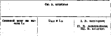
What they innervate and where the nerve ends: a) skin of the radial margin of the dorsal surface of the thumb to the base of the nail, b) skin of the ulnar margin of the dorsal surface of the thumb to the base of the nail, c) skin of the radial margin of the dorsal surface of the index finger to the nail phalanx, d) skin of the ulnar margin of the dorsal surface of the index finger to the nail phalanx, e) skin of the radial margin of the dorsal surface of the third finger to the nail phalanx. Topography of the nerve and its branches: rad. longus, descends along the palmar surface of the forearm, lying radially from the arteria radialis and along the m. brachio-radialis, passes above the radiocarpal joint to the dorsal surface, pierces the fascia superficialis antibrachii, gives off a) r. anastomoticus ulnaris to the ramus dorsalis manus from the nervus ulnaris and breaks up into b) nn. digitales dorsales for the skin of the dorsal surface of the first two fingers and the radial half of the third. 1) Variations in the area of distribution of the cutaneous branches are shown in Fig. 67-70 (Vol. XX, p. 769-780). 2) In rare cases, the n. thoraco-dorsalis may branch off from the radial nerve, usually starting directly from the fasciculus posterior. 3) Nervus cutaneus antibrachii dorsalis can sometimes be the same thickness as the trunk of the radial nerve itself. 4) In the region of the shoulder, one or two thin branches may branch off from the radial nerve to the m. brachialis internus (75% according to Frohse). Character of the nerve and its function. Variations: See n. mandibularis. 1) Plexus cardiacus, 2) ganglion cervicale medium et inferius, 3) cervical part of the trachea, 4) cervical part of the esophagus, 5) a) anterior branch—to the muscles: a) m. crico-arytaenoideus lateralis, b) m. thyreo-arytaenoideus and m. vocalis, c) m. thyreo-epiglotticus, d) m. ary-epiglotticus, b) posterior branch: a) n. laryngeus superior, b) to the muscles: m. crico-arytaenoideus posticus, m. arytaenoideus, c) mucous membrane below the vocal cords. Having started from the vagus nerve and its cervical part on the right, somewhat above the origin of the a. subclaviae, and on the left somewhat above the arcus aortae, the nerve descends downwards, giving off on this path: 1) rr. cardiaci inferiores for the cardiac plexus, 2) rr. anastomotici to the middle and lower cervical ganglia of the sympathetic nerve, 3) rr. tracheales to the cervical part of the trachea and 4) rr. oesophagei superiores for the cervical part of the esophagus; after this, it forms a loop on the right around the arteria subclavia, and on the left around the arcus aortae, and already under the name n. laryngeus inferior it ascends up the neck, lying in the groove between the esophagus and the trachea, and divides here into its r. anterior and posterior. Ramus anterior ends in the following muscles of the larynx: a) m. crico-arytaenoideus lateralis, b) m. thyreo-arytaenoideus et m. vocalis, c) m. thyreo-epiglotticus and d) m. ary-epiglotticus. Ramus posterior anastomoses with the n. laryngeus superior and gives rr. musculares for a) m. crico-arytaenoideus posticus and b) mm. arytaenoideus transversus et obliquus and c) rr. sensitivi for the mucous membrane below the vocal cords. Mixed. 1. Innervates the following groups of muscles: a) abducting the vocal cords and dilating the glottis—m. crico-arytaenoideus posticus; b) adducting the vocal cords and closing the glottis—m. crico-arytaenoideus lateralis, m. arytaenoideus transversus et obliquus; c) relaxing the vocal cords—m. vocalis; d) dilating the entrance to the larynx and raising the epiglottis—musculus thyreo-epiglotticus, and e) narrowing the entrance to the larynx and lowering the epiglottis—musculus ary-epiglotticus. 2. Conducts sensory stimuli from the mucous membrane of the larynx below the vocal cords. See n. vagus. See n. splanchnicus minor et minimus. See n. sympathicus. See n. acusticus. See n. acusticus. I. Plexus meningeus anterior et posterior; II. N. sympathicus; III. Plexus sacralis et pudendus (partly). Having started from the spinal cord at the level of approximately Li, the nerve gives off 1) r. meningeus, which, remaining in the vertebral canal, takes part in the formation of the plexus meningeus anterior et posterior; 2) ramus communicans, which begins. Mixed. I. Transmits sensory stimuli: a) from the skin of almost the entire leg except for the area of distribution of the nerves of the lumbar [plexus]. No. Name Latin and Russian. Synonyms. Origin. Corresponds to which segment. With which nerves it anastomoses. Which branches arise from it. 1) n. glutaeus superior, 2) n. glutaeus inferior, 3) n. piriformis, 4) n. cutaneus femoris posterior, 5) n. ischiadicus, 6) n. pudendus (partly); IV. R. posterior gives: 1) r. medialis, 2) r. lateralis nn. clunium mediorum, 3) rr. articulares. Sacralis quartus (fourth sacral nerve). Spinal cord at the height of Li, Sin and Sv. I. R. meningeus; II. R. communicans; III. R. anterior. Sacralis quintus (fifth sacral nerve). See Nerve plexuses (plexus coccygeus). Sacralis secundus (second sacral nerve). Syn.: n. bigeminus, s. biradiatus. Sacralis tertius (third sacral nerve). Similar in all respects to n. sacralis primus. Spinal cord at the height somewhat below Li, Sin and Siv. I. R. meningeus; II. R. communicans; III. R. anterior: 1) n. ischiadicus, 2) n. pudendus; IV. R. posterior. Saphenus (major) (saphenous n.). Syn.: n. cutaneus femoris internus major. N. femoralis. 1) N. cutaneus femoris posterior, 2) n. cutaneus surae medialis, 3) n. peronaeus superficialis. 1) Ramus infrapatellaris (see), 2) nn. cutanei cruris mediales (see). What they innervate and where the nerve ends. Topography of the nerve and its branches. Character of the nerve and its function. Variations. 1-6) see corresponding nerves; IV. 1) M. multifidus, skin over the posterior part of the sacrum and the tip, 2) skin over the medial part of m. glutaei maximi, 3) articulatio sacro-iliaca. Already in the foramen sacrale anterius and enters on the anterior surface of the sacrum into the composition of the sympathetic nervous system; 3) r. anterior, which exits from the foramen sacrale anterius, along the anterior margin of the m. piriformis, immediately takes part in the formation of the plexus sacralis, lying on the m. piriformis, and gives its fibers mainly into the nervus ischiadicus, but also into the nn. piriformis, glutaeus superior et inferior, cutaneus femoris posterior and partly into the nervus pudendus; 4) ramus posterior exits from the foramen sacrale posterius primum and takes part in the formation of the plexus sacralis posterior, which is located on the posterior surface of the sacrum and on the ligamentum sacro-tuberosum; from the latter arise: 1) branches for the m. multifidus, for the skin of the posterior part of the sacrum, 2) nn. clunium medii for the skin over the medial part of the m. glutaei maximi and 3) branches for the sacroiliac joint. Plexus, b) from the skin of the gluteal region, c) into the joint of the leg and the sacroiliac joint. II. Takes part in the innervation of the following groups of muscles: a) extensors of the thigh, rotating it outward—mm. glutaei, maximus, medius et minimus, b) rotating it outward—mm. piriformis, obturator internus, gemelli, quadratus femoris, c) abducting the thigh in the hip joint—m. tensor fasciae latae, d) extensors of the thigh in the hip joint and flexors of the leg in the knee joint—mm. biceps femoris, semitendinosus et semimembranosus, e) all groups of muscles of the lower leg and f) the foot. III. Transmits sensory stimuli from the membranes of the spinal cord and the spine in the sacral region. IV. Takes (some) part in the innervation of the muscles and skin of the perineum. Plexus meningeus anterior et posterior; II. N. sympathicus; III. Plexus pudendus. See pudendus plexus. See pudendus plexus. See Nerve plexuses (plexus coccygeus). I. Plexus meningeus anterior et posterior; II. N. sympathicus; III. Plexus sacralis et pudendus; See these nerves, as well as n. sacralis I. In all respects similar to n. sacralis I, but only its r. anterior exits through the foramen sacrale anterius III from under the posterior margin of the m. piriformis; the nerve takes part in the formation of: a) plexus sacralis, giving its upper fibers into the n. ischiadicus, and b) plexus pudendus, giving its lower fibers mainly into the n. pudendus. Mixed. I. Transmits sensory stimuli from the area of distribution of the nn. ischiadici and pudendi (see). II. Takes part in the innervation of the following groups of muscles: a) extensor of the thigh in the hip joint—m. glutaeus maximus, b) extensors of the thigh in the hip joint and flexors of the leg in the knee joint—mm. biceps femoris, semitendinosus et semimembranosus, c) all groups of muscles of the lower leg and foot. III. Innervates the skin and muscles of the perineum. IV.
Transmits sensory impulses from the spinal cord and spine in the sacral region 1) Skin of the medial side of the knee, 2) see the corresponding nerve. Starting from the nervus femoralis very high on the thigh, immediately under the ligamentum inguinale (Pouparti), the nerve lies first on the lateral, and then on the anterior side of the a. femoralis, descends with it into the canalis adductorius, passes between the m. vastus medialis and the m. adductor magnus, and then under the musculus sartorius, reaches under its tendon to the medial side of the knee, gives off its branch - r. infrapatellaris for the skin of the medial side of the knee, becomes superficial, descends together with the v. saphena magna to the lower leg, divides into its nn. cutanei cruris mediales, which are directed parallel to the branches of this vein in the skin of the medial surface of the lower leg to the metatarsi region. 1. The region of its distribution may vary depending on the degree of development of neighboring cutaneous nerves, as shown in Fig. 87-88 (Vol. XX, pp. 776-782). 2. Two types are observed: Type I, when there is a single trunk and gradual departure of peripheral branches without significant connections with neighboring nerves; Type II, when a double or looped trunk is present with early departure of branches and a sharply expressed tendency to form connections between branches and neighboring nerves (n. obturatorius and n. peronaeus). Sensory. Carries sensory impulses from the medial surface of the lower leg. No. Name Latin and Russian. Synonyms. Origin. To which segment it corresponds. With which nerves it anastomoses. Which branches depart from it. m Saphenus minor (lesser saphenous nerve). See n. femoralis. Scrotales anteriores (anterior scrotal nerves). Syn.: scrotales transversales. Scrotales posteriores (posterior scrotal nerves). See n. ilio-inguinalis. See pudendus plexus. Spermaticus externus (external spermatic nerve). Syn.: n. pudendus externus, s. genitalis, s. genito-cruralis. See n. genito-femoralis. Sphenoidalis lateralis (lateral sphenoidal nerve). Sphenoidalis medialis (medial sphenoidal nerve). A branch connecting the ganglion oticum and ganglion semilunare. A branch connecting the ganglion oticum with the ganglion spheno-palatinum through the n. canalis pterygoidei. Spheno-palatini (nervi) (sphenopalatine nerves). Syn.: pterygo-palatini, s. palatini descendentes, s. plexus spheno-palatinus. Spinales (spinal nerves). N. maxillaris. Several parallel nerves. See nn. cervicales dorsales (thoracales), lumbales, sacrales et coccygei. Spinalis accessorius (cervical part of the accessory nerve). See n. accessorius. Spinosus (spinous nerve). Syn.: n. recurrens n. inframaxillaris, s. rami tertii n. trigemini. N. mandibularis. Splanchnicus imus (lowest splanchnic nerve). Synonyms: splanchnicus tertius, s. minimus. Splanchnicus major (greater splanchnic nerve). Syn.: splanchnicus superior, s. supremus. X-XI thoracic nodes of the sympathetic trunk. (V) VI-IX thoracic nodes of the sympathetic trunk. What they innervate and where the nerve ends. Topography of the nerve and its branches. Character of the nerve and its function. Variations. See n. femoralis. See n. ilio-inguinalis. See pudendus plexus. See n. genito-femoralis.

Ganglion spheno-palatinum and through it along the course of the nn. palatini.
Starting from the n. maxillaris while still in the foramen rotundum, the nerves pass into the fossa pterygo-palatina and enter the ganglion spheno-palatinum as its radix sensitiva. See Nerve ganglia (ganglion spheno-palatinum) and nn. palatini. See nn. cervicales dorsales (thoracales), lumbales, sacrales et coccygei. See n. accessorius. The dura mater of the base of the brain, the greater wing of the sphenoid bone, and the mucous membrane of the cellulae mastoideae. Having separated from the n. mandibularis at the lower base of the skull, the nerve passes together with the a. meningea media through the foramen spinosum into the cranial cavity and spreads in the dura mater, in the mass of the sphenoid bone, and in the cellulae mastoideae. Sensory. Carries sensory impulses from the dura mater, from the sphenoid bone (greater wing), and cellulae mastoideae. Plexus renalis. This inconstant nerve, having started independently from the X-XI nodes of the sympathetic trunk or rr. communicantes, passes into the plexus renalis. Sympathetic. Carries its fibers into the plexus renalis. Ganglion coeliacum and through it into numerous secondary paired and unpaired sympathetic nodes of the abdominal cavity. Having started with several roots from the (V) VI-IX thoracic nodes of the sympathetic trunk of the sympathetic nervous system or from the corresponding rr. communicantes and then merged into one trunk, the nerve descends along the vertebral bodies, penetrates together with the v. azygos through the slit of the diaphragm between the crus intermedium and mediale into the abdominal cavity, where the ganglion coeliacum lies adjacent to it, gives off fibers for the plexus aorticus and takes part in the formation of the plexus coeliacus s. Solaris, from which, in its turn, numerous secondary paired and unpaired plexuses depart to all organs of the abdominal cavity. 1. Nervus splanchnicus major can enter directly into the solar plexus or after first passing through a ganglionic mass in its vicinity. 2. Two types can be observed: a) magistral, when the number of roots can be 2-4 and when they emerge at the level of the II-X thoracic vertebrae; b) diffuse type - number of roots 4-7, origin at the level of the V-VI thoracic vertebrae. Sympathetic.
No. Name Latin and Russian. Synonyms. Origin. To which segment it corresponds. With which nerves it anastomoses. Which branches depart from it. Splanchnicus minor (lesser splanchnic nerve). Syn.: splanchnicus inferior. X-XI thoracic nodes of the sympathetic trunk. 456 Stapedius (stapedial nerve). Syn.: nervulus stapedius. See n. facialis. 457 Staphylinus n. palatini anterioris (r.) (uvular branch of the anterior palatine nerve). One branch from the anterior palatine nerve to the floor of the nasal cavity. 458 Sternales (rr.) (sternal branches). Branches departing from the upper intercostal nerves to the sternum. 459 Stylo-hyoideus (stylohyoid nerve). Syn.: r. stylo-hyoideus, s. styloideus, s. mastoideus posterior. Branch of the n. digastricus. See n. facialis. 460 Stylo-pharyngeus (stylopharyngeus nerve). Syn.: r. stylo-pharyngeus n. glosso-pharyngei, s. circumflexus. See n. glosso-pharyngeus. 161 Subclavius (subclavian nerve). Plexus brachialis. Cv. 1) R. muscularis, 2) ramus. 462 Sublingualis (nerve of the sublingual gland). See n. lingualis. 463 Submaxillares (nn.) (submaxillary nerves). Syn.: rr. submaxillares ganglii submaxillaris. See Nerve ganglia (ganglion submaxillare). 464 Suboccipitalis (suboccipital nerve). Syn.: infraoccipitalis, s. r. posterior nervi cervicalis I. See n. cervicalis primus. Subscapulares: superior, medius et inferior (subscapular nerves: superior, middle, and inferior). See n. subscapularis superior, medius, inferior. 466 Subscapularis inferior (lower subscapular nerve). Syn.: n. thoraco-dorsalis.
s. circumflexus. See n. glosso-pharyngeus. 161 Subclavius (subclavian nerve). Plexus brachialis. Cv. 1) R. muscularis, 2) ramus. 462 Sublingualis (nerve of the sublingual gland). See n. lingualis. 463 Submaxillares (nn.) (submaxillary nerves). Syn.: rr. submaxillares ganglii submaxillaris. See Nerve ganglia (ganglion submaxillare). 464 Suboccipitalis (suboccipital nerve). Syn.: infraoccipitalis, s. r. posterior nervi cervicalis I. See n. cervicalis primus. Subscapulares: superior, medius et inferior (subscapular nerves: superior, middle, and inferior). See n. subscapularis superior, medius, inferior. 466 Subscapularis inferior (lower subscapular nerve). Syn.: n. thoraco-dorsalis.
Plexus brachialis. Cvi-viii. Rr. musculares. Subscapularis medius (middle subscapular nerve). Plexus brachialis. Cv-vi. Rr. musculares. What they innervate and where the nerve ends. Topography of the nerve and its branches. Character of the nerve and its function. Variations. Ganglion coeliacum and through it into numerous secondary sympathetic nodes of the abdominal cavity. Having started from the X-XI thoracic nodes of the sympathetic trunk of the sympathetic nervous system or from the corresponding rr. communicantes, the nerve independently or together with the n. splanchnicus major descends along the vertebral bodies, passes through the diaphragm laterally from the n. splanchnicus major, reaches the ganglion coeliacum, and passes mainly into the plexus renalis. In the plexus Solaris, the nerve can enter either with all its branches or only with a part; in that case, the rest enter the nearby ganglionic mass. See n. facialis. Sympathetic.
See n. glosso-pharyngeus 1) M. subclavius, 2) n. phrenicus. Having started from the plexus brachialis, from its primary upper trunk. The nerve goes into the fossa supraclavicularis, laterally from the n. phrenicus, to which it gives a small branch and ends in the m. subclavius. Motor. Fixes the clavicle. See n. lingualis. See Nerve nodes (ganglion submaxillare). See n. cervicalis primus. See n. subscapularis superior, medius, inferior. M. latissimus dorsi. Having started from the posterior trunk of the brachial plexus, the nerve is placed in the axillary fossa, in its depth, dorsal to other short nerves of the plexus, lies on the m. subscapularis in the mass of adipose tissue, reaches here along the margo axillaris scapulae to the m. latissimus dorsi and ends in the muscle with numerous muscular branches. The nerve can start from the n. axillaris or more rarely from the n. radialis. Motor. Innervates the latissimus dorsi muscle, which rotates the arm inward, lowers the raised arm down, and throws it onto the back with the palm outward. M. subscapularis, m. teres major. Having started from the posterior trunk of the brachial plexus, the nerve in the depth of the axillary fossa is placed dorsally from other short nerves, passes along the m. subscapularis, gives its branches to the lateral bundles of this muscle, and ends in the m. teres major. Motor. Innervates the muscles that rotate the arm inward, adduct, and lower the raised arm. No. Latin and Russian name. Synonyms. Origin. Which segment it corresponds to. With which nerves it anastomoses. Which branches depart from it. Subscapularis superior (upper subscapular nerve). Plexus brachialis. Cv-vi. Rr. musculares. Superficialis n. plantaris lateralis (r.) (superficial branch of the lateral plantar nerve). See n. plantaris lateralis. Superficialis n. radialis (r.) (superficial branch of the radial nerve). Syn.: n. cutaneus, s. dorsalis, s. n. radialis superficialis. See n. radialis. Superior n. laryngei superioris (r.) (upper branch of the superior laryngeal nerve). Syn.: r. externus n. laryngei superioris. See n. laryngeus superior. 472 Superior n. oculomotorii (upper branch of the oculomotor nerve). See n. oculomotorius. Supraclaviculares: anteriores, medii et posterior (supraclavicular nerves: anterior, middle, and posterior). Syn.: suprasternales anteriores, medii et posterior, s. cucullares (Schwalbe), s. acromiales, s. superficiales scapulae (Langenbeck), s. claviculares, s. sternales. Plexus cervicalis. Ciii, iv. 1) Nn. supraclaviculares anteriores (6-8), 2) nn. supraclaviculares medii (3), 3) nn. supraclavicularis posterior. Supraorbitalis (supraorbital nerve). Syn.: frontalis externus, s. major. See n. ophthalmicus. Suprascapularis (suprascapular nerve). Syn.: scapularis medius, s. superior. See n. ophthalmicus. Plexus brachialis. Cv-vi. 1) Rr. musculares, 2) rr. articulares. Supratrochlearis (supratrochlear nerve). Syn.: frontalis internus, s. minor. See n. ophthalmicus. Suralis (sural nerve). Syn.: communicans surae, s. n. tibialis, s. magnus, s. cutaneus longus cruris et pedis, s. Under this name is understood a small section of the nerve located on the posterior surface of the lower leg in its lower third, above the m. gastrocnemius; it represents a continuation of the n. cutaneus surae medialis (see), which receives this name after its connection with the ramus.
What they innervate and where the nerve ends. Topography of the nerve and its branches. M. subscapularis. Having started from Cv and Cvi, the nerve lies as the shortest of all subscapular nerves in the depth of the axillary fossa, is directed into the most medial bundles of the m. subscapularis along the margo vertebralis scapulae, where it branches into its rami musculares. Character of the nerve and its function. Variations. Motor. Innervates the subscapularis muscle, which rotates the arm inward, lowers and adducts the raised arm. See n. plantaris lateralis. See n. radialis. See n. laryngeus superior. See n. oculomotorius. 1) Skin over the sternum in its upper part, articulatio sterno-clavicularis, 2) skin of the lateral part of the chest to the fourth rib, 3) skin of the shoulder and over the anterior part of the m. deltoideus, gives a branch to the n. accessorius. Having started from Ciii and Civ near the foramen intervertebrale, the nerves pass along the medial surface of the musculi sterno-cleido-mastoidei outward, exit from under its posterior edge below the other cutaneous nerves of the cervical plexus, descend down along the posterior edge of the muscle, covered by the musculus platysma and fascia superficialis colli, and are distributed as follows: 1) nn. supraclaviculares anteriores cross the sterno-cleido-mastoid muscle and are directed to the incisura sterni, are distributed in the skin over the extremitas sternalis claviculae, and also give a branch to the sterno-clavicular joint; 2) nn. supraclaviculares medii go down, cross the clavicle from its middle to the processus acromion and are distributed in the skin of the chest to the IV rib; 3) n. supraclavicularis posterior descends down along the lateral edge of the musculi trapezii and ends in the skin of the upper scapular region and over the m. deltoideus. Sensory. 1. Carries sensory irritations from the sterno-clavicular joint. 2. Carries sensory irritations from the upper part of the chest and shoulder. See n. ophthalmicus. 1) M. supraspinatus, m. infraspinatus, 2) articulatio humeri. Having started from the brachial plexus, from its upper primary trunk, on its dorsal surface, the nerve, being the most lateral and upper (except for the nervus dorsalis scapulae), goes down into the fossa supraclavicularis along the lateral edge of the plexus, and then turns in the lateral direction and backward along the m. omo-hyoideus, reaches together with it the incisura scapulae, enters under the ligamentum transversum scapulae superius into the fossa supraspinata, lies under the musculus supraspinatus, gives it its muscular branches, is directed in the lateral direction to the neck of the scapula, approaches under the ligamentum transversum scapulae inferius, penetrates into the fossa infraspinata and ends in the musculus infraspinatus, having also given a branch to the articular capsule of the shoulder. Mixed. I. Sends sensory irritations from the capsule of the shoulder joint. II. Innervates the muscles: 1) m. supraspinatus, which abducts the arm, 2) m. infraspinatus, which rotates the arm outward, adducts and lowers the raised arm. See n. ophthalmicus. No. Latin and Russian name. Synonyms. Origin. Which segment it corresponds to. With which nerves it anastomoses. Which branches depart from it. 477
saphenus brevis inf., s. externus, - >
s. minor, s. saphenus tibialis, £
s. r. communicans surae, s. B
communicans tibialis anastomoticus peronaeus (see this N. and n. peronaeus communis); n. suralis gives off rr. calcanei laterales (see) and again receives another name - n. cutaneus dorsalis (pedis) lateralis (see). 478 Sympathicus (nervus, s. truncus) (sympathetic nerve). Syn.: n. gangliosus, s. intercostalis maximus, s. sympathicus magnus, s. maximus, s. sympatheticus, s. trisplanchnicus, s. vasomotorius (Stilling, Henle). rr. communicantes of all trunk nerves. For anastomoses see the following column. I. Cervical part: A. Ganglion cervicale superius: 1) n. caroticus internus, 2) n. jugularis, 3) n. cardiacus superior, 4) r. anastomoticus, 5) r. anastomoticus, 6) nn. carotici externi, 7) rr. pharyngei, 8) rr. laryngei; B. Ganglion cervicale medium: 9) rr. carotici, 10) n. cardiacus medius; C. Ganglion cervicale inferius: 11) n. cardiacus inferior, 12) n. cardiacus imus (not recognized by all); II. Thoracic part: Ganglia thoracalia 13) nn. bronchiales, 14) nn. aortales, 15) n. splanchnicus major, 16) n. splanchnicus minor, 17) n. splanchnicus minimus, s. imus (inconstant); III. Abdominal part: Ganglia lumbalia 18) rr. postganglionares; IV. Pelvic part: Ganglia sacralia 19) rr. postganglionares. What they innervate and where the nerve ends. Topography of the nerve and its branches. Character of the nerve and its function. Variations: 1) Plexus caroticus internus, 2) one branch to ganglion jugulare n. vagi, and another to ganglion petrosum n. glosso-pharyngei, 3) plexus cardiacus, 4) ganglion nodosum n. vagi, 5) n. hypoglossus, 6) to all branches from arteria carotis externae, 7) plexus pharyngeus, 8) plexus laryngeus, 9) plexus caroticus communis, 10) plexus cardiacus, 11) plexus cardiacus, 12) plexus cardiacus, 13) plexus bronchiales, 14) plexus aortae thoracalis, 15) plexus coeliacus, 16) plexus coeliacus, 17) plexus renalis, 18) plexus coeliacus, mesentericus superior et inferior, 19) plexus hypogastricus (pelvicus). Having started from the rr. communicantes of the spinal nerves, the sympathetic nerve represents on each side: a) a boundary trunk - truncus sympathicus, which consists of 20-25 nodes, of interganglionic fibers - rr. interganglionares, connecting these nodes of each side with each other, and transverse fibers - rr. transversi, connecting the nodes of the opposite side with each other; b) from these nodes go postganglionic fibers - rr. postganglionares; c) to the peripheral ganglia of the sympathetic nervous system: ganglia ciliare, spheno-palatinum, submaxillare and oticum, located in the head, ganglion coeliacum, mesentericum superius et inferius, ganglia renale dextrum et sinistrum, aortico-abdominalis, located in the abdominal cavity, and ganglion pelvicum, located in the pelvis; d) from these nodes, in turn, fibers go to the nerve plexuses in all organs, starting from the oral cavity, neck, thoracic and up to the abdominal cavity, to the vessels, to the skin, to the muscles and to the glands. In the cervical section, the nerve lies on the fascia praevertebralis, with the ganglion cervicale superius located on the transverse processes of Cii and Ciii and giving off its branches: 1) nervus caroticus internus, which entwines the internal carotid artery and all branches departing from it and forms plexuses bearing the names of these vessels; in addition, the plexus caroticus internus gives the following branches: a) radix sympathica ganglii ciliaris, b) rr. orbitales, c) ramus to hypophysis cerebri, d) nervus petrosus profundus major to ganglion spheno-palatinum, e) nn. carotico-tympanici superior et inferior to plexus tympanicus; 2) n. jugularis, which immediately divides into two branches; of these, one goes to the ganglion jugulare nervi vagi, and the other to the ganglion petrosum nervi glosso-pharyngei; 3) n. cardiacus superior - to the cardiac plexus; 4) r. anastomoticus - to the ganglion nodosum n. vagi; 5) r. anastomoticus - to the nervus hypoglossus; 6) nn. carotici externi, which entwine the external carotid artery and form plexuses along all branches departing from it, and between them also: radix sympathica for ganglion submaxillare from plexus arteriae maxillaris externae and for ganglion oticum radix sympathica from plexus meningeus arteriae meningeae mediae; 7) rr. pharyngei - for plexus pharyngeus and 8) rr. laryngei - for plexus laryngeus. Ganglion cervicale medium - inconstant: sometimes completely absent, and sometimes replaced by two nodes; lies on the truncus thyreo-cervicalis at the level of Cvi and gives: 9) rr. carotici for plexus caroticus communis and 10) n. cardiacus medius for plexus cardiacus. Ganglion cervicale inferius often fuses with the 1st thoracic, lies above the head of the 1st rib on the transverse process of Cviii behind the subclavian artery and gives: 11) n. cardiacus inferior and 12) n. cardiacus imus (inconstant) for the cardiac plexus. In the thoracic cavity, the nerve lies on the lateral surface of the spine on the articulatio costo-vertebralis under the pleura and gives: 13) nn. bronchiales for plexus bronchialis, 14) nn. aortales for the plexus around the thoracic aorta, 15) n. splanchnicus major, which begins from the V-IX nodes, lies on the anterior surface of the vertebral column, descends through the diaphragm and enters the plexus coeliacus; to it also approaches 16) n. splanchnicus minor. Plexus coeliacus is located on the anterior surface of the spine at the level of Li on the sides of the a. coeliaca, receives fibers from the vagus nerve and gives secondary plexuses around all abdominal organs, both paired and unpaired, namely: a) plexus phrenicus, b) plexus gastricus superior et inferior, c) plexus hepaticus, d) plexus lienalis, e) plexus mesentericus superior, f) plexus mesentericus inferior, g) plexus renalis, h) plexus spermaticus (ovaricus). In the pelvic cavity, the N. also lies on the anterior surface of the spine, receives rr. postganglionares of the corresponding sacral sympathetic nodes and forms the plexus pelvicus with secondary plexuses around the organs of the small pelvis, namely: plexus colicus, vesicalis and vaginalis. See Autonomic nervous system. No. Name in Latin and Russian. Synonyms. Temporales (rr.) (temporal branches). Syn.: rr. temporales n. facialis, s. rr. temporo-frontales, s. nn. faciales temporales. Origin. Which segment it corresponds to. With which nerves it anastomoses. Which branches depart from it. Temporales superficiales (rr.) (temporal branches of the auriculo-temporal nerve). Syn.: rr. temporales subcutanei. See n. facialis. See n. auriculo-temporalis. 481 Temporalis profundus anterior et posterior (anterior and posterior deep temporal nerve). See n. masticatorius. Temporo-facialis (temporo-facial nerve). Branch from n. facialis to glandula parotis. Tensor vaginae femoris (nerve of the muscle that tenses the fascia of the thigh). Branch from n. glutaeus superior to m. tensor fasciae latae. Tensoris tympani (nerve of the muscle that tenses the eardrum). N. pterygoideus internus and ganglion oticum. R. muscularis. Tensoris veli palatini (nerve of the muscle that tenses the soft palate). N. pterygoideus internus and ganglion oticum. R. muscularis. Tentorii (nerve of the cerebellar tentorium). Syn.: n. recurrens (Arnoldi), s. tentorii cerebelli, s. r. sinualis, s. n. sinualis. Terminalis radialis (terminal radial branch). See n. ophthalmicus. See n. medianus. Terminalis ulnaris (terminal ulnar branch). See n. medianus. Thoracales (thoracic nerves). Syn.: nn. thoracici, s. dorsales, s. costales, s. intercostales, s. spinales dorsales, s. subcostales. Spinal cord approximately from the level of the end of the spinous process of Dvi TO THE HEIGHT of the spinous process of Dxi. Di-xii. 1) Rr. meningei, 2) rr. communicantes, 3) rr. anteriores, 4) rr. posteriores, s. rr. dorsales give: a) rr. mediales, b) rr. laterales. 490. Thoracales anteriores: primus et secundus (anterior thoracic nerves: first and second). Plexus brachialis. Cv-viii and Di. Primus: 1) r. muscularis, 2) r. anastomoticus. Secundus: 1) r. muscularis, 2) r. muscularis. What they innervate and where the nerve ends. Topography of the nerve and its branches. Character of the nerve and its function. Variations.
See n. facialis. See n. auriculo-temporalis. See n. masticatorius. M. tensor tympani: Starting with two branches: 1) from n. pterygoideus internus and 2) from ganglion oticum (the latter receives its motor fibers—radix motoria—also mainly from n. pterygoideus internus), the nerve enters the semicanalis tensoris in the pyramid of the temporal bone and ends in the m. tensor tympani. Motor. Innervates the muscle that tenses the eardrum. M. tensor veli palatini: Starting with two branches: 1) from n. pterygoideus internus and from the ganglion oticum—its anterior edge (the latter receives its radix motoria mainly from the same nerve), the nerve descends from the foramen ovale together with the n. pterygoideus internus and ends in the m. tensor veli palatini. Motor. Innervates the muscle that tenses the soft palate. See n. ophthalmicus. See n. medianus. See n. medianus. 1) Plexus meningeus anterior et posterior, 2) n. sympathicus (nervus splanchnicus major et minor) (see), 3) a) plexus brachialis—DI part, b) n. intercosto-brachialis (DII), c) nn. intercostales I-XII (see), 4) a) skin of the back along the midline and muscles: mm. rotatores, multifidus, semispinalis, spinalis dorsi, b) m. longissimus and m. ilio-costalis. Each of the thoracic nerves, while still in the vertebral canal, gives off its branch 1) r. meningeus, which returns back into the vertebral canal and takes part in the formation of the plexus meningeus anterior and posterior, and then the nerve, having exited the intervertebral canal, gives: 2) r. communicans, going to the boundary chain of the sympathetic nervous system; 3) r. anterior, which passes into the intercostal space—n. intercostalis (see); of these, only the first and partly the second pass into the brachial plexus and participate in the innervation of the skin and muscles of the arm; 4) ramus posterior, which, having exited between the transverse processes of the vertebrae, divides: a) into a medial branch (thicker in the eight upper nerves and thinner in the rest) and b) into a lateral branch (thicker in the four lower nerves and thinner in the rest). Mixed. 1) Carry sensory stimuli from the skin of the trunk and upper limb. 2) Innervate the muscles of the chest and back (see nn. intercostales). 3) Follow the course of the n. sympathicus into the organs of the abdominal cavity, into the muscles and skin. 4) Bring sensory stimuli from the membranes of the spinal cord and from the walls of the spine. 1) M. pectoralis major, 2) n. thoracalis anterior secundus; 1) m. pectoralis major, 2) m. pectoralis minor. Starting: the first—from the upper secondary trunk of the brachial plexus, the second—from the lower secondary trunk, from their anterior surface, while still in the pars supraclavicularis of the brachial plexus, the nerves approach under the clavicle in front of the a. and v. subclavia and enter: the first into the musculus pectoralis major from its inner surface, and the second into the m. pectoralis major and m. pectoralis minor, also from their inner surface. Motor. Innervate the pectoral muscles.
25b № Name in Latin and Russian. Synonyms. Origin. Which segment it corresponds to. With which nerves it anastomoses. Which branches depart from it. Thoracales posteriores (posterior thoracic nerves). Under this name are understood the n. dorsalis scapulae and n. thoracalis longus. 492 Thoracalis longus (long thoracic nerve). Syn.: thoracicus medius, s. posterior, s. lateralis, s. thoracico-dorsalis, s. respiratorius externus inferior. Plexus brachialis. CV-VI (VII). R. muscularis. Thoracico-dorsalis (thoracodorsal nerve). Syn.: n. marginalis scapulae, s. subscapularis inferior. See nn. subscapulares. Thyreo-hyoideus (r.) (branch of the hypoglossal nerve to the thyrohyoid muscle). Tibialis (tibial nerve). Syn.: n. popliteus internus, s. posticus. Properly a branch from the ansa hypoglossi, i.e., from the n. cervicalis (see n. hypoglossus). N. ischiadicus. 1) N. peronaeus, 2) n. femoralis, n. cutaneus femoris posterior, 4) peripheral branches among themselves. 3) I. Before division into terminal branches: 1) n. cutaneus surae medialis (see): a) rr. calcanei laterales (see), b) n. cutaneus dorsalis lateralis (see), 2) rr. articulares, 3) rr. musculares, n. canalis musculo-peronaei, 4) r. popliteus, 5) rr. calcanei mediales (see); II. Branches of the n. plantaris medialis (see): 6) rr. musculares, r. medialis, 7) r. muscularis, 8) n. plantaris hallucis medialis, 9) nn. digitales plantares communes I-III, 10) nn. lumbricales; III. Branches of the n. plantaris lateralis: 11) rr. musculares, r. superficialis, 12) n. plantaris lateralis digiti V: a) rr. musculares, b) rr. lumbricales, 13) n. digitalis plantaris communis IV (see): r. profundus, 14) nn. interossei, 15) rr. musculares 257
HUMAN NERVES. What they innervate and where the nerve ends. Topography of the nerve and its branches. Character of the nerve and its function. Variations. M. serratus anterior. Starting with two or three roots from the posterior surface of the plexus brachialis while still in the pars supraclavicularis and having formed into one trunk, the nerve descends along the lateral wall of the chest along the linea axillaris between the musculus subscapularis and musculus serratus anterior, lying tightly on the latter and giving its branches to the teeth of the musculi serrati anterioris. Motor. Innervates the m. serratus anterior. See nn. subscapulares. 1) Skin over the lateral malleolus, skin of the lateral edge of the foot, 2) articulatio genu, 3) a) m. gastrocnemius, b) m. plantaris, c) m. tibialis posterior, d) m. soleus, e) m. flexor digitorum longus, f) m. flexor hallucis longus, g) for the periosteum and foramen nutritium of the fibula, 4) a) m. popliteus, b) a. tibialis, c) articulatio talo-cruralis, d) tibia, periosteum, e) bones of the lower leg, f) membrana interossea (n. interosseus cruris) (see), 5) skin of the medial surface of the heel, 6) a) m. flexor digitorum
brevis, 6) m. abductor hallucis, 7) m. flexor hallucis brevis, 8) and 9) vessels of the plantar side of the toes, 10) mm. lumbricales, 11) a) m. quadratus plantae, b) m. abductor digiti V, 12) a) m. flexor digiti V, m. opponens digiti V, b) mm. lumbricales III, 13) skin of the plantar side of the toes, 14) mm. interossei, 15) a) m. adductor hallucis, b) m. flexor hallucis brevis, c) m. flexor digiti V, d) m. opponens digiti V. Having started from the sciatic nerve approximately at the middle of the thigh, but still remaining with the common peroneal nerve for some time in one common sheath and having given off a whole series of branches in this space (see sciatic nerve), namely: muscular branches for the obturator internus muscle, gemelli muscles, quadratus femoris muscle, semitendinosus muscle, semimembranosus muscle, adductor magnus muscle, and the biceps femoris muscle (long head), the tibial nerve in the popliteal fossa occupies a position between the common peroneal nerve, lying more laterally, and the popliteal vessels, lying more medially, and gives off a whole series of branches here: 1) medial sural cutaneous nerve (see this nerve and sural nerve), which descends between the common heads of the gastrocnemius muscle, lies under the crural fascia along the small saphenous vein, anastomoses with the peroneal anastomotic branch, receives the name sural nerve, pierces the crural fascia, gives off its lateral calcaneal branches (see), and then again under another name—lateral dorsal cutaneous nerve (see)—spreads in the skin of the lateral edge of the foot; 2) articular branches (usually two)—for the knee joint; 3) muscular branches for all muscles of the posterior group of the lower leg: a) gastrocnemius muscle, b) plantaris muscle, c) tibialis posterior muscle, d) soleus muscle, e) flexor digitorum longus muscle, f) flexor hallucis longus muscle, and g) musculoperoneal canal nerve, which goes along the musculoperoneal canal together with the peroneal vessels, reaches the fibula, and gives off nerves to the periosteum and to the nutrient foramen of the fibula; 4) popliteal branch, which descends along the popliteus muscle on its posterior surface and gives off very many small branches: a) for the popliteus muscle, b) for the tibial artery, c) for the tibia, d) for the periosteum, e) for the ankle joint, and f) for the interosseous membrane; 5) medial calcaneal branches for the skin of the medial surface of the heel and the posterior section of the foot. Further, the nerve itself descends along the lower leg, first between both heads of the gastrocnemius muscle, then descends under the soleus muscle, lies between it and the tibialis posterior muscle, and reaches the medial side of the ankle joint together with the posterior tibial artery, and near the medial malleolus divides into both its terminal branches: the medial and lateral plantar nerves (for their description, see the corresponding nerves). 1. Variations in the area of distribution of the cutaneous branches are shown in Figs. 84-91 (Vol. XX, pp. 775-782). 2. Both heads of the gastrocnemius muscle usually receive independent branches, but in the mass of the tibial nerve, one can always distinguish a dorsal bundle, which includes the medial sural cutaneous nerve, both branches to the gastrocnemius muscle, and to the soleus muscle. 3. The site of origin of the articular branch of the knee also varies. 4. The popliteal branch usually gives off the tibial diaphysial nerve and the posterior tibial epiphysial nerve for the corresponding parts of the tibia and for the superior tibiofibular joint. 5. The articular branch of the foot (crural) can arise independently from the trunk or from its branches. Mixed. I. Transmits sensory stimuli: a) from the skin of the posterior surface of the lower leg, b) from the sole and plantar surface of the toes, c) from the joints: knee, ankle (from the posterior side), and foot (from its plantar side). II. Innervates muscle groups: a) posterior muscles of the lower leg, flexing the leg at the knee joint—triceps surae muscle; plantar flexing the foot—triceps surae, plantares, flexor hallucis longus, and flexor digitorum longus; flexing the toes—flexor digitorum and flexor hallucis longus muscles; adducting and supinating the foot—tibialis posterior muscle; b) all plantar muscles of the foot.
No. Name in Latin and Russian. Synonyms. Origin. To which segment does it correspond? With which nerves does it anastomose? Which branches arise from it? Tonsillar branches of the glossopharyngeal nerve (branches of the glossopharyngeal nerve to the tonsil). Syn.: tonsillar nerves. See glossopharyngeal nerve. Tonsillar branches of the lingual nerve (branches of the lingual nerve to the tonsil). Syn.: branches of the faucial isthmus. See lingual nerve. Inferior tracheal branches (inferior tracheal nerves). Syn.: anterior bronchial branches. See vagus nerve. Superior tracheal branches (superior tracheal nerves). Syn.: tracheal branches of the recurrent nerve. See inferior laryngeal nerve and vagus nerve. Trapezius branch (trapezoid branch). See fourth cervical nerve. Trigeminal nerve: major portion, or sensory portion; minor portion, or motor portion (trigeminal—sensory portion and motor portion). Syn.: fifth, or fifth pair of cranial nerves. The major portion begins in the semilunar ganglion; its cells send peripheral collaterals as the nerve itself, and central collaterals end: a) in the sensory nucleus of the trigeminal nerve in the tegmentum of the pons and b) in the spinal tract nucleus of the trigeminal nerve, which continues through the entire rhomboid fossa and into the spinal cord. The minor portion begins: a) in the nucleus of the descending root of the trigeminal nerve, lying in the posterior part of the corpora quadrigemina on the sides of the cerebral aqueduct (Sylvii), and b) in the locus coeruleus nucleus. Besides the anastomoses of its three constituent nerves, there exist macroscopically almost indistinguishable sympathetic anastomotic fibers from the carotid plexus to the Gasserian semilunar ganglion. The nerve gives off three branches: I—ophthalmic nerve, II—maxillary nerve, III—mandibular nerve, which are so independent that for their descriptions, see the corresponding nerves. Trochlear nerve (trochlear nerve). Syn.: superior oculomuscular nerve, or pathetic nerve, or fourth pair of cranial nerves. Begins in the nucleus of the trochlear nerve, located in the corpora quadrigemina on the sides and on the floor of the cerebral aqueduct (Sylvii) caudally from the oculomotor nerve nucleus; its collaterals rise to the dorsal surface of the quadrigeminal lamina, cross under the posterior colliculus of the corpora quadrigemina, and emerge on the sides of the frenulum of the anterior medullary velum. 1) Cavernous plexus, 2) ophthalmic artery. Tubal branches (nerve of the auditory tube). Syn.: Eustachian tube, auditory canal, or tympanic plexus of the tube. See glossopharyngeal nerve. See auriculotemporal nerve. Tympanic branch (nerve of the eardrum). Syn.: branch of the tympanic membrane. See glossopharyngeal nerve. Tympanic nerve (tympanic, or Jacobson's nerve). Syn.: Jacobson's nerve. See glossopharyngeal nerve.
9«2 What they innervate and where the nerve ends Topography of the nerve and its branches Character of the nerve and its function. Variations See n. glosso-pharyngeus See n. lingualis See n. vagus See n. laryngeus inferior and n. vagus See n. cervicalis quartus Having started with two independent roots: 1) portio sensitiva, s. major and 2) portio motoria, s. minor, the nerve exits with separate roots from the brain on the ventral surface of the pons along the linea trigemino-facialis, which represents the boundary of the pons and crura cerebelli ad pontem, lies on the anterior surface of the pyramid of the temporal bone in the impressio trigemini; here the portio major passes into the ganglion semilunare (Gasseri), being located here between two leaflets of the dura mater in the cavum semilunare (Meckeli), in immediate proximity to the processus clinoideus posterior, at the lateral wall of the sinus cavernosus. From the semilunar ganglion, from its anterior edge, all three N. depart: the most medial-nervus ophthalmicus (see), the middle-n. maxillaris (see) and the most lateral-n. mandibularis; into the path of the latter nerve enter, without entering the ganglion semilunare and lying ventrally from it, all fibers from the portio minor Mixed. Motor fibers enter: a) into the nervus mandibularis and in it constitute almost entirely the whole n. masticatorius (except for fibers of the n. bucciflaterii) and b) a part into the nervus lingualis (fibers of the n. mylo-hyoidei). Distribution of sensory fibers-see the corresponding nerves obliquus oculi superior Having exited on the dorsal surface of the brain from the quadrigeminal plate along the sides of the frenulum of the superior medullary velum and having passed along the lateral surface of the crus cerebri to the ventral surface of the brain, the nerve in the form of a thin thread pierces the dura mater-porus trochlearis behind the processus clinoideus posterior, laterally from the nervus oculomotorius, and lies adjacent to the sinus cavernosus; here it receives sympathetic fibers from the plexus cavernosus,- further, the nerve, lying adjacent to the nervus ophthalmicus and having received sensory fibers from it, passes through the fissura orbitalis superior into the cavity of the orbit, bends here over the place of origin of the m. levator palpebrae superioris and enters the m. obliquus oculi superior Motor. Rotates, lowers and abducts the eye See n. glosso-pharyngeus See n. auriculo-temporalis See n. glosso-pharyngeus l. Name Latin Russian. Synonyms Origin. To which segment it corresponds With which nerves it anastomoses Which branches depart from it Ulnaris (r.) (ulnar branch) See n. cutaneus antibrachii medialis Ulnaris (ulnar nerve). Syn.: | cubitalis; r. dorsalis manus= ulnaris dorsalis; r. volaris manus=n. ulnaris volaris; r. profundus-ulnaris volaris profundus; r. superficialis-ulnaris volaris superficialis Plexus brachialis. Cviii and Di 1) N. musculo-cutaneus, 2) n. medianus, 3) n. radialis, 4) n. cutaneus brachii medialis, 5) peripheral twigs among themselves A. Branches of the forearm: 1) rr. articulares, 2) rr. musculares, 3) r. cutaneus palmaris; B. Terminal branches: 4) r. dorsalis manus: n. digitalis: a) r. dig. dorsalis digiti V ulnaris, b) r. digitalis dorsalis digiti V radialis, c) n. digitalis dorsalis digiti IV ulnaris, d) r. digitalis dorsalis digiti IV radialis, e) n. digitalis dorsalis digiti III ulnaris, 5) r. volaris manus: r. superficialis: a) r. muscularis, b) r. lumbricalis, c) n. digitalis volaris digiti V ulnaris, d) n. digitalis volaris communis IV: r. digitalis volaris digiti V radialis, r. digitalis volaris digiti IV ulnaris, r. profundus: a) r. anastomoticus, b) rr. articulares, c) r. interosseus, d) rr. lumbricales, e) rr. musculares, f) rr. perforantes 608 Ulnaris cutanei antibrachii medialis (r.) (ulnar branch of the medial cutaneous nerve of the forearm) See n. cutaneus antibrachii medialis Urethralis nervi perinei (r.) (urethral branch of the perineal nerve) Twig to the bulbus urethrae-see n. pudendus Utricularis (nerve of the auditory sac). Syn.: funiculus superior nervus vestibuli, s. nervus saccularis major, s. superior n. vestibuli See n. acusticus 511; Vaginales (nerves of the vagina) See pudendus plexus Vagus (vagus N.). Syn.: pneumogastricus, s. par decimum nn. encephali, n. parasympathicus Motor fibers begin from the nucleus dorsalis and nucleus ventralis, s. ambiguus, 1) Nervus facialis, 2) n. glosso-pharyngeus 3) n. accessorius, 4) n. hypoglossus, 5) ganglion I. Head part: 1) r. meningeus, 2) r. auricularis: What they innervate and where the nerve ends Topography of the nerve and its branches Character of the nerve and its function. Variations See n. cutaneus antibrachii medialis 1) Articulatio cubiti, 2) a) m. flexor carpi ulnaris, b) m. flexor digitorum profundus (caput ulnare), 3) skin of the palmar surface of the forearm and eminentiae hypothenaris; 4) skin of the dorsal surface of the corresponding fingers; 6) r. superficialis: a) m. palmaris, b) m. lumbricalis IV, c) skin of the palmar ulnar edge of the V finger; d) skin of the palmar surface of the IV and V fingers facing each other; r. profundus: a) r. dorsalis manus, b) ligaments of the wrist, c) mm. interossei volares dorsales, d) mm. lumbricales III and IV, e) m. adductor pollicis, m. flexor digiti V, m. opponens digiti V, m. abductor digiti V, f) for the dorsal surface of the spatia interossea Having started from the brachial plexus, its medial (lower) trunk, the nerve descends along the arm, lying in the sulcus bicipitalis medialis on the posterior and medial side of the a. axillaris, and then the a. brachialis and along the anterior surface of the m. triceps brachii, its caput mediale; further N. approaches the elbow joint, bending superficially around the humerus along the sulcus n. ulnaris between the processus olecranon and epicondylus medialis humeri; it gives off here 1) rr. articulares for the elbow joint. Further N. passes between both heads of the m. flexor carpi ulnaris already along the palmar surface of the forearm, goes then between this muscle and the musculus flexor digitorum profundus along the a. ulnaris, on its medial side and gives off: 2) rr. musculares for the flexors of the forearm: a) m. flexor carpi ulnaris and b) m. flexor digitorum profundus (its caput ulnaris) and 3) ramus cutaneus palmaris, which departs in the middle of the forearm in the form of a thin twig, pierces the fascia antibrachii between the m. flexor carpi ulnaris and musculus flexor digitorum profundus and ends in the skin of the palmar surface of the forearm in its lower third and in the skin of the hypothenar eminence. 4) In this same part begins also the r. dorsalis manus, which in the lower third of the forearm passes to the dorsal side of the forearm, pierces the fascia antibrachii and above the head of the ulna breaks up into nn. digitales dorsales digiti V, IV and III (ulnaris) for the dorsal surface of 2 1/2 ulnar fingers. As for b) ramus volaris manus, it in the region of the os pisiforme under the fascia antibrachii divides into r. superficialis and r. profundus. Ramus superficialis gives: a) r. muscularis for the m. palmaris brevis, b) r. lumbricalis for the musculus lumbricalis and divides into nn. digitales volares V, IV (ulnaris) for the palmar surface of 1 1/2 ulnar fingers. Ramus profundus gives: a) a thin r. anastomoticus to the r. dorsalis manus, located in the region of the os pisiforme, b) rr. articulares for the ligaments of the wrist, c) ramus interosseus for all dorsal and palmar interosseous muscles, d) rr. lumbricales for the III and IV lumbrical muscles, e) rr. musculares for all muscles of the hypothenar eminence and for the m. adductor pollicis and finally f) rr. perforantes, penetrating to the dorsal surface of the interosseous spaces. 1. The regions of distribution of the cutaneous branches of the fingers can vary, as is shown in fig. 67-70 (vol. XX, art. 759-760). 2. Rarely there is in the region of the arm a Villar's anastomosis with the n. medianus. 3. The n. ulnaris for some distance can go together with the n. collateralis ulnaris from the n. radialis, and then it may seem that the medial head of the triceps muscle receives branches from the n. ulnaris. 4. Although in rare cases, but still there are one or two twigs of the n. ulnaris for the triceps muscle Mixed. I. Carries sensory stimuli: a) from the skin of fingers V and IV (partly) from their palmar side and V, IV and III (partly) from their dorsal side, and also from the skin of the hypothenar eminence. II. Innervates muscles: a) flexor of the hand, abducting it to the ulnar side (m. flexor carpi ulnaris); b) flexor of the fingers (flexor digitorum profundus); c) muscles of the hand: adductor of the thumb (musculus adductor pollicis), all three muscles of the little finger, the short palmar muscle, all interosseous and one lumbrical See n. cutaneus antibrachii medialis See n. acusticus See pudendus plexus 1) The dura mater, Having started from the medulla oblongata in the sulcus lateralis behind the oliva with 12-16 separate root fibers, going through the foramen jugulare to the ganglion jugulare, the nerve goes already as one Mixed: sensory, secretory, visceromotor, motor. I. Transmits sensory i. No. Name Latin and Russian. Synonyms Origin. To which segment does it correspond? With which nerves does it anastomose? Which branches originate from it, located on the floor of the rhomboid fossa of the medulla oblongata? Sensory fibers begin in the ganglion jugulare cervicale superius et inferius: a) r. anastomoticus, b) r. cutaneus, 3) r. anastomoticus, 4) r. anastomoticus superior, 5) r. anastomoticus; II. Cervical part: 6) r. anastomoticus inferior, 7) rr. anastomotici, 8) rr. pharyngei superior et inferior: a) rr. musculares, b) r. sensitivus, c) r. lingualis, 9) n. laryngeus superior: a) r. anastomoticus, b) r. anastomoticus, c) r. anastomoticus, d) r. externus: rr. musculares, r. sensitivus, r. cardiacus superior, e) r. internus: rr. epiglottici, rr. inferiores, rr. pharyngei, r. anastomoticus, 10) rr. cardiaci superiores, among them n. depressor cordis, 11) n. recurrens: a) rr. cardiaci inferiores, b) rr. anastomotici, c) rr. tracheales superiores, d) rr. oesophagei superiores, 12) n. laryngeus inferior: a) r. anterior: rr. musculares, b) r. posterior, r. sensitivus; III. Thoracic part: 13) rr. tracheales inferiores, 14) rr. bronchiales anteriores, 15) rr. bronchiales posteriores, 16) rr. oesophagei, 17) rr. pericardiaci; IV. Abdominal part: 18) rr. gastrici (vagus sinister), rr. gastrici (vagus dexter), 19) rr. hepatici, 20) rr. coeliaci (vagus dexter). What do they innervate and where does the nerve begin? Topography of the nerve and its branches. Character of the nerve and its function 2) a) r. auricularis n. facialis, b) skin of the convex surface of the auricle,
alis, b) skin of the convex surface of the auricle,
3) n. glosso-pharyngeus,
4) ganglion cervicale superius,
5) n. accessorius,
6) ganglion cervicale superius, 7) n. hypoglossus, 8) plexus pharyngeus: a) mm. constrictores pharyngis: superior, medius et inferior, m. levator veli palatini, m. uvulae, m. pharyngo-palatinus, m. stylo-pharyngeus, b) tongue together with n. hypoglossus and plexus caroticus externus,
9) a) plexus pharyngeus, b) plexus caroticus externus, c) ganglion cervicale superius, d) m. constrictor pharyngis superior, m. crico-thyreoideus, mucosa above the vocal cords, glandula thyreoidea,
depressor cordis,
e) epiglottis, posterior wall of the larynx, n. laryngeus superior, 10) plexus cardiacus, 11) a) plexus cardiacus, b) ganglion cervicale inferius, c) plexus trachealis, d) plexus oesophageus, 12) a) n. laryngeus superior: m. crico-arytaenoideus lateralis, m. thyreo-arytaenoideus, m. vocalis, m. thyreo-epiglotticus, m. ary-epiglotticus, b) m. crico-arytaenoideus posticus, mm. arytaenoidei, mucosa below the vocal cords,
13) plexus trachealis, 14) plexus bronchialis anterior,
15) plexus bronchialis posterior,
16) plexus oesophageus anterior (vagus sinister) et posterior (vagus dexter),
17) plexus pericardiacus, 18) plexus gastricus anterior, plexus gastricus posterior, 19) plexus hepaticus, 20) plexus coeliacus for the spleen (r. lienalis). The trunk goes to the ganglion nodosum, having in front of it the n. glosso-pharyngeus, behind it the n. accessorius and vena jugularis interna, and medially the nervus hypoglossus; in this space between both nuclei (about 1 cm), the nerve gives off its branches: 1) ramus meningeus, returning to the brain and spreading in the dura mater along the sinus transversus and occipitalis. 2) Ramus auricularis, which, having received a twig from the ganglion petrosum nervi glosso-pharyngei, heads into the fossa jugularis in front of the bulbus superior venae jugularis internae and enters the canaliculus mastoideus, crosses with the nervus facialis, reaches the fissura tympano-mastoidea and divides here into its: a) ramus anastomoticus for the ramus auricularis posterior n. facialis and b) ramus cutaneus for the skin of the convex surface of the auricle and the lower posterior wall of the external auditory meatus; 3) ramus anastomoticus to the n. glosso-pharyngeus, which lies next to it; 4) r. anastomoticus superior to the ganglion cervicale superius and 5) ramus anastomoticus to the nervus accessorius, which also lies next to it. Then, in the region of the ganglion nodosum, the nerve receives the ramus internus nervi accessorii and now gives off in the cervical section—first of all from the ganglion nodosum itself—the following branches: 6) ramus anastomoticus inferior to the ganglion cervicale superius and 7) rr. anastomotici to the nervus hypoglossus, and further: 8) rr. pharyngei superior et inferior to the pharyngeal plexus, which is located on the lateral wall of the pharynx, on the m. constrictor pharyngis medius, receives twigs from the n. glosso-pharyngeus (rr. pharyngei) and from the nervus sympathicus (rr. pharyngei); the plexus itself gives twigs to the muscles of the pharynx, its mucosa, and into the path of the n. hypoglossus (r. lingualis n. vagi). 9) N. laryngeus superior also begins in the ganglion nodosum, descends along the medial wall of the a. carotis interna downwards and forwards, receives on this path its anastomotic twigs from a) plexus pharyngeus, b) ganglion cervicale superius and gives off branches to the plexus caroticus internus, and then near the membrana thyreoidea divides into two branches: c) r. externus, which descends downwards, gives rr. musculares for the m. constrictor pharyngis inferior and m. crico-thyreoideus, a twig to the mucosa, a twig to the thyroid gland and the nervus depressor cordis and d) r. internus, which together with a twig (r. laryngeus) from the arteria thyreoidea superior pierces the membrana thyreo-hyoidea, penetrates inside the larynx and gives off twigs: rr. epiglottici to the mucosa of the epiglottis and to the root of the tongue (ramus lingualis), rr. inferiores into the mucosa of the larynx below the vocal cords, rr. pharyngei into the posterior wall of the larynx and r. anastomoticus to the n. laryngeus inferior—Haller's anastomosis. The nerve itself descends along the neck along the groove between the a. carotis interna, and lower down the a. carotis communis and v. jugularis interna and in front of the nervus sympathicus and gives off in this space: 10) n. depressor cordis and other rr. cardiaci superiores into the cardiac plexus, 11) nervus recurrens; these nerves descend into the thoracic cavity, lying on the right in front of the a. subclavia, and on the left in front of the arcus aortae, form a loop around them, return along the neck upwards in the groove between the esophagus and the anterior wall of the spine and give off here: a) rr. cardiaci inferiores into the cardiac plexus, b) rr. anastomotici to the ganglion cervicale inferius, c) rr. tracheales superiores into the trachea, d) rr. oesophagei superiores into the esophagus. Then the N. receives a new name—12) n. laryngeus inferior, rises along the groove between the spine and the esophagus and gives off: e) r. anterior for all muscles of the larynx except the m. crico-thyreoideus to the crico-arytaenoideus posticus and f) r. posterior for this muscle and the mm. arytaenoidei, for the mucosa below the vocal cords, and Haller's anastomosis. In the thoracic section, the nerve adheres to the esophagus and descends with it into the abdominal cavity; in the thoracic section it gives off: 13) rr. tracheales inferiores, 14) rr. bronchiales inferiores to the plexus bronchialis anterior, 15) rr. bronchiales posteriores to the plexus bronchialis posterior, 16) rr. oesophagei and 17) rr. pericardiaci. Finally, in the abdominal section, the left N. forms: 18) rr. gastrici for the plexus gastricus anterior, 19) rr. hepatici; the right N. 18) rr. gastrici for the plexus gastricus posterior and 20) rr. coeliaci for the plexus coeliacus, and through it for the spleen, pancreas (in some cases for it irritation: a) from the external auditory meatus and auricle, b) root of the tongue and epiglottis, c) larynx, pharynx and internal organs, d) from the dura mater. II. Sends secretory fibers for the mucosa of the stomach and for the pancreas. III. Sends vasomotors for the vessels. IV. Inhibitory fibers for the heart. V. Motor fibers to the muscles of the palate, pharynx, larynx.
JV» Name in Latin and Russian. Synonyms. Beginning. To which segment does it correspond? With which nerves does it anastomose? Which branches originate from it? Vesicales interiores plexus pudendi (lower nerves of the pudendal plexus to the urinary bladder). See pudendus plexus 5141
Vesicales: inferiores et superiores plexus vesicalis (lower and upper nerves of the vesical plexus to the urinary bladder). See Nerve plexuses (plexus vesicalis n. sympathici) B15
Vestibularis (vestibular N.). Syn.: n. vestibuli, s. radix posterior, s. vestibularis nervi acustici. See n. acusticus
Vidianus (Vidian nerve). Syn.: n. canalis pterygoidei
See n. canalis pterygoidei. Volares: communes et proprii (rr.) (palmar nerves, common and proper). See nn. digitales volares: communes et proprii. Volaris (r.) (palmar). See n. cutaneus antibrachii medialis. Volaris manus (r.) (palmar branch of the hand). See n. ulnaris. Volaris n. cutanei antibrachii medialis (palmar branch of the medial cutaneous nerve of the forearm). See n. cutaneus antibrachii medialis. Zygomatici (rr.) (zygomatic branches). Syn.: rr. orbitales, s. palpebrales, s. nn. faciales superiores, s. nn. malares. See n. facialis. Zygomatico-facialis (r.) (zygomaticofacial nerve). Syn.: r. facialis n. subcutanei malae, s. malaris, s. orbitalis. See n. zygomaticus. Zygomatico-temporalis (r.) (zygomaticotemporal nerve). Syn.: ramus temporalis n. subcutanei malae. See n. zygomaticus. Zygomaticus (zygomatic nerve). Syn.: subcutaneus malae, s. orbitalis (Henle), s. malaris, s. orbitarius, s. temporo-malaris (Quain). N. maxillaris. N. lacrimalis: 1) R. anastomoticus, 2) r. zygomatico-temporalis, 3) r. zygomatico-facialis. What they innervate and where the nerve ends. Topography of the nerve and its branches. Character of the nerve and its function. Variations of the pancreas, small intestines, adrenal glands, kidneys (r. renalis) are independent small branches from the n. vagi (rr. pancreatici n. vagi), of the small intestines, adrenal glands, and kidneys. 1. The Nervus vagus may enter not directly into the plexus solaris, but, bypassing it, into the plexus mesentericus superior. 2. It may also, in the rarest cases, without entering the plexus solaris, end in the walls of the stomach. 3. Both vagus nerves may enter the solar plexus only from one side, most often the left. See pudendus plexus. See Nerve plexuses (plexus vesicalis n. sympathici). See n. acusticus. See n. canalis pterygoidei. See nn. digitales volares: communes et proprii. See n. cutaneus antibrachii medialis. See n. ulnaris. See n. cutaneus antibrachii medialis. See n. facialis. See n. zygomaticus. See n. zygomaticus. 1) N. lacrimalis, 2) skin of the anterior region of the temple, 3) skin of the cheek and the outer corner of the eye. Having started from the trunk of the n. maxillaris while still deep in the fossa pterygo-palatina, the nerve passes through the fissura orbitalis inferior into the orbit, receiving here: 1) ramus anastomoticus cum n. zygomatico from the n. lacrimalis, approaches the lateral wall of the orbit and enters the canalis zygomaticus, dividing there into both its branches: r. zygomatico-temporalis and r. zygomatico-facialis. 2) Ramus zygomatico-temporalis exits from the canalis zygomatico-temporalis into the temporal fossa, passes through the temporal muscle and temporal fascia, and branches in the skin of the temple. 3) R. zygomatico-facialis passes through the canalis zygomatico-facialis, exits onto the cheekbone, and branches in the skin of the cheek and the outer corner of the eye. Sensory. Carries sensory stimuli from the skin of the temple and cheeks. V. Gnidets, NERIUM OLEANDER L.
Related articles
Mentioned in
Cite this page
“Human Nerves.” Soviet Medical Encyclopedia. English translation of Bolshaya Meditsinskaya Entsiklopediya, 1st ed. (Moscow, 1928–1936), ed. N. A. Semashko. https://sovietmedicalencyclopedia.com/article/human-nerves-2/