Recurrent Nerve
Historical document, translated for reference. It reflects medical knowledge of the 1920s–30s and is not medical advice.
Summary
This article from the 1928-1936 Great Medical Encyclopedia describes the anatomy, pathology (including Semon-Rosenbach law), and clinical presentation of paralysis of the recurrent laryngeal nerve.
Encyclopedia article (1928–1936)
RECURRENS NERVUS (laryngeal inferior, recurrent nerve), primarily a motor nerve innervating all muscles of the larynx with the exception of m. crico-thyreoidei (n. laryngeus superior); a branch of the vagus nerve. It originates on the right before the appearance of the subclavian artery (Fig. 1), and on the left anterior to the end of the aortic arch (Fig. 2). The right recurrent nerve forms a loop around the subclavian artery, and the left around the aortic arch, after which both ascend between the trachea and the esophagus to the larynx; penetrating the inferior constrictor muscle of the pharynx, they enter the larynx, where they divide into terminal branches—anterior and posterior. Along its path, the recurrent nerve gives off many branches (Fig. 3): tracheal branches, superior esophageal branches, superior and inferior cardiac branches, which participate in the formation of the cardiac plexus; anastomoses with the inferior cervical ganglion. The anterior terminal branch terminates in the lateral cricoarytenoid muscle, thyroarytenoid muscle, vocal muscle, thyro-epiglottic and aryepiglottic muscles. The posterior branch, after anastomosing with the communicating branch of the superior laryngeal nerve, terminates in the posterior cricoarytenoid muscle and the arytenoid muscle. Pathology of the recurrent nerve. Among the etiological factors, as with diseases of other nerves, infection, trauma (in the form of compression of the nerve by the thyroid gland, lymph glands, aortic aneurysm, or a tumor in the formations surrounding the recurrent nerve) can be noted.
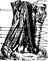
Figure 1. Course of the recurrent nerve on the right side: 1 - fourth cervical nerve; 2 - superior cardiac nerve; 3 - phrenic nerve; 4 - inferior thyroid artery; 5 - middle cervical ganglion; 6 - brachial plexus; 7 - subclavian loop (Vieussens); 8 - middle cardiac nerve; 9 - esophageal branches; 10 - recurrent nerve; 11 - tracheal branches; 12 - inferior laryngeal nerve; 13 - anterior branch; 14 - posterior branch; 15 - communicating branch with the inferior laryngeal nerve; 16 - external branch and internal branch (of the superior laryngeal nerve).
infection, trauma, etc., can be noted. In infection and intoxication, the recurrent nerve is affected together with other branches of the vagus nerve and with other cranial nerves; the paralysis is mostly bilateral. In acute and chronic diseases of the brainstem, the nuclei of the vagus nerve giving rise to the recurrent nerve also suffer; the process is also predominantly bilateral (see Polioencephalitis, Bulbar paralysis). In all organic lesions of the recurrent nerve, paralysis of the dilator
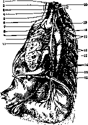
Figure 2. Arrangement of the recurrent nerve on the left side: 1 - middle cervical ganglion; 2 - middle cardiac nerve; 3 - first thoracic ganglion; 4 - subclavian loop (Vieussens); 5 - inferior cardiac nerve; 6 - superior cardiac nerve; 7 - superior cardiac branch of the vagus nerve; 8 - cardiac plexus; 9 - inferior cardiac branch of the vagus nerve; 10 - anterior bronchial branch; 11 - posterior bronchial branches; 12 - lesser splanchnic nerve; 13 - greater splanchnic nerve; 14 - anterior esophageal plexus; 15 - sympathetic trunk; 16 - esophageal branches; 17 - communicating branches; 18 - recurrent nerve; 19 - left vagus nerve; 20 - inferior cervical ganglion.
of the glottis (posterior cricoarytenoid muscle) occurs first—the Semon-Rosenbach law—and only in further development
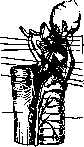
are other muscles innervated by the recurrent nerve affected, through the synergistic action of which narrowing, i.e., closure of the glottis, occurs. According to the Semon-Rosenbach law, the
Figure 3. Muscles of the pharynx and larynx innervated by the recurrent nerve: 1 - lateral thyrohyoid ligament; 2 - superior horn of the thyroid cartilage; 3 - hyothyroid membrane; 4 - branch for the transverse arytenoid muscle; 5 - posterior cricoarytenoid muscle; 6 - branches of the inferior laryngeal nerve to the posterior cricoarytenoid muscle; 7 - esophagus; 8 - trachea; 9 - branch to the lateral cricoarytenoid muscle; 10 - lateral cricoarytenoid muscle; 11 - thyroarytenoid muscle; 12 - branch to the thyroarytenoid muscle; 13 - thyroid cartilage; 14 - superior laryngeal nerve (internal branch). dilators enter into function first and only later the constrictors. Since the recurrent nerve is the main motor nerve of the larynx, innervating both the dilators and the constrictors of the glottis, in its diseases both muscle groups are paralyzed, of which only one (cricothyroid muscle) is innervated by the superior laryngeal nerve and plays such an insignificant role in the movements of the vocal cords that its activity, as opposed to all other muscles, is not taken into account. As for the muscle that widens the glottis (posterior cricoarytenoid muscle), its paralysis is the first sign of recurrent nerve disease. The Semon-Rosenbach law is currently universally recognized for all progressive diseases affecting the trunk of the recurrent nerve itself, whereas in nuclear and radicular diseases its significance is disputed (Körner). Upon examination of the larynx, one can see, along with paralysis of the muscles closing the glottis, incomplete abduction of one or both vocal cords during breathing. In this case, the cord remains close to the midline; it assumes a position intermediate between adduction and abduction, the so-called cadaveric position, and no longer moves toward the medial line during phonation, whereas the cord on the healthy side crosses the midline, and the arytenoid cartilages cross. At the moment of inhalation, the vocal cord on the affected side remains at rest, and only the healthy one assumes the abduction position. In bilateral paralysis of the recurrent nerve, both vocal cords assume the cadaveric position and cannot be moved out of this position during speech and breathing, due to which aphonia and significant respiratory disturbance are observed. When the brainstem is affected, simultaneously with paralysis of the laryngeal muscles, paralysis of the soft palate and pharyngeal muscles is observed, making swallowing also difficult. When pressure on the nerve is removed or the inflammatory process improves, the function of the muscles that open the glottis returns first, followed by the muscles that narrow it. Treatment. In the majority of cases of recurrent nerve disease, treatment is symptomatic, except for paralyses of syphilitic origin, for which treatment with mercury is used. In bilateral paralysis of the laryngeal muscles, tracheotomy must be performed.
Kiselev.

REMAK, Robert (1815-1865), one of the greatest German neurologists. In 1838 Remak graduated from the Berlin medical faculty.
R. Remak. Due to prevailing antisemitism, Remak was for a long time not admitted to teaching and only in 1847 began to lecture, being the first Jewish privatdozent in Prussia. Remak's main contribution lies in the study of the fine structure of the nervous system, particularly the autonomic nervous system. He showed that the fibers of the sympathetic nerve lack a medullary sheath. Such non-medullated fibers, at the suggestion of Kölliker, were named "Remak fibers" in honor of Remak. The fibrils comprising them were likewise named "Remak" fibrils. Another contribution of Remak lies in the field of clinical neurology: he was the first to introduce the direct current into electrotherapy.
Related articles
Mentioned in
Cite this page
“Recurrent Nerve.” Soviet Medical Encyclopedia. English translation of Bolshaya Meditsinskaya Entsiklopediya, 1st ed. (Moscow, 1928–1936), ed. N. A. Semashko. https://sovietmedicalencyclopedia.com/article/recurrent-nerve-2/