Microscopic Technique
Historical document, translated for reference. It reflects medical knowledge of the 1920s–30s and is not medical advice.
Summary
This article explains the proper handling and maintenance of microscopes, including cleaning procedures, protection from dust and sunlight, and warnings against disassembling optical components. It also covers the preparation of specimens for microscopic examination.
Encyclopedia article (1928–1936)
Microscopic Technique, in the narrow sense, is the technique of microscopy and contains rules for handling the microscope and auxiliary apparatus for the rational use of the optical properties and mechanical design of these instruments; in the broader sense, M. t. also includes the technique of preparing objects for microscopic examination, i.e., histological technique (see).
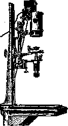
The first condition when working with a microscope should be the most pedantic observance of cleanliness; therefore, before starting work, it is necessary to inspect and, if necessary, thoroughly clean the optical and mechanical parts of the microscope. The same must be done after finishing work with the microscope. When cleaning optical glasses, it is necessary to use a clean old linen or silk rag, washed many times, so that no mineral particles that could irreparably damage the polished surface of the optical glasses can be found in it. Objectives must never be unscrewed under any circumstances, as this may disrupt the accuracy of centering and the distances between individual members of the system. When wiping the objective, its surface must be moistened with exhaled air; if this does not remove the contamination, then the rag can be moistened with gasoline or xylene, but certainly not with alcohol, and then thoroughly wiped dry. The mechanical parts of the microscope are best wiped with gasoline or clean kerosene; they should not be lubricated with any oil. In any case, even the mechanical parts of the microscope should not be unscrewed without extreme necessity, and it is better to entrust the necessary cleaning to an experienced mechanic or send the instrument to the factory. During non-working hours, the microscope should be carefully protected from dust, covering it with a glass cover or hiding it in the wooden case that comes with the microscope. The microscope should never be left under direct sunlight, especially in warm weather and in a southern climate, as this may cause the optical parts to overheat and the glasses cemented with Canadian balsam to separate from each other, which will ruin the objective and it will inevitably have to be sent to the factory for recementing.
When possible, the choice of a place for the workbench should always favor a window facing north, because under such conditions natural daylight can be used almost all the time, which is more convenient for the eyes, and in the southern latitudes of the USSR makes it possible to use natural light all day. Illumination by direct sunlight is not suitable for a microscope, as it causes a number of inconveniences: entoptic phenomena in the eye, sharp diffraction rings, and blinds the eyes. It is best to use diffused daylight from a brightly lit white wall or a light cloud. When it is impossible to use daylight, and often specifically when examining objects, a light source is used: a kerosene lamp, the flame of a gas burner, an electric light bulb. All such light sources produce a yellow flame, and the colors of the preparation change their hues significantly. To avoid this under artificial light, depending on the quality of the lamp, it is necessary to insert a more or less dark cobalt glass into the special holder of the illumination device, transparent or frosted (if the light is very harsh). Among artificial light sources for the microscope, the best are special lamps with incandescent bulbs (half-watt) or the "Lilliput" arc lamp. The latter provides a very good light, close in shade to daylight and requiring only the lightest cobalt glass (see Microscope). Special lamps connected to them are attached to some instruments. When using the microscope, a very important condition must be considered to be the absence of bright illumination of the workbench (especially with a monocular microscope), because brightly lit objects on the workbench, giving a bright image in the other eye, distract attention from the microscopic picture even for an experienced microscopist. Therefore, the surface of the workbench should be painted black or covered with dark paper. It is very convenient to make sliding shelves with rims in the workbench for placing preparations in them—they do not collect dust, and there are no objects on the table causing reflections. The chair in front of the table (preferably an armchair) should be of such a height that sitting is comfortable, because with intensive work one has to sit at the microscope for hours. For the convenience of microscopy, the stand has a hinge for tilting; therefore, it can always be tilted so that the observer's body is in a restful position. When microscoping with a monocular microscope, the second eye must certainly be kept open, and the beginner should from the very beginning develop the habit of ignoring the visual images in the free eye. This is of very great importance, because with the open free eye, convergence and accommodation of the eyes are eliminated; consequently, the working eye remains at rest, and the sharpness of the contours is set by the micrometric screw without any participation of the eye muscles. Only under this condition is prolonged microscopy possible without eye fatigue. It is very useful to learn to look with both eyes alternately. If this for some reason proves unattainable, it is better to learn to look with the left eye, then it is more convenient to draw the preparation by acting with the right hand and looking at the paper and pencil with the right eye. When using high-power systems, as well as when examining with a dark field of view, it is useful to concentrate the beam of rays from the lamp using a collecting lens placed separately or connected to the lamp itself, or using the so-called bull's-eye condenser (see Microscope). To illuminate the microscope faster, it is best to remove the eyepiece and, looking from above, catch the image of the light source produced by a single objective, and, turning the mirror, bring it to the center of the field of view; then, putting the eyepiece back in place, one can be sure that the field of view of the microscope is illuminated, and one only has to slightly adjust the mirror so that uniform illumination of the entire field of view is obtained. When setting the mirror, the following rule should be followed: when illuminating with a beam of parallel rays of light, i.e., from a distantly located light source, a flat mirror is taken, and with a light source located close to the microscope, i.e., when illuminating with a beam of diverging light rays, a concave mirror is taken. A flat mirror does not change the path of the rays, and therefore the condenser, designed in such a way that parallel rays are brought together by it at a distance approximately equal to the usual thickness of the microscope slide, will gather the rays in the plane of the preparation and give the best illumination; if the glass thickness is not quite suitable, it is easy by moving the rack and pinion or the screw of the illumination device to bring the image of the light source to the proper place. With artificial lighting, the light source should be placed so that it falls in the main focus of the concave mirror; then the rays will go from it in a parallel beam, and the lighting conditions will be analogous to the previous one. In some cases, even with daylight, one has to take a concave mirror: when the window frame is so thick that it is drawn in the field of view of the microscope, or when such objects are located in front of the window which cannot be eliminated and which are projected into the field of view of the microscope. Compliance with the specified rule regarding the mirror is especially important when using high-power systems—dry and immersion. In the absence of a condenser for light concentration, it is better to put a concave mirror. The objective and eyepiece should be set according to the purpose of the study; when studying the general plan of the structure of an organ (and microscopic study should begin with this), an objective with a large focus and a wide field of view should be taken. The eyepiece should also be taken weak so as not to narrow the field of view of the objective. For an especially large field of view, special eyepieces with a large front lens are arranged; by their diameter they do not fit the usual sliding tube of the microscope, and for their use this tube has to be unscrewed and the entire eyepiece screwed into its place. When moving to the study of small details of the preparation, a high-power objective is substituted, while the eyepiece is preferably left weak, unless the eye is able to clearly distinguish the details of the picture drawn by the objective. Only for very small details can high-power eyepieces be taken, and even then, if the objective has a very good correction in the sense of eliminating spherical and chromatic aberrations. In this regard, apochromats deserve special attention, which can be used with the strongest eyepieces. However, the choice of an eyepiece to a strong degree depends on the individual characteristics of the investigator. Some prefer strong eyepieces. When using high-power objectives with a large aperture (above 1.0), to fully utilize the resolving power of the objective, it is necessary to place a drop of cedar oil between the condenser and the coverslip; objectives with a high aperture of 1.30 and 1.40 also require a condenser with an aperture of 1.40. Having received new objectives, they should be checked regarding the degree of correction and the magnitude of the aperture. To test the correction of the objective, Abbe's test plate (Abbe's Testplatte), manufactured by Zeiss, is used. It is a microscope slide on the silvered surface of which wide light stripes with jagged edges are drawn, covered with coverslips of various thicknesses, from 0.10 to 0.25 mm, or by one narrow and long glass, the thickness of which from left to right varies from 0.10 to 0.25 mm (new model). To check the spherical and chromatic correction, let us set the test plate, choosing the thickness of the coverslip that is engraved on the objective mount, usually 0.17 mm, and the tube length adopted by the given firm. With full correction, the black stripes will be visible completely sharply and without any colored fringes. With oblique illumination in achromats, a yellow-green fringe will be visible on one side, and violet, purple, and pink on the other, but the edge will remain sharply outlined. If spherical aberration is not completely eliminated, the edge will appear blurry. It may turn out that this blurriness disappears at a different thickness of the coverslip; in this case, the indication of the coverslip thickness on the objective is erroneous, and it should be corrected. To a weak degree, correction inaccuracy can be compensated by lengthening or shortening the microscope tube; to a greater extent, this is achieved by rotating the correction collar ring, if any is available. In case an apochromat is available, even with oblique illumination there should be no colored fringes along the edge. To set oblique illumination in large lighting apparatuses, there is a rack and pinion that allows the diaphragm to be moved out of the center. The degree of oblique illumination is easily controlled by removing the eyepiece and looking into the microscope from above; at the same time, the illuminated circle in the field of view of the microscope should be visible at the edge of the field of view. To determine the aperture of the objective, a special device built by Abbe and bearing the name of apertometer serves. It is a thick plate of plate glass, the rear edge of which is ground at an angle of 45°. The front edge, ground at a right angle, forms a semicircle, the center of which is occupied by a silvered circle with a transparent center.
Two pointers move along the edge, with their points directed toward each other. Divisions are marked here on both sides of the middle line, showing the numerical values of the aperture. Let us place the apertometer on the microscope stage (without a mirror) and illuminate its front edge with a lamp as shown in the figure (Fig. 1). Let us slide the points of the pointers until they touch at the middle line. With the usual arrangement of the eyepiece and objective, let us set the silver circle of the apertometer with its transparent center so that both points are visible right in the middle. Now, without changing the setting, let us take out the inner drawtube of the microscope and screw into its lower end a special system attached to the apertometer, thus forming an inner or auxiliary microscope. Raising and lowering the inner tube, we will find a sharp image of the inner circle of the apertometer and the ends of the pointers. Since the rear edge of the apertometer is cut at an angle of 45°, complete internal reflection occurs here, and the rays travel as if the entire apparatus were located along the axis of the microscope. Now we will begin to evenly push the pointers of the apertometer apart to the sides until their ends touch the edges of the field of view. By looking at the divisions of the apertometer, we directly read off the aperture of the objective under test. The thickness of the microscope slide plays no special role in ordinary diascopic examination, but in dark-field examination it must be precisely matched to the focus of the condenser, since the intersection of the rays must necessarily occur in the plane of the preparation, without which many reflexes will appear, distorting the picture. The thickness of the coverslip, on the contrary, strongly affects the image quality, causing the phenomenon of aberration (see Microscope), which is why, especially with high-power systems, one should always select glasses of corresponding thickness. For a complete characterization of an objective, one should check to what extent it is capable of transmitting fine details of structure, i.e., its resolving power. Test objectives for this purpose are Nobert or Grayson plates with systems of lines drawn with a diamond on glass. The systems are designated by numbers, and in the densest system the lines are spaced from one another by an interval equal to 0.2 µ. Also very convenient are sets of diatom frustules manufactured by Möller. In them, diatoms of varying fineness are arranged in a single row, starting from Triceratium favus to Amphipleura pellucida, a total of
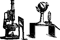
FIG. 1. 20 numbers. Moving from the coarsest diatom to finer ones, it is possible to determine the quality of the system under test and its resolving power. A good oil immersion apochromat should resolve all diatoms (the latter under oblique illumination). (For the limits of resolution for various objectives, see Microscope.) Monocular microscopy causes significant fatigue even in accustomed microscopists. The introduction of a double tube into the manufacturing technique of microscopes introduces a very significant improvement in this regard. When setting up a preparation with a binocular attachment or tube (preferably), one should be guided by the same rules as in monocular microscopy; if there is a difference in the refraction of the eyes, each eyepiece should be set so that the image is clearly visible to each eye individually, for which a worm gear is arranged in one of the eyepieces, bringing the eye lens closer or moving it away. It is also necessary to precisely set the distance between the centers of the eyepieces corresponding to the distance between the centers of the observer's pupils. Under these conditions, stereoscopicity is obtained, insofar as it is possible with a single objective, and complete rest for the eyes. Less convenient for binocular microscopy with a single objective are the Reichert and Leitz binocular attachments and the Zeiss "bitukni". The Reichert and Leitz attachments, inserted in place of the eyepiece, lengthen the microscope tube, which creates inconvenience for the position of the observer's body and significantly weights the microscope tube. The Zeiss "bitukni" is more convenient because it stands obliquely and therefore provides more convenience for the position of the body, and in addition it is screwed in instead of the drawtube and does not lengthen the microscope tube as much. The Reichert attachment is equipped with two systems of terminal collecting lenses facing the objective: one for the compound microscope and the other for using the attachment as a binocular magnifier, for which a special heavy tripod is attached to it upon request, making it possible to use it without a microscope tripod (as a dermatoscope, dissecting magnifier, etc.). When examining opaque objects, to study their surface with low-power systems, illumination is used by means of a concentrated beam of light passed through a collecting lens located above the microscope stage. It is more convenient to use vertical illuminators (opak-illuminators). Since ordinary objectives are mounted in such a way that when they are screwed onto the revolving nosepiece their focal planes coincide, they are usually unsuitable for microscopy with a vertical illuminator, and objectives with a short mount should be used for this purpose, since only under this condition is the proper image brightness obtained. If one has to study a surface not covered with a coverslip, one should also take an objective in which the calculation is made for the absence of a coverslip. For examination with top light, it is necessary to have a light source bright enough, capable of being set at the same level as the vertical illuminator and closed on all sides except the one facing the instrument. Some firms, for example Reichert, include a light source in the form of a compact low-voltage bulb in the instrument itself. Having set the light source so that the beam of rays goes horizontally straight into the small mirror of the vertical illuminator at a certain combination of objective and eyepiece, one can then change the objectives, provided the difference between their foci is not very large. Otherwise, the setup has to be made anew. This is especially necessary if the objects of study differ greatly in thickness. But setting up the illumination with vertical lighting is quite troublesome, and in recent years tripods have been released in which, by means of a rack and pinion, the microscope stage can be lowered and raised, then fixed with a clamp, which is very convenient. A number of special condensers and ultramicroscopes serve for examination in a dark field. The latter instrument, which sends light into the preparation at a right angle to the microscope axis, is almost completely inapplicable for the study of histological and bacteriological preparations; it has found application in colloid chemistry and in the study of solutions. For microscopic purposes proper, only special condensers are suitable (see Microscope). When starting an examination in a dark field, it is necessary to place a drop of cedar oil under the slide, between it and the illuminator, to make the medium between the glasses homogeneous and avoid the deflection of rays that have passed through the condenser and travel so obliquely to the surface that the phenomenon of total internal reflection is possible. After that, having set up the preparation, it is necessary to choose the proper position for the mirror and for the distance between the slide and the condenser. To do this, having illuminated the microscope mirror so that its middle is brightly lit, the proper position of the mirror and illuminator is set with a low-power objective and eyepiece. It is necessary that in the middle of the field of view of the low-power system a light circle is clearly visible, as shown in the accompanying figures (Figs. 2 and 3). Having thus set the illumination, one can
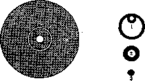
Figure 2.
Figure 3.
Figure 2. Illumination in the field of view. Figure 3. 1-incorrect position of the mirror; 2-incorrect position of the condenser; 3-correct illumination. One can then proceed to the objective with which it is planned to conduct observations. Precise adjustment to a high degree contributes to the clarity of the image and eliminates the strongly interfering reflections and circles of light scattering. With precise adjustment and correspondence of the objective aperture to the condenser used, the image becomes so clear that it is possible to examine and even photograph in living condition the cell organelles (e.g. mitochondria). The effect of dark field and a very clear image can be obtained, especially with weak systems, without a special condenser. For this, a central diaphragm is placed in the holder of the illuminator's diaphragm instead of the iris diaphragm, allowing only marginal rays to pass into the objective, i.e., blocking the absolute maximum (see Microscope). With a suitably chosen central opaque disk and weak objectives, an image is obtained whose purity leaves nothing to be desired. The same effect is achieved by blackening the central part of the objective's front lens or by inserting a central diaphragm into the objective in place of the image of the light source (as in experiments with Abbe's plate). The latter two methods of obtaining dark field vision, however, are not convenient, and it is much better to use appropriate condensors. Research in dark field vision gives very much when examining fresh and living objects. Thus, it is possible to detect microorganisms in a liquid and observe their movement, study tissue cultures, make ordinary photomicrographs, and also produce motion pictures. Research under conditions of light polarization-see Polarization. In experiments with ultraviolet rays, all the above-mentioned equipment, as well as the condenser, must be made of quartz, as glass does not transmit ultraviolet rays. For studying fresh and living objects and for experiments with them, there are a number of devices: hanging drop (see), moist chamber, microaquarium, electric chamber, and for cells of warm-blooded animals-warming stages and warming microscope. To set up a moist chamber, used for studying any films from the body of animals, e.g., the frog's mesentery, a cork disk or plate with a hole is taken, the edges of which are stretched with a film, covered with a coverslip. To prevent drying, a slow current of liquid is led to the preparation or it is surrounded by strips of filter paper moistened with physiological solution or Ringer's solution, isotonic with respect to the object being studied. The microaquarium is a small flat reservoir (glass). For this purpose, it is best to attach on a ground slide using Canadian balsam or Mendeleev's putty a quadrangular or circular low frame with a polished upper edge, on which a coverslip is applied, also greased with fat or vaseline. Holes should be made in the frame, through which thin tubes for the inflow and outflow of liquid are passed. Since the microaquarium is usually set up for studying moving objects, the medium used varies depending on the speed of movement. When studying slow movements, ordinary aqueous solutions of salts or organic compounds, as well as natural body fluids of the animal being studied, can be taken; if the movements are very fast, it is useful, and sometimes necessary, to make the medium more viscous to slow down the movement. For this purpose, a greater or lesser amount of mucus is mixed into the water, e.g., tragacanth, agar, or a neutral gelatin solution. In such a medium, movement is greatly slowed down, making it possible to study its various phases. For studying the effect of electric discharges, conductors soldered into the wall are added to one of the above-mentioned devices for connection with simple (preferably platinum) or non-polarizable electrodes. The above-mentioned devices for studying living objects can be placed on a warming stage or in a Zeiss warming cabinet together with the microscope. The best and most convenient stage is the Leitz warming stage, which makes it possible to regulate the temperature as needed-to heat with electric current or to cool with liquid CO2 (for which a well-functioning reduction valve is essential, allowing current to pass under very low pressure). The above-mentioned equipment makes it possible to use vital stains (see) and to conduct experiments either directly by hand or with the help of a micromanipulator (see). Fresh material for studying isolated elements can be directly teased apart on a slide with dissecting needles. Covering it with a coverslip, test solutions can be passed from the side by applying well-cut pieces of filter paper to the edge of the coverslip. By sucking the liquid from under the coverslip with such a paper, the test solution or desired dye can be introduced. The material to be fixed is examined either in the teased state or on sections, stained according to the purpose of the research. (Methodology of isolation, fixation, preparation of sections and staining of specimens-see Histological technique.) When studying bacteriological material, special stains are used, the recipes and methodology of which are described in microbiology reference books. When studying unstained specimens, the elements of which are visible only due to the difference in refractive indices, it is necessary to use a narrow diaphragm to obtain the sharpest contours of the diffraction image. In addition, the medium for mounting the specimen should have a low refractive index, because otherwise the entire difference in refractive indices disappears and the details will not be visible. Such mounting media are water, diluted glycerin (pure or acidified), levulose, glycerin-gelatin. Specimens mounted in such a medium must be for storage framed with asphalt varnish or special alcohol varnishes sold ready-made. Asphalt varnish, sold in a thick state, must first be diluted with xylene or turpentine to the consistency of liquid syrup. When wishing to preserve an interesting specimen for a long time, a frame of such width is applied with a brush or stick along the edges of the coverslip so that it completely covers the edges of the coverslip and the adjacent part of the slide; it is better to make the frame wider and less elegant than to underfill the edges and risk drying out the interesting specimen. When studying brightly stained specimens, the illuminator's diaphragm should be taken wide, however, not so wide that the contours of the specimen are blurred. Since brightly stained specimens are usually mounted in Canadian balsam or dammar resin dissolved in xylene, which evaporates quickly, the application of a frame is unnecessary and only needlessly dirties the specimen. When studying a specimen, it is sometimes necessary to sketch interesting parts of it. In the simplest case, this can be done directly by eye, and some micro-painters always draw this way, but a non-artist can use this method only for sketching outlines, plans, and notes. For more careful sketching, drawing eyepieces or Abbe's special drawing apparatus (see Microscope) are used, as well as a camera lucida attached to the eyepiece. These devices make it possible to simultaneously see both the microscopic image and the tip of the pencil on paper. When using them, only the outlines are drawn on the paper, while the detailed drawing is more conveniently done along the outlined contours without the apparatus. The apparatus according to Vasiliev is very convenient for sketching. With the help of a flat mirror in a frame, placed obliquely, with an inclined microscope stand, the specimen can be projected onto paper and its contours accurately drawn (see Microscope). A more complex apparatus for the same purpose is Edinger's embryograph. For sketching the microscopic image projected onto paper, it is necessary to shield extraneous light with screens, and it is even better to work in a dark room; then all details are well visible and easy to sketch. The most objective way of reproducing a microscopic image is considered to be photomicrography (see). Finished specimens should be stored in special boxes or folders that protect them from dust and the action of light, from which dyes can fade. Each specimen must be provided with a label with a number or a description of the treatment, staining, and origin of the material. When conducting microscopic research, it is very useful to keep a work journal in which is noted when and what was taken for research, how it was fixed, how the sequential processing was carried out up to staining and the medium in which the section is mounted, as well as what was found in such a specimen. The prepared material can be stored for varying times depending on the properties of the material itself and its treatment. In alcohol, specimens can be stored for a very long time (years), especially after fixation with formalin, but old material usually stains worse than fresh material.
The material is preserved better if 10-15% pure glycerin is added to the alcohol. Objects embedded in paraffin can be preserved without any changes for an indefinitely long time. Celluloid embedding is not suitable for long-term storage, especially if the pieces are mounted on wooden or cork bases. Celluloid blocks, when stored in alcohol, after some time become acidic due to the decomposition of celluloid, and in addition, tannins are extracted from the wood or cork, as a result of which the sections either do not stain at all or give a distorted coloration. Blocks (without wood) are preserved longer if 10-15% pure glycerin is added to the alcohol. However, celluloid blocks can also be made stable and capable of withstanding indefinitely long storage if, immediately after the celluloid has hardened, they are impregnated with turpentine in 70% alcohol. For this purpose, blocks from 70% alcohol are transferred to 90% alcohol, changing it two or three times; after that, they are suspended in a tall cylinder in a mixture of two parts absolute alcohol and one part chloroform. The next day the mixture is changed, and then, after transferring the embedded material to turpentine, it is left in an open dish for a day and on the following day transferred to a fresh portion of turpentine. After 2-5 days, depending on the size of the pieces, they are removed and stored in a jar with a good cork, placing under and over them a wad of cotton wool soaked in turpentine. When cutting such pieces, the microtome knife is also moistened with turpentine, and the sections can be conveniently stored between sheets of thin smooth paper soaked in turpentine. Throughout the entire described process of transferring to turpentine, it is necessary to ensure that the blocks remain completely transparent at all times.
Related articles
Mentioned in
Cite this page
“Microscopic Technique.” Soviet Medical Encyclopedia. English translation of Bolshaya Meditsinskaya Entsiklopediya, 1st ed. (Moscow, 1928–1936), ed. N. A. Semashko. https://sovietmedicalencyclopedia.com/article/microscopic-technique/