Inguinal Region
Historical document, translated for reference. It reflects medical knowledge of the 1920s–30s and is not medical advice.
Summary
This article from the 1928–1936 Great Medical Encyclopedia describes the anatomy, fascial layers, blood supply, and muscular structure of the inguinal region. It provides detailed descriptions of the boundaries, the inguinal ligament, and the structures forming the inguinal canal.
Encyclopedia article (1928–1936)
INGUINAL REGION (regio inguinalis) is located at the lower part of the abdomen and represents a rectangular triangle, the sides of which are: below—Poupart's ligament, above—a part of the line interspinalis superior, medially—a line running along the outer edge of the rectus abdominis muscle. Within these limits, the most important and studied is the inguinal triangle (see Inguinal canal), separated from the thigh by a deep fold called the groin. The skin of the inguinal region is delicate, mobile, covered with hair starting from the middle of the inguinal fold and passing onto the pubis; in men, the hair extends beyond the inguinal region, rising up to the navel. The hairy part of the skin is abundantly supplied with sebaceous and
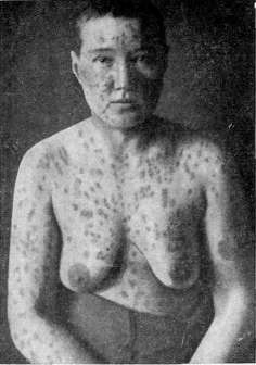
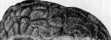
Figure 3.
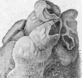
Figure 1.
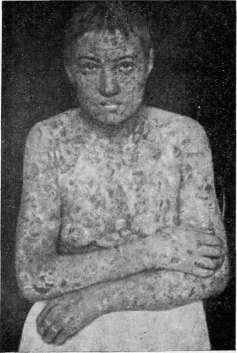

Figure 4.
Figure 2.
Figure 1. Pemphigus foliaceus (initial stage). Figure 2. Pemphigus foliaceus (the same case several years later). Figure 3. Pacchionian granulations. Figure 4. Pericarditis - cor villosum. Figure 5. Infiltration of the renal pelvis wall (a) by leukocytes in pyelitis; b - epithelium, partially desquamated and accumulated in the lumen (c); d -
part of the renal parenchyma, and sweat glands. The subcutaneous fascia consists of two layers that separate well from each other in individuals with developed subcutaneous fat. The first layer—the superficial fascia of the abdomen (fascia subcutanea)—continues into the subcutaneous fascia of the thigh and scrotum, where it acquires smooth muscle fibers (tunica dartos); within its thickness lies the superficial epigastric vein of Haller (vena subcutanea abdominis Halleri), running from above and medially and emptying into the saphenous vein. The second fascial layer—the deep lamina of the subcutaneous fascia (lamina profunda fasciae subcutaneae, s. lamina Thompson)—is separated by loose cellular tissue from the aponeurosis of the external oblique abdominal muscle. Within its thickness, and sometimes between it and the aponeurosis, runs the superficial epigastric artery, anastomosing with branches of the deep circumflex iliac artery and perforating branches of the inferior epigastric artery. In the lower-medial part at the level of the lower edge of the external inguinal ring runs almost transversely the external pudendal artery, which is easily injured during hernia repair with a low incision. The arteries are accompanied by corresponding veins emptying into the saphenous vein. The thickness of the subcutaneous fatty layer reaches 4-5 cm in obese people; it is especially developed in the region of the inguinal triangle, closer to the mons veneris, and sharply thins toward the inguinal fold. The fatty tissue separates both fascial plates and the aponeurosis; this must be kept in mind during hernia repair, as the deep lamina can easily be mistaken for the aponeurosis of the external oblique muscle. Lymph nodes are absent; lymph vessels drain into the inguinal nodes located in Scarpa's triangle. Some authors acknowledge the presence of 2-3 small lymph nodes above Poupart's ligament. The skin is innervated by branches of the 12th intercostal nerve, ilioinguinal nerve, and genitofemoral nerve, which do not have a precise localization. The muscular part of the abdominal wall of the inguinal region consists of the lower ends of the external and internal oblique and transverse muscles. From the level of the anterior superior iliac spine, the external oblique muscle transitions into a tendinous aponeurosis, the fibers of which run in an oblique direction from above downward and from without inward; folding into bundles-ribs of various thickness and width, they in places closely adhere to each other, while in other places they are separated by connective tissue layers, due to which the aponeurosis has the appearance of a perforated plate. The bundles are fastened by arched, transversely running fibers that form a continuation of the aponeurosis of the external oblique muscle of the opposite side (collateral fibers, arcuate fibers). Depending on the combination of these fibers, the aponeurosis can be strong, dense, weakly developed, fan-shaped, or separated into fibers. In women, the aponeurosis is denser than in men; the tendinous bands are wider and thicker, closely adjacent to each other, and there are more arched fibers. The lower edge of the aponeurosis is turned inward and upward in the form of a trough and, having merged with the fascia lata, forms the inguinal ligament (ligamentum inguinale, ligamentum Pouparti), which is attached on one side to the anterior superior iliac spine and on the other to the pubic tubercle, where it expands in the form of a triangle: one side of it goes deep to the bone (lacunar ligament of Gimbernat), bounds the vascular lacuna with a sharp edge and transitions into the iliopectinaceous arch of Cooper; the other side goes in the form of a cord forward along the anterior surface of the pubic bone to the midline; the third side forms the reflected inguinal ligament, or Colles' ligament (Fig. 1). In the region of the inner end of Poupart's ligament, directly above it, the fibers of the aponeurosis split, forming a cleft—the external, or subcutaneous, opening of the inguinal canal (superficial inguinal ring), through which the spermatic cord exits
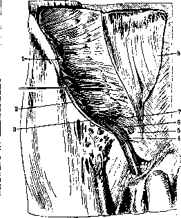
Figure 1. Muscles of the abdominal wall in the region of the inguinal canal: 1 and 7 - internal oblique muscle; 2 - external oblique muscle; 3 - cremaster muscle; 4 - reflected ligament of Colles; 5 - rectus muscle; 6 - pubic part of the internal oblique muscle; 8 - aponeurosis of the external oblique muscle.
tic or round ligament. The tendinous bundles forming this opening constitute the external, or inferior crus (crus, s. columna lat.) and the internal, or superior crus (crus, s. columna int.). The lateral crus is stronger and thicker and, merging with Poupart's ligament, is attached partly to the pubic tubercle and partly continues forward to the pubic symphysis. The upper margin of this crus is rather sharp and slightly concave in its lower part; part of the fibers, turning under the spermatic cord, pass into the fibers of the reflected inguinal ligament of Colles. The internal crus is broader than the lateral, is attached to the pubic bone at a varying distance from the pubic tubercle (sometimes up to 2 cm) and, continuing downwards and medially, becomes part of the superior suspensory ligament of the penis or clitoris (lig. suspens. sup. penis s. clitoridis). In this crus, when the aponeurosis is weak, a long cleft is sometimes formed through which the hypogastric branch of the iliohypogastric nerve passes; sometimes this cleft continues downwards to the pubic bone, and then two subcutaneous inguinal openings are formed--lateral and median, each of which can serve as a site for hernia emergence. The third, or posterior crus, also called the reflected inguinal ligament (lig. ing. reflex. Collesi, s. lig. Collesi), begins at the junction of Poupart's ligament with the lateral crus, the fibers of which, turning, hide under the spermatic cord, bounding the inguinal ring posteriorly (Fig. 1). Beginning from the outer edge of Poupart's ligament, it runs along its groove, located on Gimbernat's ligament, thus having a part in common with it; further it goes posteriorly and upwards to the anterior wall of the sheath of the rectus abdominis muscle and on the midline passes into the aponeurosis of the opposite side. The lateral margin gradually merges with the posterior wall of the inguinal triangle; it is partially covered by the internal crus of the cremaster muscle, and it can be well traced when the ring is wide, if the spermatic cord and the cremaster are lifted up. The crura bounding the canal, as well as the aponeurosis, are fastened by arciform fibers coming from the aponeurosis of the opposite side. These fibers (fibrae arciformes, f. intercrurales) run in a transverse direction and round off the angle at the place where the crura diverge, giving the ring a various shape (see Inguinal Canal). Not being limited only to the region of the crura, they throw themselves across the entire cleft in the form of a plate (fascia intercruralis), which upon passage of the spermatic cord stretches out and spreads onto it, forming Cooper's fascia (fascia cremasterica Cooperi), which constitutes the first tunic of the spermatic cord. Some anatomists believe that Cooper's fascia represents a continuation of the deep lamina of the subcutaneous fascia, which, attaching to the edges of the external inguinal opening, spreads onto the spermatic cord during the descent of the testis into the scrotum. The internal oblique abdominal muscle and the transversus abdominis muscle arise from the groove of the outer two-thirds of Poupart's ligament; the transition into tendinous expansion along Spigelian line occurs in some almost near the very edge of the rectus abdominis muscle, in others--not reaching it by 2-4 cm. Interweaving with each other, both muscles, having approached the inguinal triangle, arch over the spermatic cord, and the internal oblique muscle with its lower edge (pars pubica) tightly embraces it, so that the space not occupied by muscles will be only the place where the spermatic cord passes (Fig. 2). From the internal oblique muscle depart muscle fibers--the cremaster muscle, s. levator testis, which continues downwards onto the spermatic cord and forms a muscular sheath for it and the testis. Sometimes it runs as a continuous layer from the edge of the internal oblique muscle, sometimes it begins with two crura: the external--from the groove of Poupart's ligament and the edge of the internal oblique muscle, and the internal--from the anterior wall of the sheath of the rectus abdominis muscle and covering the aponeurotic falx and part of the reflected ligament of Colles (Fig. 2). The transversus abdominis muscle lets the spermatic cord or round ligament pass through itself; its muscle fibers are well developed until the bend over the spermatic cord; further they pass into a tendinous expansion, participating together with the edge of the internal oblique muscle in the formation of the upper margin of the inguinal triangle. But below this margin it continues in the form of a thin aponeurotic plate, which merges medially with the outer edge of the sheath of the rectus abdominis muscle, inferiorly--with the posterior lip of Poupart's ligament, laterally bounds the medial edge of the internal inguinal ring and thus constitutes the posterior wall of the inguinal triangle and canal. This plate has different thicknesses in different places. In the place where it bounds the internal and lower edges of the internal inguinal opening--a cleft in the transversus abdominis muscle (Tandler considers this cleft to be the internal inguinal opening),--the plate is reinforced by a ligament (lig. interfoveolare Hesselbachi, s. lig. Hesselbachi), which is formed from tendinous fibers.
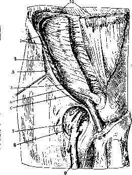
Figure 2. Muscles of the abdominal wall: 1 - m. obliquus int.; 2 - m. transversus; 3 - m. obliquus ext.; 4 - pars pubica m. obliqui int.; 5 - lig. inguinale; 6 - m. cremaster; 7 - margo falciformis; 8 - v. femoralis; 9 - v. saphena magna; 10 - m. obliquus ext. of fibers coming from the posterior lip of Poupart's ligament; in it are distinguished the lower crus, bounding the internal inguinal opening from below, and the ascending crus, bounding it from the inside; higher up the fibers are lost in the tendinous expansion of m. transversi (see figure 4 to the article Inguinal Canal). Ligamentum interfoveolare, together with the inferior epigastric artery lying directly behind it, constitutes the boundary between the external and internal inguinal fossa. Behind this ligament, sometimes somewhat medially, passes a thin muscular bundle (m. interfoveolaris), also beginning from the posterior lip of Poupart's ligament and disappearing in the muscular layers of m. transversi. The inner part of the posterior wall of the inguinal triangle is reinforced by tendinous fibers formed from fibers descending along the lateral margin of m. recti, from the aponeurosis of m. transversi, as well as from fibers coming from the anterior sheath of m. recti (falx aponeurotica inguin., s. lig. Henle). This ligament, ending in a sharp edge (proc. falciformis) or gradually merging with the posterior wall of the inguinal triangle, is covered anteriorly in part by the fibers of lig. reflexi Collesi and the internal crus of m. cremasteris; the lower end merges with lig. Cooperi. Between lig. interfov. and falx aponeurot. there is a thin plate, to which the external inguinal ring adjoins anteriorly, and posteriorly the peritoneum of the middle inguinal fossa, through which direct inguinal hernias emerge. The strength of the posterior wall of the inguinal triangle depends on the various relationships between lig. interfoveol., the thinned middle part, and falx aponeurot. The outer edge of the cleft in the transverse muscle (internal inguinal ring) is ventrally muscular, and posteriorly and laterally (the spermatic cord passes obliquely through this muscle) tendinous, reinforced by the fibers of fasciae transversae. Behind the muscles passes the transverse fascia (fascia transversa), which, having covered m. transversus, is attached laterally to the posterior lip of Poupart's ligament; in the region of the internal inguinal opening, it is carried along by the spermatic cord passing through the abdominal wall and encases it in the form of a sheath common to the spermatic cord and testicle (tunica vaginalis commun. funiculi sperm. et testis); the site of transition is somewhat drawn into the canal and is called lig. infundibuliformis. Further medially, fascia transversa adjoins the posterior aponeurotic wall of the inguinal triangle, where it is not always well expressed and represents loose cellular tissue. Tandler denies the presence of a transverse fascia in this place; in his opinion, the fascia ends at the lateral edge of the internal inguinal opening in the transverse muscle. The place of transition of fasciae transversae into fascia infundib. is considered by the majority of anatomists to be the internal opening of the inguinal canal; according to Tandler, it is located in the thickness of the transverse muscle. The preperitoneal tissue is described as one or two layers. Its thickness depends on the amount of fat, which gradually increases toward the middle. In the region of the inguinal triangle, 3 regions are distinguished: 1) the region of the abdominal opening of the inguinal canal, 2) the region of the inferior epigastric artery, and 3) the prevesical space. At the level of the abdominal opening and somewhat higher, the preperitoneal layer is poorly developed, as a result of which the peritoneum forms a depression here (fovea inguin. lat.). Medially, the fat layer thickens and forms a fold protruding into the abdominal cavity, within the thickness of which the inferior epigastric artery and the diverging elements of the spermatic cord run. The preperitoneal fat of the region of the posterior wall of the inguinal triangle is in contact with the fatty tissue of the prevesical space, being separated from it by fascia praevesicalis. The peritoneum of the inguinal region in the lateral part is even; the transitional fold is located higher or lower depending on the development of the preperitoneal fat. In the medial part, due to the folds protruding into the abdominal cavity, 3 depressions are formed - the inguinal fossae (see Abdominal Wall). Of the vessels of the inguinal region, the inferior epigastric artery, accompanied by 2 veins, requires description; it begins from the median side of the external iliac artery at a distance of 1-2 cm from Poupart's ligament, sometimes at the level of it, crosses the external iliac vein, after which it turns upwards, forming an arch. It lies in the preperitoneal tissue medial to the internal inguinal opening, behind lig. interfoveolare, forms the external peritoneal fold and serves as the boundary between the external and middle inguinal fossa. The cremasteric artery departs from it into the composition of the spermatic cord. Innervation occurs mainly due to the branching of the iliohypogastric and ilioinguinal nerves. Both nerves above the anterior superior iliac spine pass between m. transversus and m. obliq. int. and then, piercing the latter, lie between the oblique muscles. The iliohypogastric nerve runs along the edge of m. obliq. int. and, having given off a cutaneous branch above the external inguinal ring, emerges through a special cleft in the medial crus of the inguinal ring (r. hypogastricus). The ilioinguinal nerve, passing in the region of the inguinal canal somewhat lower than the previous branch, gives a branch to the spermatic cord; the terminal branches go to the skin of the pubis and scrotum. Both nerves have many variations, replacing each other. M. Ginzburg. INGUINAL CANAL (canalis inguinalis) is located in the inguinal triangle, bounded below by the lower 2/3 of Poupart's ligament, on the inside by the lateral edge of the rectus abdominis muscle, above by a horizontal line connecting this same edge with the upper boundary of the lower two thirds of Poupart's ligament. In essence, there is no canal in an adult, but there
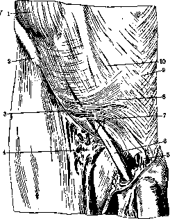
Figure 1. Abdominal wall in the region of the inguinal canal: 1 - m. obliquus ext.; 2 - spina iliaca ant. sup.; 3 - lig. inguinale; 4 - fascia cribrosa; 5 - lig. suspensorium penis; 6 - funiculus spermaticus; 7 - crura annuli inguinalis subcutanei; 8 - fibrae collateralis; 9 - linea alba; 10 - aponeurosis m. obliqui externi.
is only a cleft between the muscles, in which the spermatic cord in men or the round ligament in women is placed. If this formation is removed from a frozen corpse, the cleft will indeed have the appearance of a canal; the latter is also formed when the processus vaginalis is not obliterated. Therefore, when describing the inguinal canal, the boundaries of the spermatic cord are given. The external opening (annulus inguin. ext., s. subcutan.) and the internal or abdominal opening (annulus inguin. int., s. abdominalis) are distinguished, and finally the canal itself, in which the anterior and posterior walls, upper and lower margins are described. The external ring is located in the lower-inner corner of the inguinal triangle, in the aponeurosis of the external oblique muscle, and is bounded by the lateral and medial crura (crura, s. columna lat. et med.) (Fig. 1), below and partly posteriorly by the third crus (lig. inguin. reflexum Collesi, s. lig. Collesi) (Fig. 2). The external opening has a triangular shape, but arcuate fibers (fibrae intercrurales) passing in the aponeurosis round off the acute angle, and the opening takes the form of an oval or polygon. A transversely oval shape is observed in cases where the free edge of the median crus is significantly displaced from the pubic tubercle. The size of the subcutaneous opening is subject to large fluctuations. Tillaux determines the major length of the oval to be 2-2.5 cm. According to Yaltinsky, the size of the transverse diameter ranges from 1.2 to 3.2 cm, and the longitudinal from 2.2 to 3 cm; according to Venglovsky, the longitudinal diameter is 1.5-6 cm, the transverse - 1.1-2.5 cm. In women, the subcutaneous opening is much smaller, almost half; the shape in most cases is almost round, less often oval. The opening can be divided by a tendinous strand extending from the medial crus into 2 parts.
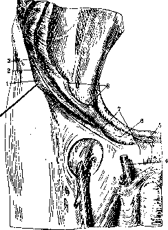
Figure 2. Section of the anterior abdominal wall parallel to Poupart's ligament: 1 - m. obliquus ext.; 2 - m. obliquus int.; 3 - m. transversus; 4 - funiculus spermaticus; 5 - m. rectus; 6 - lig. reflexum Collesji; 7 - annulus inguinalis subcutaneus; 8 - annulus inguinalis abdominis.
The internal ring is located in the region of the lateral angle of the inguinal triangle, within its posterior wall, at the border of the inner and middle third of Poupart's ligament, 1-1.5 cm above it. Being located in the thickness of m. transversi, in its dorsal tendinous part, it has the shape of a longitudinal or oval cleft (Figs. 3 and 4), bounded laterally and superiorly by the tendinous part of m. transversi, reinforced by the fascia transversa fused with its edge (Tandler); medially and inferiorly the opening is strengthened by a ligament - lig. interfoveolare Hesselbachi. Fascia transversa, being drawn through the ring by the spermatic cord into the inguinal canal in the form of fascia infundibuliformis, passes into tunica vaginalis communis funiculi sperm. et testis. This funnel-shaped depression is considered by some anatomists to be the internal opening of the canal. The inguinal canal is formed due to the fact that the internal oblique and transverse muscles pass over the spermatic cord, and the free edge (pars pubica) of the internal oblique
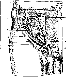
Figure 3. Annulus inguinalis anteriorly: 1 - m. obliquus int.;
2 - m. transversus; 3 - annulus inguinalis abdominis;
4 - m. cremaster; 5 - lig. inguinale;
6 - annulus inguinalis subcutaneus; 7 - funiculus spermaticus.
lus spermaticus; 8 - aponeurosis m. obliqui externi. The internal oblique muscle, when well developed, closely adjoins the spermatic cord all the way to its exit from the canal. The transversus muscle lies somewhat higher, originating from the
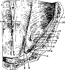
Fig. 4. Annulus inguinalis abdominis from behind: 1 - linea semicircularis Douglasi; 2 - falx inguinalis; 3 - lig. Gimbernati; 4 - lig. Cooperi; 5 - vasa femoralia; 6 - lig. ilio-pectineum; 7 - lig. inguinale; 8 and 11 - crus horizontal et crus verticale lig. Hesselbachi; 9 - annulus inguinalis abdominis; 10 - n. femoralis; 12 - fascia ilio-psoica; 13 - m. ilio-psoas; 14 - m. transversus.
cord and forms the weak posterior wall of the inguinal triangle. The canal has an oblique direction from behind forward and from above downward; its length in men is 4-4.5 cm, while in women it is somewhat longer depending on the greater width of the pelvis. Since the internal ring lies higher than the subcutaneous one (1-1.5 cm), the canal does not run parallel to the inguinal ligament, but at a certain angle to it. Boundaries of the inguinal canal: in front - fascia intercolumnaris within the limits of the external ring, above and laterally - the aponeurosis of the external oblique muscle, and further - the muscle fibers of the internal oblique and transversus muscles; the latter forms a tendinous ring - the internal inguinal aperture - in the dorsal part, while in the ventral part the muscle fibers embrace the spermatic cord anteriorly, laterally, and superiorly. The lower boundary is the groove of the medial part of Poupart's ligament, and as the spermatic cord departs upward - the fibers of the cremaster muscle and the muscle fibers of the transversus muscle. The upper boundary is the edge of the internal oblique muscle with the cremaster muscle extending from it and passing onto the spermatic cord. The posterior boundary is the posterior wall of the inguinal triangle, which, according to many anatomists, consists of the fascia transversa; some modern anatomists (Tandler, Spalteholz, Krause) believe that the posterior wall is formed by the continuation of the tendinous expansion of the transversus muscle below its clearly visible edge, merging with the posterior lip of Poupart's ligament throughout its extent medial to the internal inguinal ring and with the lateral edge of the sheath of the rectus muscle (Fig. 4). This tendinous plate has varying thickness in different places; on the outside it is reinforced by the ligament, lig. interfoveolare Hesselbachi; medially, at the inner corner of the triangle, it is strengthened by a ligamentous-aponeurotic plate - falx aponeurotica inguinalis (see Inguinal Region). The middle, most vulnerable part of this plate adjoins anteriorly the external inguinal ring, and posteriorly, covered by the thinned fascia transversa and preperitoneal fat, - the bottom of the middle inguinal fossa. In most people with moderately and poorly developed musculature, especially in the presence of a hernia protrusion, such a perfect structure of the inguinal canal is disrupted by the fact that the edge of the internal oblique and transversus muscles departs from the spermatic cord, forming a gap of various shapes and sizes, also called the inguinal canal, the inguinal triangle, or the inguinal slit. Boundaries of such an expanded inguinal canal: below - Poupart's ligament, above - the edge of the internal oblique and transversus muscles, from the middle - the edge of the rectus muscle; anterior side - the aponeurosis of the external oblique muscle, posterior - the aforementioned aponeurotic plate reinforced by lig. interfoveolare and falx aponeur. or, as usually described, the fascia transversa; the spermatic cord occupies the lower lateral section in this gap. The different shape of the inguinal canal or gap depends on the variability of the upper and middle boundaries. Yashchinsky distinguishes three shapes: triangular, fusiform, and slit-like; in the triangular shape, the lateral edge of the rectus muscle serves as the base, and the apex faces laterally; the length of the gap from the middle of the base to the apex averages 5.5 cm, with a base averaging 3.5 cm. In the fusiform shape, the average length is 7 cm, and the width is 3-7 cm. Venglovsky distinguishes oval and triangular shapes. In most cases in healthy people, the oval shape is encountered; the dimensions of the inguinal canal in this shape are as follows: length 1.2-2.2 cm, height 4.5-7.7 cm. The triangular shape is observed much less frequently: height (along the lateral edge of the rectus muscle) 1.6-4 cm, length (along Poupart's ligament) 4-6 cm. The main protective role falls not on the length, but on the height of the gap: the smaller the height, the stronger the inguinal region. In women, the inguinal gap is in most cases slit-like or oval, rarely triangular, and is filled by the round ligament running in company with the ilioinguinal nerve. The fascia transversa and preperitoneal fat adjoin the posterior wall of the inguinal canal; in the region of the internal inguinal ring, the fascia transversa is absent, since it passes into the sheath of the spermatic cord. Its elements (vas deferens and vasa spermatica) occupy the posterior periphery of the cord and the funnel and diverge along the posterior wall of the abdomen (vasa spermatica upward along the ilio-psoas muscle, vas deferens downward to the lesser pelvis, crossing the inferior epigastric artery), and therefore hernias falling into the funnel lateral to the artery will pass anteriorly and laterally to the spermatic cord in a common sheath with it and preserve these relationships throughout their extent. From the side of the peritoneum, the external inguinal fossa (fovea inguinalis lateralis) adjoins the region of the internal inguinal ring, lying lateral to the lateral fold [see separate plate (vol. III, pp. 99-100), Fig. 1], in the thickness of which the inferior epigastric vessels pass, and this fossa serves as the origin of indirect inguinal hernias. Medial to this fold is the middle inguinal fossa (fovea inguinalis media), adjoining the posterior wall of the inguinal canal; the hernia protrusion (direct hernia), encountering no obstacle on its part from the poorly developed fascia transversa and the posterior wall of the inguinal canal, emerges into the inguinal triangle, where it encounters the spermatic cord in front of it, and is located medially and posteriorly to it outside the sheaths. The formation of the inguinal canal and the complex relationships between the elements of the spermatic cord and the abdominal wall are associated with the process of development of the gonads. During their descent in intrauterine life, the peritoneum forms a protrusion along Hunter's gubernaculum (gubernaculum Hunteri), penetrating with it lateral to the epigastric artery through the abdominal wall into the scrotum or labia majora - the vaginal process (proc. vaginalis) in men or Nuck's canal (canalis, s. diverticulum Nucki, rudimentum proc. vaginalis muliebris) in women. The testis, having descended into the scrotum behind the vaginal process, drags along its path the encountered layers of the abdominal wall, which form the common sheaths for the vaginal process and the spermatic cord. The fascia transversa is drawn out into the tunica vaginalis communis funiculi spermatici et testis; the internal oblique muscle - in the form of the cremaster muscle; on the external abdominal oblique muscle, the fascia intercolumnaris protrudes, which forms Cooper's cremasteric fascia (fascia cremasterica Cooperi). Only the transversus muscle, being the least yielding, forms a complete ring; some anatomists believe that this muscle also participates in the formation of the cremaster muscle (see Testis). The vaginal process begins to obliterate at the end of the fetus's intrauterine life, turning into the vaginal ligament (lig. vaginale, s. habenula). The cause of obliteration is the presence of transverse fibers of the cremaster muscle forming transverse folds (Krymov). However, the vaginal process is not obliterated always and not throughout its entire extent; it can remain open for life entirely or in a limited area along the course of the spermatic cord. Krymov distinguishes 3 forms of its remnant: the first - the vaginal process is entirely open and communicates with the cavity of the tunica propria of the testis; the second - obliteration occurred at the bottom, while from above the vaginal process is open for a greater or lesser length; the third - complete obliteration from above and an open vaginal process from the side of the tunica propria testiculi. Its remnants may have constrictions in one or many places. In women, Nuck's canal, obliterating, turns into a ligament (ligamentum Nucki), along the course of which, with uneven obliteration, lumens remain - cystes ligamenti Nucki. Lit. - see lit. to art. Hernias.
Related articles
Mentioned in
Cite this page
“Inguinal Region.” Soviet Medical Encyclopedia. English translation of Bolshaya Meditsinskaya Entsiklopediya, 1st ed. (Moscow, 1928–1936), ed. N. A. Semashko. https://sovietmedicalencyclopedia.com/article/inguinal-region/