Macrogyria and Macroscopy
Historical document, translated for reference. It reflects medical knowledge of the 1920s–30s and is not medical advice.
Summary
This article defines macrogyria as a brain development anomaly characterized by abnormally wide gyri, and macroscopy as the examination of objects with the naked eye. It details the pathological and clinical aspects of macrogyria and the methodological importance of macroscopic examination in clinical and pathological practice.
Encyclopedia article (1928–1936)
MACROGYRIA (from the Greek makros—large and Latin gyrus—convolution), an irregularity in the development of the brain characterized by the presence of abnormally wide convolutions; some authors use the term pachygyria to denote a brain with such convolutions. The etiology and pathogenesis of such brain malformations are not yet fully clear: they may be a congenital change or depend on secondary exogenous causes affecting the brain of the fetus during various periods of its development. Macrogyria can also be observed in the brain of an adult as a result of excessive stretching of the convolutions by cerebrospinal fluid (hydrocephalus) or by neoplasms lying under the cortex (for the etiology, pathogenesis, pathological anatomy, and clinical picture of these macrogyrias, see the corresponding articles). Macroscopically, the brain may be either normal in size and weight or, conversely, very small in size (see Microcephaly) and atrophied; its convolutions are very wide and normally convex; the number of sulci is very insignificant (see figure). For the most part, primary sulci exist and a small part of the secondary ones, while tertiary ones are completely absent, due to which the brain is very poor in convolutions and resembles the brain of a fetus at the
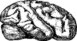
6th–7th month of intrauterine life. Upon microscopic examination of a macrogyric brain, one may not detect any abnormalities in the structure of the cortex; more often, however, deviations are encountered: an embryological type of cortical structure, the absence of proper differentiation of layers, a change in the shape, size, and position of cortical cells, and the presence in the white matter of the convolutions of a layer of gray matter (Mittelschicht) that has, as it were, separated from the other layers. Clinically, macrogyria may not manifest itself in any way, but for the most part, in such subjects, a lowering of intelligence is observed, up to complete idiocy (see Microcephaly). E. Kononova.
MACROSCOPY (from the Greek makros—large and scopeo—I look, I examine), macroscopic examination, or examination of various objects with the naked eye; it is contrasted with microscopy, i.e., examination with the help of a microscope or a magnifying glass. Macroscopy in clinical and pathological-anatomical practice represents the first and most important moment in objective examination. Macroscopy primarily includes a general, or so-called external, inspection of the object, whether it be an entire individual or a part of it (an organ, a tumor, etc.). The second stage of macroscopic examination is a detailed inspection of the particulars of the object, for example, in the process of dissecting it. A general inspection of the object is necessary for the purpose of recognizing its general structure and form, for example, the genus and species of the animal, the habitus of the subject, the coloration of the integuments; at the same time, odors, consistency, temperature, humidity, etc., perceived during the inspection are recorded. Practically, both in the clinic and on the autopsy
table, it can be considered firmly established that the indicated macroscopic inspection of the "whole," performed systematically and sufficiently carefully, can in itself already provide much, and conversely, the insufficiency or ignoring of such an inspection, a rapid transition to the analysis of particulars, often only confuses the researcher and does not yield the desired results. The second stage in macroscopy—the analysis of the particulars of the same object—must either confirm the correctness of the assumptions already made in connection with the general inspection and deepen them, or provide new data, perhaps completely refuting the indicated assumptions. Here, too, one often resorts to all kinds of macroscopic reactions (e.g., the reaction for amyloid, the Pirquet reaction, the extinction phenomenon, the Rumpel-Leede phenomenon, etc.). In pathological-anatomical practice, it is customary to check all particular macroscopic conclusions under a microscope whenever possible, and in forensic medical practice, often chemically, microspectroscopically, etc. Experience shows that even with great powers of observation, macroscopic diagnoses are often refuted microscopically; on the other hand, this is still not a reason to ignore macroscopy: as a rule, the position remains in force that in the vast majority of cases, a macroscopic diagnosis of an affliction is quite possible, and a significant share of the art in medical and pathological-anatomical research falls precisely on the ability to sort out what is visible without the help of microscopy, leaving the latter the role of an obligatory control instance (see also Biopsy, Autopsy, Diagnosis). Macroscopy is a valuable method when examining various fluids, secretions, and excretions of the organism. Thus, by the macroscopy of urine, one can to a certain extent judge the course of a gonorrheal process; by the macroscopy of sputum, the nature of a pulmonary affliction; by the macroscopy of blood, some of its diseases; by the macroscopy of vomitus, gastric juice, and feces, the diseases of the gastrointestinal tract and its glands. Lit.: Weisz E., Diagnostik mit freiem Auge-Ektoskopie, Berlin-Wien, 1924 (Russian edition—Ectoscopy—diagnosis with the naked eye, Berlin, 1925). I. Davydovsky
Related articles
Cite this page
“Macrogyria and Macroscopy.” Soviet Medical Encyclopedia. English translation of Bolshaya Meditsinskaya Entsiklopediya, 1st ed. (Moscow, 1928–1936), ed. N. A. Semashko. https://sovietmedicalencyclopedia.com/article/macrogria/