Vectors
Historical document, translated for reference. It reflects medical knowledge of the 1920s–30s and is not medical advice.
Summary
An overview of living vectors that mediate the spread of infections and invasions, distinguishing between mechanical and specific transmission, and detailing various arthropods and the diseases they transmit according to 1930s medical literature.
Encyclopedia article (1928–1936)
VECTORS, living intermediaries in the spread of infections and invasions. The spread of pathogens of invasions and infections in a large number of cases occurs through the direct or indirect mediation of animals. The latter obtain the pathogen from its location and transmit it to other animals or humans, who as a result may fall ill with the corresponding infectious or invasive disease. The notion of a "vector" must be distinguished from the notion of an "intermediate host." Vectors may spread the pathogens contained within them (or on them) repeatedly while preserving their lives; intermediate hosts infect the definitive host with the corresponding parasite in most cases when they are consumed by the definitive host. Not infrequently, a vector is at the same time an intermediate or principal host of the disseminated pathogen. The objects of transfer are mainly various fungi, bacteria, protozoa, and unrevealed viruses (for example, the pappataci fever virus); in a similar way, certain helminths are transmitted to definitive hosts (microfilariae and mosquitoes Mansonia, Chrysops, etc.); there are cases of the transfer of relatively large objects, for example, chewing lice by blood-sucking flies, false scorpions by the common house fly, etc., but as a rule, the objects of transfer are extremely small compared to the vector. The object of infection by a vector can be humans, domestic animals, and wild animals, and the latter can also be reservoirs of the corresponding virus. The transfer of pathogens can be mechanical and specific. In the first case, between the transmitted pathogen and the vector there are no biological relations, i.e., the vector is not the host of the organism it transmits; in the second, the object of transfer multiplies in the vector or undergoes a certain part of its life cycle in it. In mechanical transfer, the transmission of the invasion is possible at any moment of the period of stay of the pathogen in or on the vector, and the duration of such a period can be very short. Specific transfer is carried out no earlier than the time when the pathogen reaches an invasive stage of development in its life cycle and occupies such a place in the vector's body from which an exit outward is open to it. The realization of transfer in some cases is associated with the procedure of blood-sucking, and the pathogen is injected with the vector's saliva into the body or introduced by the mouthparts themselves; this method of transfer is called inoculative (inoculation). In other cases, the vector contaminates the host's integument or mucous membranes with its excreta, which contain pathogens of invasion or infection: such contamination is completed when the transmitted pathogens passively or actively penetrate the body (Figure 1). Contamination and inoculation can each be mechanical and specific. Horseflies (Tabanidae) and stable flies can inoculate anthrax with mouthparts contaminated during the sucking of an ulcer of a sick animal; Stomoxys nigra transmits the causative agent of surra—Trypanosoma Evansi—no later than 10 minutes after its own infection on the animal. Trypanosoma gambiense upon sucking blood by the tsetse fly enters its stomach, here Trypanosoma gambiense multiplies, through the proventriculus and proboscis passes into the lumen of the salivary glands, where the second stage of multiplication takes place with the formation of invasive trypanosomes, which with saliva are injected into human blood when the tsetse fly sucks his blood (specific inoculation) (Figure 2).
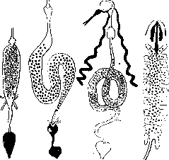
Figure 1. Scheme of the development cycles of trypanosomes in the intestinal canal of intermediate hosts-vectors: A and B - development with contaminative, C and D - with inoculative methods of infection; A - Trypanosoma Lewisi in the rat flea; B - Trypanosoma melophagium in the sheep ked; C - Trypanosoma gambiense and brucei in the tsetse fly; D - Trypanosoma rotatorium in the leech. Solid black indicates areas of development of infectious stages (metacyclic trypanosomes); black dots - areas with a predominance of the crithidial stage; white circles - areas with stages of the trypanosome type; unshaded parts do not contain developmental stages. contaminated during the sucking of an ulcer of a sick animal; Stomoxys nigra transmits the causative agent of surra - Trypanosoma Evansi - no later than 10 minutes after its own infection on the animal. Trypanosoma gambiense upon sucking blood by the tsetse fly - enters its stomach, here Trypanosoma gambiense multiplies, through the proventriculus and proboscis passes
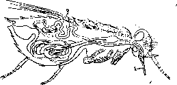
Figure 2. Trypanosoma gambiense in the tsetse fly: 1 - proboscis by which the fly infects man; 2 - trypanosomes in the salivary glands. into the lumen of the salivary glands, where the second stage of multiplication takes place with the formation of invasive trypanosomes, which are injected with saliva into human blood when the tsetse fly sucks his blood (specific inoculation) (Figure 2). Table of vectors of pathogens of infections and invasions. Vector | Transmitted organism | Disease spread by the vector | Method of pathogen transmission
pathogen
Disease of Chagas
Japanese river fever
Rat anemia
Acne | Mechanical transfer
Staphylococcus albus, Streptococcus aureus, chicken diarrhea, contamination via faeces, cholera, mechanical transfer. 7. Parasita: Epidemic typhus virus, epidemic typhus, specific transmission via a bite; Spirochaeta Obermeieri, relapsing fever (cosmopolitan); Rickettsia quintana, Pediculus capitis, Pediculus vestimenti, trench fever, Volhynian fever; various bacteria, Achorion Schoenleini, favus; Staphylococcus pyogenes, Diplococcus pemphigi contagiosi, impetigo contagiosa, mechanical transfer, impetigo (tropics); transmission by crushing lice on the skin during scratching, upon a bite. 8. Rynchota (proboscis insects): Triatoma megista and other species, Rhodnius prolixus, Trypanosoma Cruzi, Chagas disease in Brazil, specific transfer; contamination. 9. Diptera (flies): Anopheles algeriensis, malaria plasmodium, larvae of Wuchereria Bancrofti, larvae of Dirofilaria immitis, malaria, elephantiasis (?), specific transfer with saliva upon a bite and blood-sucking, intermediate host, dog filariasis; Vector, Transmitted organism, Disease spread by the vector, Mode of transfer of the agent.
agent; Anopheles bifurcatus, Anopheles maculipennis, malaria plasmodium, malaria, specific transfer; Anopheles plumbeus, Anopheles pulcherrimus, Anopheles superpictus, Anopheles hyrcanus, Culex pipiens, Mansonioides uniformis, larvae of Dirofilaria immitis, malaria plasmodium, larvae of Dirofilaria immitis, malaria plasmodium, larvae of Dirofilaria immitis, malaria plasmodium, Leucocytozoon Danilewskyi, intermediate host, malaria (?) / avian malaria, specific transfer, intermediate host, malaria, specific transfer, intermediate host, does not transmit tropical malaria, specific transfer, avian anemia, Plasmodium Danilewskyi, larvae of Dirofilaria immitis, Aedes silvestris, Aedes vexans, larvae of Wuchereria Bancrofti, larvae of Wuchereria Bancrofti, Bacillus anthracis, larvae of Dirofilaria immitis, avian malaria, specific transfer with saliva, dog filariasis, elephantiasis (?), elephantiasis (?), anthrax, filterable virus, dengue, intermediate host, mechanical transfer, intermediate host, avian malaria, Plasmodium Danilewskyi, specific transfer upon a bite; Aedes argenteus, Leptospira icteroides, yellow fever, larvae of Wuchereria Bancrofti, elephantiasis (?), intermediate host; Phlebotomus pappatasi, agent of pappataci fever, pappataci fever, Leishmania tropica, Pendra sore, specific transfer; Phlebotomus argentipes, Leishmania Donovani, kala-azar; Simulium damnosum, larvae of Onchocerca volvulus, human onchocerciasis; Culicoides Austeni, larvae of Acanthocheilonema perstans, intermediate host; Culicoides Grahami; Vector, Transmitted organism, Disease spread by the vector, Mode of transfer of the agent; Tabanus (various species), Bacillus anthracis, anthrax, mechanical transfer; Tabanus tropicus, Tabanus striatus, Tabanus lineola, Trypanosoma Evansi, surra, Tabanus glaucopis, Trypanosoma Theileri, non-pathogenic livestock trypanosomiasis, probably by contamination; Tabanus trigeminus, filterable virus, equine infectious anemia, inoculation; Haematopota pluvialis, Bacillus anthracis, larvae of Loa loa, anthrax, mechanical transfer; Chrysops silaceus, Chrysops dimidiatus, Calabar swelling, intermediate host; Chrysops caecutiens, various bacteria, septicemia, Bacillus anthracis, anthrax, tularemia, mechanical transfer; Chrysops discalis, Bact. tularense; Chrysops japonicus, filterable virus, infectious anemia of horses, inoculation; Streptococcus faecalis, Streptococcus pyogenes, erysipelas, suppuration, septicemia; Streptococcus salivarius, ordinarily non-pathogenic; Diplococcus gonorrhoeae, gonorrhea; Diplococcus intracellularis meningitidis, cerebral meningitis; Staphylococcus pyogenes, septicemia; Staphylococcus albus, abscesses; Musca domestica, Staphylococcus aureus, mechanical transfer, Staphylococcus citreus, Sarcina aurantiaca, Bacillus Koch-Weeks, acute infectious conjunctivitis; Bact. Morgani, infant diarrhea; Bacillus acidi lactici; Bacillus aerogenes capsulatus, gangrene; Bacillus anthracis, anthrax; Bacillus cuniculicidus, septicemia; Bacillus diphtheriae, diphtheria; Vector, Transmitted organism, Disease spread by the vector, Mode of transfer of the agent; Musca domestica, Bacillus paratyphi B, Bacillus pestis, Bacillus pyocyaneus, Bacillus tuberculosis, Bact. tularense, Bacillus typhi, Vibrio cholerae, trachoma virus, Bacillus dysenteriae, bacillary dysentery; Bact. enteritidis, food intoxications; Bacillus mallei, glanders; Bacillus paratyphi A, paratyphoid; plague, wound suppuration, tuberculosis, tularemia (?), typhoid fever, cholera, trachoma, mechanical transfer; Glossina palpalis, Glossina morsitans, Stomoxys calcitrans, agent of fowl cholera, fowl cholera, cysts of Entamoeba histolytica, Entamoeba coli, Giardia intestinalis, Treponema pertenue, amebic dysentery, lambliasis, helminth eggs, Trypanosoma gambiense, Trypanosoma rhodesiense, Trypanosoma cazalboui, Trypanosoma dimorphon, Trypanosoma vivax, Trypanosoma brucei, yaws, sleeping sickness, souma disease, animal trypanosomiasis; Trypanosoma cazalboui, filterable virus, filterable virus, Trypanosoma equinum, Trypanosoma Evansi, nagana, equine anemia, specific transfer; inoculation with saliva, swine cholera, mechanical transfer, Mai de Caderas, surra, mechanical inoculation; Transmitted organism, Disease spread by the vector
Mode of transfer spreads the agent; Stomoxys calcitrans, Trypanosoma soudanense, El-debab, camel trypanosomiasis; Streptococcus, septicemia, mechanical inoculation; Lyperosia irritans, Bacillus anthracis, anthrax; Borborus punctipennis, ascarid eggs, ascariasis; Bacillus anthracis, anthrax; Lucilia caesar, Bact. coli, Staphylococcus pyogenes, septicemia; Bacillus anthracis, anthrax; Sarcophaga carnaria, Bact. coli, mechanical contamination, Staphylococcus pyogenes, septicemia; Calliphora erythrocephala, Calliphora vomitoria, Vibrio cholerae, cholera, Bact. coli, colitis; Bacillus anthracis, anthrax, Staphylococcus pyogenes, septicemia; Eristalis tenax, Vibrio cholerae, cholera, mechanical transfer; Melophagus ovinus, Trypanosoma melophagium, non-pathogenic trypanosomiasis. 10. Coleoptera (beetles): Anthrenus museorum, Bacillus anthracis (spores), anthrax; Attagenus pellio, mechanical contamination via faeces; Ptinus sp., Dermestes vulpinus. 11. Hymenoptera (wasps and bees): Ants, Bacillus typhi abd., typhoid fever; Monomorium pharaonis (pharaoh ant) and other species, Bacillus pestis, plague, contamination, Bacillus anthracis (?), anthrax. 12. Aphaniptera (fleas): Dermatophilus penetrans, Bacillus tetani, tetanus, mechanical transfer; Vector, Transmitted organism, Disease spread by the vector, Mode of transfer of the agent; Xenopsylla cheopis, Bacillus pestis, plague; Ceratophyllus fasciatus, mechanical contamination; Ctenocephalus canis, Trypanosoma Lewisi, rat trypanosomiasis; Ceratophyllus tesquorum, Bacillus pestis, plague; Oropsylla silantiewi; Pulex irritans, Trypanosoma Lewisi, rat trypanosomiasis. 13. Vermes (worms): Leeches, Hemiclepsis marginata, Trypanosoma rotatorium, fish and amphibian trypanosomiasis, specific inoculation. Between these two extreme forms of transfer, there are a number of transitions. The malaria parasite undergoes a different developmental cycle and a different pathway in the body of the vector (Fig. 3). Its sexual reproduction takes place in the stomach of Anopheles, with the parasite then passing from the stomach into the body cavity of the mosquito and further into the salivary glands, from where it finds its way to the outside. It is highly probable that piroplasms also undergo a transition from the stomach into the body cavity of the tick and then into its salivary glands. That piroplasms penetrate into the eggs of the tick is indisputable, since the offspring of an infected female tick itself infects animals upon its first feeding on them (for example, Boophilus, Rhipicephalus; see Ticks). These cases of invasion transfer also pertain to inoculation of a specific character, as does the transmission of the yellow fever agent by the mosquito Stegomyia fasciata. Regarding relapsing fever spirochetes spread by ticks of the genus Ornithodoros, two methods of their transmission can be established: with saliva during blood-sucking (specific inoculation) and with coxal fluid flowing into the skin wound.
Figure 3. Development of the malaria plasmodium in Anopheles: 1 - development of the plasmodium in the stomach cavity; 2 - development on the stomach surface; 3 - transition of sporozoites into the body cavity and invasion of the salivary glands.
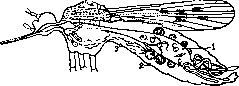
of the tick containing spirochetes (contamination). The causative agent of Chagas disease (Trypanosoma cruzi) is transmitted by the kissing bug (Fig. 4). Trypanosomes are ingested by the bug with human blood and transform into critidia (in the midgut); the latter divide and produce metacyclic trypanosomes, which accumulate in the hindgut of the bug. They are expelled with the feces and contaminate human skin. Through skin scratch marks or intact mucous membranes, the trypanosomes penetrate the body and infect humans (specific contamination). The transmission of the rat trypanosome (Trypanosoma lewisi) via fleas (Fig. 5) also occurs due to the release of metacyclic invasive forms with feces onto the rat skin. Infection occurs either when rats accidentally lick up the trypanosomes or (more frequently) when they devour infested fleas containing trypanosomes that have reached the infective stage. The contaminative transmission of cosmopolitan relapsing fever spirochetes has the characteristic
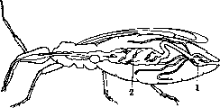
feature that this transmission is accomplished by crushing infected lice on the human body, since the spirochetes are released to the outside only when the chitinous integuments of the lice are ruptured (Fig. 6). Purely mechanical contamination is effected, for example, by the housefly, which becomes contaminated with pyogenic bacteria when crawling on suppurating ulcers of animals, upon subsequent landing on healthy animals or on humans (for example, the transmission of the trachoma virus by flies). Beetles can play a similar role. The role of vectors in epidemiology and epizootiology is extremely significant, since specific vectors are at present the sole agents for the spread
Figure A. Course of development of Trypanosoma cruzi in the bug Triatoma: 1 - metacyclic trypanosomes in the rectum, expelled with the feces of the bug onto human skin; 2 - trypanosomes in the stomach. of certain diseases (Anopheles and malaria, sandflies and pappataci fever, ticks and piroplasmoses, lice and typhus, etc.). But non-specific vectors can also play a very substantial subsidiary role in the spread of infestations and infections
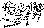
Figure 5. Course of development of Trypanosoma lewisi in the flea: 1 - trypanosomes in the stomach; 2 - metacyclic trypanosomes in the rectum. 11 of certain diseases (Anopheles and malaria, sandflies and pappataci fever, ticks and piroplasmoses, lice and typhus, etc.). Therefore, the control of vectors, in whatever special forms it may be expressed, rises fully to the level of prophylaxis of the diseases spread by them (mosquito control and malaria prophylaxis, control of Ornithodorus and prophylaxis of tick-borne relapsing fever, louse control and parasitic typhuses, etc.). It must be noted that in the USSR, vector control measures are increasingly becoming a standard part of prophylactic work (see Disinsection). Lit.: see literature for the articles Insects, Parasitology, Protozoology, Entomology, and Epidemiology.
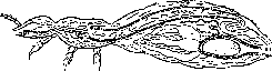
Figure 6. Spread of relapsing fever spirochetes in the body of a louse. Spirochetes can escape to the outside from its body cavity only when legs or antennae are broken or when the integuments are ruptured. (housefly and typhoid fever, etc.). Therefore, the control of vectors, in whatever special forms it may be expressed, rises fully to the level of prophylaxis of the diseases spread by them (mosquito control and malaria prophylaxis, control of Ornithodorus and prophylaxis of tick-borne relapsing fever, louse control and parasitic typhuses, etc.). It must be noted that in the USSR, vector control measures are increasingly becoming a standard part of prophylactic work (see Disinsection). Lit.: see literature for the articles Insects, Parasitology, Protozoology, Entomology, and Epidemiology.
Related articles
Mentioned in
Cite this page
“Vectors.” Soviet Medical Encyclopedia. English translation of Bolshaya Meditsinskaya Entsiklopediya, 1st ed. (Moscow, 1928–1936), ed. N. A. Semashko. https://sovietmedicalencyclopedia.com/article/vectors/