Trichostrongyloidosis
Historical document, translated for reference. It reflects medical knowledge of the 1920s–30s and is not medical advice.
Summary
This article describes the parasitic roundworm infections caused by the family Trichostrongylidae, which primarily affect the gastrointestinal tracts of animals but can also infect humans. It details the morphology of several species, their geographic distribution—noting their discovery in the USSR—and the clinical presentation of human infection, which ranges from mild to severe anemia.
Encyclopedia article (1928–1936)
Trichostrongyloidosis, helminthic diseases caused by the parasitism in the human intestinal tract of roundworms belonging to the family Trichostrongylidae Leiper, 1912, representatives of which are characterized by small size, a thread-like body, and a simple mouth, in most cases without a buccal capsule; males have a bursa with well-developed lateral lobes. The following representatives of 3 genera of Trichostrongylidae are found in humans. 1. Trichostrongylus instabilis (Railliet, 1893). Male 4.5–5.5 mm long with a width before the bursa of 0.078–0.093 mm. Spicules (Fig. 1) are slightly unequal; their length is 0.118–0.135 mm. They have the shape of a curved boat with a thick root process at the proximal end and a triangular outgrowth at the distal end. The gubernaculum (0.065–0.076 mm long) in a lateral position has the appearance of a wavy curved line with 2 waves. Female 5–6 mm long with a width in the vulvar region of 0.089–0.100 mm. The vulva has the appearance of a slit located along the length of the parasite in the middle of the ovijector (Fig. 2); it is provided with cuticular papilla-like projections on the anterior and posterior edges. Eggs 0.077–0.088 x 0.040–0.048 mm. It parasitizes cattle, small ruminants, horses, rodents, humans, and monkeys in the duodenum, and in ruminants in the abomasum. In recent years, it has been repeatedly confirmed in humans. In the USSR, it has been detected in Moscow and other regions, especially in Armenia. 2. Trichostrongylus orientalis Jimbo, 1914. The dimensions of males and females are close to T. instabilis. The spicules are not curved. The gubernaculum has the shape of a shuttle with a hollow middle part. In the female, the vulvar slit is located across the body. Eggs 0.075–0.091 x 0.039–0.047 mm. It parasitizes humans and porcupines in the small intestine. Distributed in Japan and Europe. In the USSR, it has been detected in Armenia in humans and in Azerbaijan in a porcupine. 3. Trichostrongylus probolurus (Railliet, 1896). The dimensions are approximately the same as those of the previous species. The spicules are of almost equal length and have sharply defined, elaborate triangular outgrowths; their dimensions are 0.125–0.128 mm and 0.129–0.134 mm. Gubernaculum 0.072–0.079. The vulva has the appearance of a longer slit than in T. instabilis, located longitudinally and not bounded by papillae. Eggs 0.076–0.080 x 0.043–0.046 mm. It parasitizes sheep, camels, gophers, and humans, localizing in the duodenum. Distributed in Europe, in particular in the USSR, where it has been detected in humans in Armenia; furthermore, it has been confirmed in humans in North Africa and the USA. 4. Trichostrongylus vitrinus Looss, 1905. The dimensions are the same. The spicules are even, straight, gradually tapering to a spear-like pointed distal end; their length is 0.160–0.178 mm. The gubernaculum (0.080–0.090) has the appearance of a spindle-shaped shuttle. The vulva is an oblique longitudinal slit at a distance of 1.15–1.30 mm from the posterior end. Eggs 0.082–0.090 x 0.046–0.050 mm. Widely distributed in ruminants, found in the duodenum of humans and gophers. Distributed in Egypt; quite widely distributed in the territory of the USSR, including in humans (Armenia). 5. Trichostrongylus extenuatus (Railliet, 1898). The dimensions are the same. The spicules are unequal: 0.094–0.104 mm and 0.119–0.128 mm. Eggs 0.086–0.092 x 0.037–0.042 mm. It parasitizes ruminants, humans, and gophers. In humans, it has been detected exclusively within the USSR (Armenia). 6. Haemonchus sp. is widely distributed in ruminants. In one case, it was described in Brazil in a human suffering from a severe form of anemia. 7. Mecistocirrus digitatus (Linst., 1906). Detected only once in China in a human. All representatives of the family Trichostrongylidae develop via a direct cycle. Segmented eggs released by the female are expelled with feces into the external environment. Depending on the species of parasite and on the conditions of temperature, humidity,
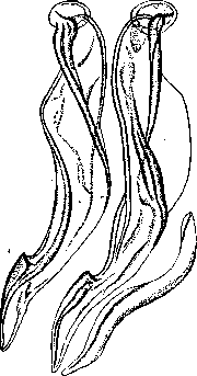
Figure 1. Trichostrongylus instabilis—spicules and gubernaculum.
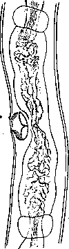
etc., the hatching of larvae from the egg and their molting occur at different times. The larvae, enclosed in a sheath, are infectious. Being swallowed by a suitable host, they reach their site of localization in the digestive tract, where they develop. Trichostrongylosis in humans was until recent years considered an extreme rarity and was confirmed in isolated cases only in certain tropical countries, such as Egypt, China, Japan, and India. Having been discovered for the first time on the territory of Europe in 1923 in Moscow by Podyapolskaya (Trichostrongylus instabilis), they were increasingly confirmed in various regions. In Moscow and the surrounding areas, over 1.5 years, human Trichostrongylosis was detected in 134 cases using methods of examining excrement for helminth eggs (Davydov and Vinogradov). At the present time, Trichostrongylosis is known in many regions of the USSR, and its distribution is apparently most widespread in Armenia (works of Kalantaryan).
The clinical picture and therapy of Trichostrongylosis are almost unstudied. The clinical picture apparently depends to a significant extent on the intensity of the infestation. With a mild infestation, clinical symptoms are weak (appetite disorders and other gastrointestinal disorders, weakness, irritability, etc.) or may even be completely absent. With intense infestations, clinical symptoms can be very severe, resembling the clinical picture of ancylostomiasis—pronounced anemia, sharp decline in strength leading to cachectic states. With the help of repeated courses of treatment with thymol (Kalantaryan), complete clinical recovery can be achieved. However, even after numerous courses, isolated eggs still remain in the excrement. In view of the fact that humans are facultative hosts, prophylaxis should be directed along the lines of combating Trichostrongylosis in domestic animals.
Related articles
Cite this page
“Trichostrongyloidosis.” Soviet Medical Encyclopedia. English translation of Bolshaya Meditsinskaya Entsiklopediya, 1st ed. (Moscow, 1928–1936), ed. N. A. Semashko. https://sovietmedicalencyclopedia.com/article/trichostrongyloidosis/