Stoffel Operation
Historical document, translated for reference. It reflects medical knowledge of the 1920s–30s and is not medical advice.
Summary
The Stoffel operation is a surgical procedure involving the resection of motor nerves or their branches to treat spastic conditions like Little's disease. Developed in 1912, it aimed to interrupt aberrant reflex arcs by reducing the number of motor impulses reaching spastic muscles.
Encyclopedia article (1928–1936)
STOFFEL OPERATION (Stoffel), the transection of motor nerves of the extremities either within the nerve trunk itself (Stoffel I) or of the motor branches before they enter the muscle (Stoffel II). The idea of the method is to interrupt a defective reflex arc. The operation is indicated in Little's disease, spastic hemiplegias, hypertonias, and in certain forms of athetosis. The operation yields the best results in lesions of the lower extremities. Historically, the ideological prerequisites for this method were myotomy, tenotomy, Vulpius tendon lengthening, and Lorenz myorexis, which aimed to act upon the muscle—the executor of the movements of the reflex arc. Operations on the latter often led to the generalization of the spastic state, which provided the rationale for Foerster to propose the resection of the posterior sensory roots of the spinal cord to reduce irritation—an operation first performed by Tietze. Stoffel proposed his operation in 1912. Having thoroughly elaborated the internal topography of the cable-like trunks, the mode of nerve branching, and their distribution within the muscles themselves, he decided to reduce the number of motor nerve impulses entering the muscle (Fig. 1). For
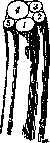
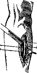
Figure 1.
Figure 2. Figure 1. Peripheral trunk of the radial nerve: 1 - pathway for the superficial ramus; 2 - pathway for the brachioradialis muscle; 3 - pathways for the radial wrist extensor muscles; 4 - pathway for the deep ramus; 5 - pathway for the supinator muscle. Figure 2. Exposure of the ulnar nerve in the upper half of the forearm: 1 - ulnar nerve; 2 - ulnar wrist flexor muscle; 3 - superficial finger flexor muscle; 4 - ulnar artery. this purpose, according to the scheme developed by Stoffel, the corresponding nerve is exposed (Fig. 2), and a partial resection (from a third to a half of the bundle) of the motor fibers is performed in it (Stoffel I). A suture is applied to the perineurium, and sutures to the soft tissues. This operation yielded numerous failures—recurrences of spastic states, easily explained by nerve regeneration. In addition, the works of a number of authors showed that nerve trunks do not run in a cable-like fashion, but rather in loops, and the pathways innervating the same muscle groups run not at a single point, but at different points of the nerve cross-section, sometimes far apart from each other (Spitzy). All this Figure 3. Topography of the pathway for
for this purpose, according to the scheme developed by Stoffel, the corresponding nerve is exposed (Fig. 2) and a partial resection (from a third to a half of the bundle) of the motor fibers is performed in it (Stoffel I). A suture is applied to the perineurium,
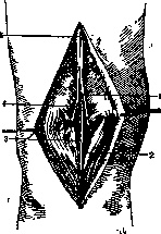
and sutures to the soft tissues. This operation yielded numerous failures—recurrences of spastic states, easily explained by nerve regeneration. In addition, the works of a number of authors showed that nerve trunks do not run in a cable-like fashion, but rather in loops, and the pathways innervating the same muscle groups run not at a single point, but at different points of the nerve cross-section, sometimes far apart from each other (Spitzy). All this caused Stoffel and other surgeons for the medial head of the gastrocnemius muscle to transition to a new modification, expressed Figure 3. Topography of the pathway for the gastrocnemius and soleus muscles and for the medial sural cutaneous nerve (according to Stoffel): 1 - pathway for the medial head of the gastrocnemius muscle; 2 - medial sural cutaneous nerve; 3 - dorsal pathway for the soleus muscle; 4 - pathway for the lateral head and gastrocnemius muscle; 5 - common pathway for the gastrocnemius and soleus rami and medial sural cutaneous nerve.
in complete or partial resection (half or a third
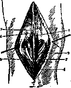
of the trunk to a given muscle group) of the motor branches before their entry into the muscle (Stoffel II) (Figs. 3 and 4). The figures below show the Stoffel operation in spastic equinus foot. The determination of motor pathways in the wound is performed using a sterile electrode: one or two of those branches whose stimulation with a weak current yields the strongest muscle contraction are resected. After closing the wound, forcible correction (redressment) of the malposition of the extremities is performed, and the achieved result is fixed with a plaster cast. Upon removal of the cast, massage, electrification, and exercises of the weaker extensors and
Figure 4. Stoffel operation for severe equinus foot: 1 - pathway for the medial head of the gastrocnemius muscle; 2 - medial head of the gastrocnemius muscle; 3 - tibial nerve; 4 - medial sural cutaneous nerve; 5 - lateral head of the gastrocnemius muscle; 6 - pathway for the lateral head of the gastrocnemius muscle; 7 - proximal segment of the dorsal pathway for the soleus muscle.
abductor muscles are initiated in order to balance their strength with the weakened antagonists. Close in idea to the Stoffel operation is the "operation of reducing the cross-section of the nerve trunk according to Vreden." The latter believed that, since a large nerve does not represent a cable with separate isolated nerve bundles, but consists of an intertwining of motor, sensory, and sympathetic fibers, excision
Figure 5. Reduction of the cross-section of the nerve trunk (according to Vreden). of a part of the nerve trunk achieves a decrease in conductivity across all types of nerve pathways. How this operation is performed can be seen from the figure given below (Fig. 5). Regarding the indications for performing this operation, modern orthopedists believe that one should begin with the subcutaneous cutting or lengthening of muscles and tendons. If, after some time, a recurrence of the contracture position develops, proving that the antagonists cannot cope, an additional weakening of the spastic-contracted muscles by the Stoffel operation is indicated. A further development of the idea of unloading spastic contractures is found in Spitzy's operation, where the excess tone of strong muscles is transferred to weak ones by the transplantation of transected nerve branches into the nerve trunks of the weakened musculature.
Related articles
Cite this page
“Stoffel Operation.” Soviet Medical Encyclopedia. English translation of Bolshaya Meditsinskaya Entsiklopediya, 1st ed. (Moscow, 1928–1936), ed. N. A. Semashko. https://sovietmedicalencyclopedia.com/article/stoffel-operation/