Sideroscope
Historical document, translated for reference. It reflects medical knowledge of the 1920s–30s and is not medical advice.
Summary
The sideroscope is a device used in ophthalmology to detect the presence and location of ferromagnetic foreign bodies in the eye. It utilizes an astatic magnetic needle that deflects in the presence of iron or steel, though its clinical utility is limited by extreme sensitivity to external interference.
Encyclopedia article (1928–1936)
SIDEROSCOPE (from the Greek sideros—iron and scopeo—I look), a device based on the properties of a magnetic needle to deflect in the presence of even the smallest particles of ferromagnetic metals. The sideroscope is sometimes used in ophthalmological practice to determine the presence and localization of iron or steel splinters in the eye. The original model of the sideroscope was proposed by Asmus in 1894 and improved by Hirschberg (Figures 1 and 2), who introduced changes that significantly simplified the use of the device. The main part of the latter is an astatic magnetic needle, freely suspended on a silk fiber inside a glass cylinder by means of three screw legs. The device is set in such a position that the needle, having a strictly vertical direction, does not touch the walls of the cylinder. To capture the most insignificant oscillations of the needle, a small flat mirror (Gauss) is attached to its middle part, onto which light is directed from a lamp. The beam of light reflected from the mirror is cast onto a screen with a special scale placed near the apparatus. When the eye is brought to the device, in the case of the presence of iron or steel in it, the needle is set in motion, which is communicated to the mirror, and thanks to the movements of the latter
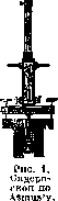
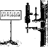
Sideroscope according to Hirschberg. The beam of light reflected from it moves on the screen along the scale. By the amplitude of the needle's oscillations, one can to a certain extent judge in which region of the eye the foreign body is located. In general, the device is distinguished by great sensitivity, and with its help, it is possible to detect splinters in the eye weighing less than 0.001 g. But the sideroscope also has significant drawbacks, as the slightest shaking of the wall on which it is mounted sets the device's needle in motion, which complicates the examination, especially with small sizes of the foreign body. The same is observed under the influence of iron or steel objects accidentally located nearby, and especially under the action of the magnetic field around the electrical wiring of the lighting network or tram wires, etc. Due to these shortcomings, the sideroscope is relatively rarely used for the diagnosis of foreign bodies, having given way to the strong electromagnet and X-ray.
A. Strashun.
Related articles
Cite this page
“Sideroscope.” Soviet Medical Encyclopedia. English translation of Bolshaya Meditsinskaya Entsiklopediya, 1st ed. (Moscow, 1928–1936), ed. N. A. Semashko. https://sovietmedicalencyclopedia.com/article/sideroscope/