Gasserian Ganglion
Historical document, translated for reference. It reflects medical knowledge of the 1920s–30s and is not medical advice.
Summary
A historical anatomical description of the Gasserian ganglion (trigeminal ganglion), detailing its location in Meckel's cave and its cellular structure. The article describes the ganglion's role as the origin of the three main branches of the trigeminal nerve.
Encyclopedia article (1928–1936)
GASSERIAN GANGLION (ganglion Gasseri s. g. semilunare), gives rise to the sensory root of the trigeminal nerve; it is located on the dorsal surface of the petrous part of the temporal bone, in a niche formed by the splitting of the dura mater (Meckel's cave); on the bone at this location, there is a small depression—the impressio trigemini; the concave edge of the ganglion faces backward, and the convex edge faces forward. In the Gasserian ganglion, there is a large number of round cells covered by a sheath formed from neuroglial cells, which transitions directly into the Schwann sheath of the nerve fiber. Most cells of the Gasserian ganglion belong to the pseudo-unipolar type, equipped with only one process that divides in a T-shape; in some cells, before division, this process is very convoluted and forms a true glomerulus—the glomerulus initialis; in others, the process is straight; there are also multipolar and fenestrated cells; some processes do not extend beyond the limits of the ganglion. The peripheral processes of the cells form three large branches: the ophthalmic nerve (n. ophthalmicus), the maxillary nerve (n. maxillaris), and the mandibular nerve (n. mandibularis); these three branches emerge from the convex edge of the ganglion (see figure). The central process forms the root of the trigeminal nerve, which is very large, consisting of 50 bundles, entering the pons Varolii together with the motor root (for peripheral branching, central fiber pathways, and pathology, see Trigeminus nervus).
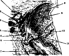
Right Gasserian ganglion (after Spalteholz): 1—n. opticus; 2—art. carotis int.; 3—n. oculomotorius; 4—n. trochlearis; 5—n. abducens; 6—n. trigeminus; 7—n. facialis and acusticus; 8—n. ophthalmicus; 9—n. maxillaris; 10—n. mandibularis; 11—ganglion Gasseri.
See Trigeminus nervus; for changes in the Gasserian ganglion in rabies—see Rabies. Literature—see Trigeminus nervus.
Related articles
Mentioned in
Cite this page
“Gasserian Ganglion.” Soviet Medical Encyclopedia. English translation of Bolshaya Meditsinskaya Entsiklopediya, 1st ed. (Moscow, 1928–1936), ed. N. A. Semashko. https://sovietmedicalencyclopedia.com/article/gasserian-ganglion/