Dermatomes
Historical document, translated for reference. It reflects medical knowledge of the 1920s–30s and is not medical advice.
Summary
This article defines dermatomes as segments of the skin innervated by specific spinal nerve roots. It explains their distribution patterns across the body and highlights their clinical significance in diagnosing spinal cord lesions.
Encyclopedia article (1928–1936)
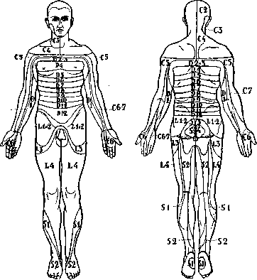
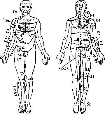
DERMATOMES, segments of the skin into which the entire surface of the human body is divided in connection with its innervation by various spinal cord roots. There are a number of diagrams illustrating this division of the skin surface into segments or dermatomes; the diagrams of Kocher and Seiffer are considered the most accurate (Kocher, Seiffer; see figures 2-5). The lines separating the dermatomes are located in a transverse direction on the trunk and in a longitudinal direction on the limbs; the roots entering the brachial plexus innervate the upper limbs, those in the lumbosacral plexus innervate the lower limbs; the roots of the thoracic segments innervate the trunk, and the lower sacral roots innervate the perineum, anus, and genital organs. Each dermatome receives sensory fibers generally from three roots—from one main, primary root, which is reinforced by the roots lying above and below it (see figure 1); therefore, when one root is damaged, sensory disturbances may not be observed in the corresponding dermatome. Such a projection of spinal cord segments onto the skin is of great importance in diagnostics, as it allows for the precise localization of a pathological process in the spinal cord.

Figure 1. Distribution of sensory fibers in the skin. Diagram of the overlapping of the roots lying above and below the main root.
C67
se 7
Figures 2 and 3. Diagram of the distribution of posterior spinal root fibers in the skin on the anterior and posterior surfaces of the body according to Kocher.
Figures 4 and 5. The same according to Seiffer. 73 8
Related articles
Mentioned in
Cite this page
“Dermatomes.” Soviet Medical Encyclopedia. English translation of Bolshaya Meditsinskaya Entsiklopediya, 1st ed. (Moscow, 1928–1936), ed. N. A. Semashko. https://sovietmedicalencyclopedia.com/article/dermatomes/