Blood
Historical document, translated for reference. It reflects medical knowledge of the 1920s–30s and is not medical advice.
Summary
This article defines blood as the vital fluid filling the circulatory system, composed of plasma and formed elements like erythrocytes, leukocytes, and platelets. It details the blood's role as the body's internal environment, facilitating gas exchange, nutrient transport, and waste removal through complex physicochemical processes.
Encyclopedia article (1928–1936)
BLOOD, a liquid filling the arteries, veins, and capillaries of the organism and consisting of a transparent, pale-yellowish plasma and formed elements suspended in it: red blood corpuscles, or erythrocytes, white ones, or leukocytes, and blood platelets, or thrombocytes. In the phylogenetic series of animals, blood is first separated, albeit imperfectly, from the interstitial fluid simultaneously with the appearance of the circulatory system—in echinoderms and annelids; in the body of the human embryo, blood appears at the 3rd–4th week simultaneously with the vascular primordia and within them. Both these primordia and the primary formed elements of blood originate from the mesoderm (see Hematopoiesis). Despite the fact that many authors still continue to speak of "blood tissue" with a liquid intercellular substance, the term "tissue" cannot by any means be considered applicable to blood, which differs from any other tissue mainly in that the site of formation of its morphological elements and the constituent parts of the plasma is not in the blood itself, but outside of it. Together with lymph (which, by the way, no one calls a tissue), blood represents the internal environment for all elements of our body, which all organs and tissues replenish with the products of their vital activity, and some organs, such as the bone marrow, spleen, lymph glands, etc. (see Hematopoiesis), also with the formed elements produced within them. The formed elements that have entered the blood continue to live and function in it until the structural changes conditioned by their activity or age reach such a degree that they make possible their capture and destruction by erythrolytic and leukolytic organs (see Hemolysis). The physiological breakdown of erythrocytes and leukocytes in the bloodstream, admitted by some authors, still remains unproven, so that in this respect, too, blood differs from all tissues of the organism. Blood differs from other parts of the internal environment, i.e., lymph and interstitial fluid, not only in composition and location within vessels, but also in its continuous and rapid circulation through the vessels, conditioned by the activity of the heart (see Blood Circulation). Flowing through the capillaries, blood comes into contact through their walls with a significant portion of the body's cells; where cells do not come into direct contact with the capillaries, the connection between them and the blood is established through the mediation of the interstitial fluid bathing them. In the lungs, where the capillaries are covered only by the thinnest layer of alveolar epithelium, blood comes into close contact with the air located in the alveoli. Throughout the entire extent of the vast capillary network, a continuous exchange of substances occurs between the tissues and the blood through the capillary walls. Gases, water, and inorganic and organic compounds, which are products of the digestion of food substances, internal secretion of tissues, dissimilation processes, and the vital activity of bacteria that have entered the organism or inhabit it, etc., are continuously entering it from the air and from the tissues. All these substances are carried by the blood flow to all parts of the body and, in the corresponding areas, pass again from the blood into the tissue cells, for which they serve, depending on the circumstances, as nutritional material, stimulating, inhibiting, or poisonous agents, or as material accumulating as reserves, or finally for excretion from the organism. In combination with the aforementioned rapidity of blood turnover, this forces us to recognize it, along with the nervous system, as the most important factor maintaining the unity of the organism and conditioning the interaction of its countless constituent elements, which has reached an amazing perfection of coordination in the process of natural selection. The same exchange of substances between blood and tissues, in combination with blood circulation, conditions, besides the function of connection, all other functions of the blood, which are most closely intertwined with each other and with the functions of all kinds of organs and tissues of the body, and in which one can only very conditionally distinguish the functions of nourishing tissues, removing products of dissimilation, supplying oxygen, and maintaining the physicochemical properties of the tissue environment necessary for cell vital activity, its active reaction, and osmotic pressure. In more or less highly organized animals, all nutrient substances perceived by them from the external environment are delivered from the digestive tract to the tissues exclusively through the mediation of the blood. Its role is not reduced in this to merely the mechanical transport of digestion products to the places of their consumption: changes in its chemistry and physicochemical properties are active factors both during the passage of these products into the blood and during the subsequent passage from the blood into the tissues. Thus, the depletion of blood in sugar, an increase in the content of lactic acid, and shifts in the electrolyte system cause chemical processes in the tissues of a specific part of the body that proceed in the direction of bringing the chemical constants of the blood closer to the average norm (breakdown of glycogen with the release of sugar into the flow of outflowing blood, resynthesis of lactic acid with a decrease in its quantity in the outflowing blood, etc.). The situation is exactly the same with those substances which, being products of the vital activity or breakdown of some organs, are necessary for other organs to perform their specific functions: incretory or excretory functions, intermediate metabolism, etc.; here, too, the exchange occurs with the most active participation of the physicochemical mechanisms of the blood. Finally, numerous nutrient substances, having entered the blood from the intestine, before passing into the tissues, undergo various transformations in its very flow; partly under the influence of enzymes and formed elements of the blood, partly through the mediation of more or less labile compounds with its constituent parts, the products of digestion are converted into a state that allows for their further assimilation by the cells of the organism, or are freed from certain toxic properties. Among the organs to which the blood delivers substances necessary for their specific function are also those through which all kinds of "production waste," products of dissimilation and breakdown, the accumulation of which in the organism could entail harmful consequences for it, are removed from the organism. Here, the blood plays, along with a nutritional role, a cleansing role, and in the performance of this function, the matter is not reduced to merely mechanical transport, but is accompanied by a number of the most complex processes of a physicochemical nature occurring in the blood itself. The colloidal state of blood proteins and their osmotic pressure, the state of the buffer properties of the blood, the ratio of electrolytes in it, and much more are of decisive importance for the activity of any secretory and excretory organ, including the kidneys and sweat glands, etc. Through the mediation of the blood, the entire gas exchange of our organism also occurs. Basically, this exchange is conditioned by differences in the tension of O2 and CO2 in the alveolar air and in the tissues on one hand, and in arterial and venous blood on the other. But the process of exchange itself does not occur by way of simple dissolution of gases in the blood in some parts of the circulatory system and their release in others, but by way of their entry into labile chemical compounds with the constituent parts of the blood. In the lungs, O2 combines in this way with the Hb of erythrocytes, forming oxyhemoglobin; in the tissues, part of the CO2 entering the blood is bound by the bases of the plasma, with which it forms bicarbonates, but part also remains in a free state according to the formula [bicarbonate]/[CO2] = K (constant). And since an increase in the content of CO2 in the blood (as well as other acids entering from the tissues) changes the aggregate state of hemoglobin and lowers its ability to bind O2 (Barcroft), then during the passage of blood through the capillaries, conditions are created that significantly facilitate the splitting off of O2 from oxyhemoglobin and its passage into the tissues. The reverse process takes place in the lungs, where the content of free CO2 in the blood drops sharply, the ability of Hb to bind O2 increases thanks to this, and thereby the formation of oxyhemoglobin is facilitated; a number of other factors also act in this same direction (the shifts of chlorine ions from plasma into erythrocytes and back, occurring under the influence of fluctuations in free CO2, the thickening of blood in the arterial part of the capillaries and its thinning in the venous part, etc.). All this shows that in the function of gas exchange, just as in other functions of the blood, all its constituent parts take an active part and that all the constituent elements of this process are in the closest connection with each other and in constant interaction. The normal functioning of any living cell is conditioned by a certain physicochemical state of the environment surrounding it, relatively small changes in which already entail a disturbance of the cell's vital activity. Highly differentiated cells of higher animals are especially sensitive to changes in the environment, immersed in an "internal environment" separated from the external world by numerous barriers, in which, thanks to its mobility, blood plays the main role. Hence the biological necessity of the constancy of the main constituent elements and properties of the blood, of which one can mention here the constancy of its active reaction (pH), osmotic pressure, and electrolyte composition.
Since the direct flow of substances passing through the blood, on the one hand, and a number of processes occurring in its constituent elements, on the other, act in diametrically opposite directions, a multitude of adaptations arose in the process of natural selection that automatically neutralize all these harmful influences, smoothing out individual fluctuations and limiting their range to known limits. Thus, with regard to the pH of the blood, the main regulating factor is the carbonates and other buffer mixtures contained in its composition, as well as proteins and hemoglobin, which possess buffer properties (see Buffer properties). Significant assistance is provided in this by the activity of the lungs and kidneys, which react to the slightest changes in pH and the acid-base balance of the blood with changes in ventilation and excretion. The liver also participates in this regulation (through the mediation of ammonia split off from amino acids within it), as do numerous digestive glands, which remove acidic and basic ions from the blood during their secretion, and a number of other organs. The constant level of osmotic pressure in the blood is maintained mainly by the kidneys, as well as by the lungs and sweat glands. No small role is played by the ability of loose interstitial tissue to quickly absorb and release significant amounts of water and salts into the blood; the colloids of the blood (due to their ability to bind water) and the level of blood pressure, caused by the dilation or constriction of vessels, as well as endocrine glands, etc., also participate. In approximately the same way, finally, the ratio of plasma electrolytes is regulated, which is of enormous importance for the activity of all organs of the body. In all these regulations, the autonomic nervous system plays a huge role, being extremely sensitive to fluctuations in pH and electrolytes and responding to these fluctuations with corresponding reflexes to various organs: lungs, kidneys, glands, vessels, etc. The content of formed elements in the blood of a healthy person is also distinguished by significant constancy. This constancy is especially clearly distinguished with regard to erythrocytes; if those physiological fluctuations that are observed during their counting in peripheral blood at different times of the day, after drinking, movement, etc., depend partly on the real change in the circulating mass of blood in the body (see below—blood mass), then nevertheless the number of erythrocytes under certain standard conditions (on an empty stomach, with complete bed rest) appears to be a value that is to a significant degree constant for a given individual. At the same time, like all other constants of the blood, the content of erythrocytes in it represents a dynamic result of continuously occurring, oppositely directed processes in the body—in this case, the processes of new formation and decay of erythrocytes. These processes are in the closest interaction with each other, mutually stimulating or inhibiting one another. It is known that an increase in hemolysis entails increased hematopoietic activity of the bone marrow, and stimulation of the latter entails an increase in hemolysis, that the suppression of erythrocyte decay is accompanied by a decrease in erythropoiesis and vice versa; however, due to the insufficiency of our knowledge about the decay of erythrocytes and the further fate of the products of this decay (see Hemolysis), as well as about the maturation of erythrocytes and their transition into the blood (see Hematopoiesis), the very mechanism of interaction between these processes remains as yet unclear. A certain role is played in this, apparently, by the stimulating effect exerted on the bone marrow by the products of the breakdown of hemoglobin and the stroma of erythrocytes. Endocrine glands, and in particular the thyroid and sex glands, as well as the spleen, undoubtedly participate in the regulation of the composition of red blood; the gas exchange of the body exerts a great influence on the composition of the blood, and recently the role of acid-base balance has also begun to be revealed. Finally, the importance of nutrition in maintaining the normal composition of the blood is becoming increasingly obvious, and it is no longer a question of iron alone, as was the case relatively recently, but of more complex animal and vegetable products, for example: amino acids, extractive substances, vitamins, etc. Even less studied and even more complex is the regulation of the leukocyte content in the blood. True, the total number of leukocytes is less constant than the number of erythrocytes, but all the more striking is the persistence with which a certain ratio between different types of white blood cells, the so-called leukocyte formula (see), is maintained in each individual. Analogous to what we have seen with regard to erythrocytes, the interaction between decay and regeneration also plays a major role here; however, the multiplicity of leukocyte forms forces us to assume the presence of a much more complex regulating mechanism here. Its clarification is hindered not a little by the absence of a final decision on the question of the multiple or single origin of different leukocyte types (see Hematopoiesis). Furthermore, the study of the regulation mechanism is complicated here by the active mobility inherent in white blood cells and the chemotaxis and tropisms manifested during this, as well as the significance of leukocyte reserves in the spleen, lymph glands, bone marrow, etc., which play a much greater role in relation to circulating leukocytes than the corresponding reserves of erythrocytes. Numerous facts testify to the influence on leukocytes of the chemistry of the blood, the activity of endocrine glands, and the autonomic nervous system. However, no definite regularities have yet been established here. Finally, one cannot fail to note the persistence with which the total mass of its blood is maintained in the body. In different individuals, it varies to a fairly significant degree, and moreover, both in its absolute and in its relative expression (see below—Total blood mass). But in each individual, the absolute mass of blood [i.e., the sum of circulating and reserve blood (see Blood circulation)] represents a very stable value, showing only insignificant physiological fluctuations in connection with drinking, movement, etc. It remains to a significant degree unchanged even under various pathological conditions: both during weight loss and obesity (hence the increase, or decrease, of its relative figures), during anemia (the loss of erythrocytes is compensated by an increase in plasma), and after hemorrhages. The very mechanism of these regulations remains unknown for now. The special place occupied by blood in the general economy of the body as its internal environment, and the features of its physiology described above, also determine the features of its pathology. Since blood is not a tissue and possesses neither a definite structure nor elements multiplying and developing within it, no processes associated with the existence of tissue as such find a place among its pathological changes: neither phenomena of inflammation, nor atrophy or hypertrophy, nor tumors, nor impregnation or infiltration, nor, finally, necrosis or gangrene are observed in it. Pathological changes that in one way or another resemble any of these processes (leukocytosis—inflammation, leukemia—tumor) are in reality only a reflection of corresponding processes occurring either in hematopoietic organs or in other parts of the body. Even the phenomena of degeneration observed in the formed elements of the blood arise in the vast majority of cases not during the time these elements spend in the blood, but even before their entry into it, in the hematopoietic organs. The most diverse changes in the white blood picture, all kinds of leukocytosis, lymphocytosis, shifts to the left or right (see Leukocyte formula), aleukia, agranulocytosis, lymphatic, myeloid, monocytic, myeloblastic leukemias, etc., depend not on the change or pathological growth of elements circulating in the blood, but on pathological changes in leukopoietic and lymphopoietic organs, on the strengthening or weakening of their functions under the influence of various kinds of impacts. The vast majority of anemic states also depend on disturbances in the activity of erythropoietic or erythrolytic organs caused by various reasons, whether in the direction of their strengthening or weakening; the same applies to hyperglobulia of various origins. An exception to this rule are those diseases in which the formed elements of the blood are destroyed or damaged in its very flow by harmful agents that have penetrated into it. This includes such parasitic diseases as malaria and some others (see below—Blood parasites), septicemia with hemolytic pathogens, poisoning with hemolytic poisons that damage the hemoglobin of erythrocytes (CO, KClO3, aniline). Here one can also include the hemolysis of erythrocytes in paroxysmal hemoglobinuria (see), caused, along with other factors, by the presence of a hemolytic amboceptor in the blood. A harmful or destructive effect on the formed elements circulating in the blood is also exerted by some physical factors, such as, for example, overheating (sun baths), X-rays.
Depending on their intensity and duration, these and similar factors can entail various changes in the composition of the blood, as well as physicochemical and morphological changes in the formed elements themselves; however, even in this case, we will not encounter in the blood reactions similar to those observed in tissues during analogous damage or destruction of their elements. The only active reaction that can be observed in this regard on the part of the circulating formed elements consists of the phagocytosis of infectious agents and damaged corpuscles or their fragments by leukocytes; the regeneration of formed elements occurs not in the blood itself, but in the hematopoietic organs, which are stimulated by the decay products of damaged corpuscles, a lack of oxygen, or other pathways unknown to us. If, however, an infection, toxin, poison, etc., has paralyzed the activity of these organs as well, then the composition of the blood, left to its own devices, deteriorates more or less rapidly, up to the point of a lethal outcome. Even less can one speak of endovascularly conditioned pathological changes in the plasma. If we do not count the slight influence exerted by leukocytes on the content of enzymes and immune bodies in it, then all pathological changes in both the chemical constituents of the plasma and its physicochemical properties (changes in the composition of electrolytes, lipoids, bilirubin, urea, residual nitrogen, phenols, sugar, acetone, reserve alkalinity, rare changes in pH, changes in coagulability, etc.) are determined by the activity of all kinds of organs and tissues of the body, which either release pathological products into the blood, or normal products but in pathologically altered quantities, or do not remove metabolic products from the blood in a timely manner and in sufficient quantities, or do not supply the blood with necessary substances. All this also applies to the pathological changes observed in genotypic diseases of the blood, which are in reality either diseases of the hematopoietic organs, or of organs regulating hematopoiesis, or, finally, of organs supplying the blood with any substances necessary for its normal state. Thus, for example, in hereditary hemolytic jaundice, the locus morbi is located either in the bone marrow, which releases pathological erythrocytes into the blood, or in the spleen, whose hemolytic function is excessively enhanced; pernicious anemia, a significant portion of the cases of which is genotypic, is caused by the improper function of the bone marrow; finally, the absence of blood clotting in hemophilia apparently depends on an insufficient supply of thrombokinase from the tissues. The situation is exactly the same in some cases of chlorosis, where there is an endocrinologically conditioned insufficient function of the bone marrow. In view of these peculiarities of the pathology of the blood, it can be asserted that pathological changes in the blood are merely symptoms of diseases of other organs or tissues. Diseases of the central nervous system and especially of the vegetative centers regulating metabolic processes, dysfunctions of the glands of internal secretion, diseases of the kidneys, liver, pancreas, digestive and respiratory organs, circulatory disorders due to anatomical changes or functional defects of the heart and vessels, all kinds of inflammatory processes, suppuration, etc.—in short, any pathological change in any part of the organism is reflected in one or another chemical constituent of the plasma, in its physicochemical properties, in the quantity of formed elements contained in the blood, and in their qualitative characteristics. The blood thus turns out to be, as it were, a 'mirror of the organism,' in which all physiological and pathological processes occurring within it find their reflection to a certain extent. Hence the enormous, unflagging interest with which the smallest, seemingly trivial details concerning the blood are studied (see Hematology), and in particular its physicochemical properties. It is necessary, however, to warn against the purely mechanical view of the connection between the blood and the rest of the organism, which is often observed in this regard, according to which the reflection must exactly 'copy' the morphological and physicochemical changes of the organs, as well as against conclusions about the functions of various organs based on this same point of view. Proponents of these views overlook the extreme stability of the basic properties of the blood and the versatility of its connections, as a result of which one or another impact upon it may be neutralized as such, but may instead exert an influence on less stable constituent elements or properties through various pathways. For example, the introduction of salts will not noticeably affect the osmotic pressure of the blood, but it will increase the water content in it and lower the level of erythrocytes and hemoglobin; the introduction of acids within certain limits will not entail changes in the pH of the blood, but will affect the reserve alkalinity or entail an increase in the number of erythrocytes, etc. Thanks to the antagonistic nature of the functions and regulations of the organism and their by no means absolute, although highly perfect, coordination, cases are not rare where the result of some impact turns out to be paradoxical, when, for example, the introduction of sugar lowers its level in the blood, the influx of hemolytic substances entails an increase in the number of erythrocytes, and the suppression of hemolysis leads to a decrease in this number. In short, in relation to the blood, as in relation to other analogous phenomena, 'mirror-like' should be understood not in the sense of an identity between the reflection and the reflected, but in the sense of a certain correspondence between the reflected and the changes in the reflecting object. From all that has been stated, it follows that when studying the influence of the activity of any organ on the blood, one should: 1) take into account both the qualitative and quantitative state of not only this organ, but also the blood; 2) not limit oneself to the investigation of only one possible immediate effect (e.g., the content of lactic acid in the blood during muscular work), but encompass the largest possible complex of blood elements (alkaline reserve, erythrocytes, leukocyte formula, and many others) that are in one way or another connected with the immediate product of the activity of the organ under study. And conversely, when clarifying the causes of one or another change in the blood, one should not limit oneself to searching for only one possible immediate cause (e.g., hemolysis or hemorrhage during anemia), but always keep in mind the possibility of more distant and indirectly influencing factors (in this example—qualitatively inadequate nutrition, toxic inhibition of the bone marrow, abnormalities of sexual life, etc.). Finally, one should always remember that all phenomena occurring in the organism, including those that cause changes in the blood and its elements, represent a unity of processes that mutually condition, stimulate, or inhibit one another. Therefore, when clarifying the pathogenesis of one or another pathological phenomenon in the blood, one cannot limit oneself to establishing only one of the factors determining it (e.g., the degree of hemolysis in anemia, changes in the protoplasm of erythrocytes during changes in their resistance), but should also reveal the corresponding opposing factor (the regenerative activity of the bone marrow, or the state of the nuclear substance in erythrocytes) and the nature and mechanism of the connection existing between these factors. In this, both the qualitative and quantitative peculiarities of the factors and the mutual dependence of both must be taken into account, as a result of which the action of any factor may turn out to be completely different if its intensity is different. Thus, for example, identical toxins, suppurative processes, and microorganisms can in some cases cause leukocytosis, and in others leukopenia; such phenomena should always be anticipated in advance and their study included in the plan of the corresponding research already at the stage of problem formulation. Only by observing these methodological conditions will the numerous contradictions with which the science of blood abounds, and which are one of the elements of the general crisis of medicine, find their appropriate place and resolution.
Ya. Chernyak. P. Comparative physiology of the blood of invertebrates. While the definition of the concept of blood does not present difficulties in the case of vertebrates, it becomes difficult when attempting to encompass the relationships observed throughout the entire animal kingdom, including invertebrates. The main difficulty lies in the fact that when defining the concept of blood, it is necessary to take into account, on the one hand, the interrelationships between circulating fluids and hematopoietic organs, and on the other, their relationship to the circulatory system. Furthermore, this definition is complicated by the need to clarify in each given case the boundary between blood in the proper sense and lymph on one side, and interstitial blood fluid on the other. This difficulty is caused by the fact that not all animals have a topographical separation of the circulatory and lymphatic systems, and therefore it is not possible for all of them to draw a sharp line between blood and lymph. True, in those cases where there is no separate lymphatic system or where it is represented by an open network of vessels, the place of lymph is taken by tissue fluid, which circulates entirely or in part in the lymphatic spaces and fills the body cavities.
The simplest relationships are observed in Coelenterata, in which there is not even a separation between the circulatory and digestive systems, and where the functions—not only digestive and circulatory, but also respiratory and excretory—are carried out by a common gastro-vascular system that branches throughout the entire organism. Thus, in this class of animals, it is impossible to draw a boundary between blood and digestive fluid in the broad sense of the word, since seawater circulates in the gastro-vascular system, containing cellular elements of blood, and possibly dissolved respiratory pigments, and in addition, oxygen, food particles, digestive enzymes, products of digestion and excretion, germ cells, and other cells. Thus, in Coelenterata, blood is essentially inseparable from chyme and chyle.
However, already in Echinodermata, the circulatory system is separated from the digestive one, and in connection with this, blood also acquires a more specialized and independent significance in them, although even in this group it is still impossible to draw a strict boundary between the fluids filling the circulatory system proper, as well as the water-vascular system and the general body cavity. Moreover, the water-vascular system, the main function of which is mechanical, apparently carries out—at least in part—the functions of gas exchange, as well as an excretory function. As one ascends in the animal kingdom, the separation of the circulatory system and blood from other systems and fluids of the organism and their functions becomes more complete. Simultaneously, there is a gradual development of independent respiratory and excretory systems.
The process of functional specialization of blood apparently occurs very gradually, and in some echinoderms, which possess on one hand an independent circulatory system, and on the other have hemoglobin-containing erythrocytes, the latter are contained not in the circulatory system, but in the water-vascular system, where, due to direct contact with the surrounding environment, conditions for gas exchange are more favorable than in the circulatory system itself. Thus, the water-vascular system, containing fluid colored by hemoglobin, carries out a respiratory function and is analogous to the pulmonary circulation in vertebrates, while the circulatory system, distributing products of absorption from the intestine throughout the organism, corresponds to the lymphatic system of vertebrates. At the same time, the separation of the digestive system from the circulatory system occurs first, while the connection of the latter with the general body cavity, as well as with the excretory and reproductive systems and their functions, is preserved even at relatively high levels of the phylogenetic system; for example, the general body cavity of worms of the genus Sipunculus is completely separated from the digestive system, but in the absence of a separate circulatory system, it carries out the functions of the latter, and simultaneously with the function of respiration, it also carries out the functions of excretion.
In the latter respect, the blood of Sipunculida is particularly remarkable. Besides true nucleated erythrocytes containing Hb, lymphoid cells typical for blood fluid are also found in it. In addition, it also contains peculiar urns, which are unicellular hollow cups with a ciliated edge, moving rapidly in the body cavity. The functions of these urns are quite diverse. Possessing active motility, they set in motion the entire mass of erythrocytes, which, due to their high specific gravity, would constantly settle to the bottom of the body cavity. Since Sipunculida lack a heart and any contractile vessels whatsoever, it is believed that the activity of the urns replaces them in this respect and, by keeping the erythrocytes in constant motion, facilitates their gas exchange (Cuenot). On the other hand, it has been established that a mucous substance is secreted from the opening of the urns, enveloping foreign particles that enter their sphere of action. Subsequently, all such particles are drawn into the cavity of the urns, where they are digested. Thus, the urns are definitely excretory elements, freeing the organism from waste products. In this respect, they can be considered analogous to nephrostomes, i.e., the stationary excretory funnels of other annelid worms. Insofar as the urns serve not for the expulsion of particles subject to removal from the organism, but for their digestion, they are also analogous to phagocytes (Cuenot, Cantacuzene). Finally, it must be noted that germ cells are also suspended in the same body cavity, passing through all stages of their development right there.
Only starting from mollusks and arthropods does the final separation of the circulatory system from other systems of the organism occur, its liberation from all extraneous functions, and its specialization in gas exchange. The respiratory pigments of vertebrates are colored red and are associated with erythrocytes. In invertebrates, red pigments corresponding to Hb are encountered very rarely; however, they do have pigments (yellow, brown, greenish, and blue), and furthermore, in invertebrates, they are only in rare cases associated with erythrocytes, and for the most part are dissolved in the blood serum. Furthermore, it must be noted that the colloids of the blood fluid of mollusks and arthropods are in a hydrosol state and thus correspond not so much to the serum proteins of vertebrate blood as to hemoglobin (Bottazzi; 1922). The difference in some physicochemical properties of the blood of vertebrates and invertebrates depends on this circumstance. In view of the fact that in the latter, respiratory pigments are dissolved in the blood serum, the latter is incomparably richer in protein substances than the blood plasma of vertebrates. Therefore, one cannot compare figures relating to the serum of vertebrates with corresponding figures found for invertebrates, since in many cases, such as in mollusks and crustaceans, the protein content in the liquid part of the blood alone corresponds to its content in the whole blood of vertebrates.
-- As for the mineral composition of the blood of various animals, it should be noted that in marine invertebrates, the mineral composition of the blood is quite similar to the mineral composition of seawater.
Data on the circulatory system and blood by individual classes.
Sponges (Spongiae) and Coelenterata (Coelenterata). They do not have a special circulatory system. The latter is represented by a common gastro-vascular system, which is filled with a colorless, non-clotting fluid containing a small number of granular leukocytes. In its chemical composition, their blood is close to seawater (Δ of seawater 2.2°C, Δ of gastro-vascular fluid 2.195°C). In many Coelenterata, the gastro-vascular fluid is free of protein substances and does not contain respiratory pigments. Oxygen from the water is absorbed directly by the tissue cells. In other representatives of this group, the respiratory pigment tetronerythrin has been found (Merejkowsky; 1881). Granular formed elements, which have been studied very little, are in general quite comparable to the corresponding cells of higher groups. From a phylogenetic standpoint, it is important that wandering cells appear in the cavity fluid already in those animals in which blood in the proper sense of the word does not yet exist, i.e., when blood is essentially represented by seawater not containing colloidal substances.
Echinodermata (Echinodermata). They possess, alongside the body cavity and the water-vascular system, a separate circulatory system, of which the latter two are filled with fluid of the same composition—transparent, colored, or colorless, containing 1-2% of coagulable albuminoids, and a large number of granular amoeboid cells provided with a yellow or violet pigment, which apparently does not carry out respiratory functions. The granular cellular elements apparently carry out phagocytic-excretory functions. In some echinoderms (Ophiuroidea), non-nucleated erythrocytes have been discovered, 15-20 μm in size, of a yellow-pink color, which in the mass causes the red coloration of the blood. The pigment of the erythrocytes gives two absorption bands of oxyhemoglobin, but in view of the fact that hemin crystals have not yet been obtained from it, one cannot with complete certainty identify this pigment with Hb and attribute a respiratory function to it (Foettinger, 1880; Ludwig-Hermann, 1901).
In sea urchins, along with non-pigmented amoeboid cells, there are cells with reddish-brown granulation, caused by the presence of a special pigment—echinochrome (MacMunn; 1885, 1889), which is apparently an iron-containing lipochrome, to which some authors attribute a respiratory function (Geddes; 1880). The composition of echinochrome is defined as C102H99N12FeS2O12, and it is noted that by boiling with mineral acids it can be converted into hematoporphyrin, which indicates its relationship with Hb (Griffiths; 1892). Other authors consider these data insufficiently proven (Winterstein, 1909; Bottazzi, 1922). In some holothurians (Echinodermata), in addition to non-granular and granular (oxy- and basophilic) leukocytes, there are also erythrocytes with a diameter of up to 30 µm, containing a red pigment, apparently fully corresponding to Hb both chemically and spectroscopically. From the Hb of vertebrates, this Hb differs only by a lower coagulation point (58-60°C) (Howell; 1885). From the point of view of comparative physiology, the process of blood coagulation in echinoderms appears extremely significant; in this case, a very characteristic agglutination of the formed elements of the blood is observed, which release long thread-like pseudopodia, by which they anastomose, forming voluminous "plasmodia." Complete coagulation in echinoderms, however, does not occur, but the precipitation of threads and the formation of a reticulated clot, distinguished by very strong retractility, is observed. In sea urchins, whole blood has a reddish-yellow color; after the clot precipitates, a layer of colorless plasma forms above it. In paraffined vessels, coagulation does not occur. Attempts to detect a soluble protein analogous to fibrinogen in the coelomic fluid were unsuccessful; the coagulation process in echinoderms is probably connected exclusively with cellular elements, which, upon entering the external environment, release pseudopodia, releasing at the same time a coagulating protein substance, and possibly also an enzyme contributing to this process (Bottazzi; 1922). Worms (Vermes). In the class of worms, which is very heterogeneous in its phylogenetic composition, the physiological relations between the families included in it are extremely complex. In particular, both the very structure of the circulatory system and the body cavity, as well as the physiological character of the fluids filling them, are very diverse and heterogeneous within this class. In flatworms, the structure of the circulatory system resembles the gastrovascular system of coelenterates. In round and annelid worms, along with the body cavity, there is an open circulatory system, while in others the circulatory system is completely closed. A closed circulatory system containing a red-colored fluid exists in many annelid worms, with the exception of the family Capitellidae, where there is no separate circulatory system and where the blood is the same as in other annelids and fills the ordinary body cavity. Leeches (Hirudinea) also have red blood. The non-communicating circulatory systems and body cavity present in annelid worms, including leeches, differ greatly in the character of the fluid contained in them. While the fluid of the circulatory system is colored red or green by the proteins contained in it, but with rare exceptions (Nemertini) does not contain erythrocytes, the second contains a non-pigmented fluid in which erythrocytes similar to the erythrocytes of mammals are suspended (Quatrefages; 1850); thus, in a morphophysiological respect, the contents of the circulatory system of worms are comparable to the blood of mollusks and arthropods, while their coelomic, or perivisceral, fluid corresponds to the blood of vertebrates (Milne-Edwards, 1859; Cuenot, 1891); as for leukocytes, they are equally found in both systems. It should be noted that these relations developed very gradually. This follows from the fact that in some genera of the family Terebellidae, in the presence of colored blood, the coelomic fluid does not contain erythrocytes; finally, in the lower representatives of this same family, there are no erythrocytes in the body cavity, but at the same time, there is no special circulatory system with a colored blood fluid. Leukocytes are represented in worms by several types of lymphocyte-like cells—mucous, fat-containing (oleocytes), vacuolated lymphocytes (cellules adiposphéruleuses). In the body cavity of all worms, there also float so-called chloragogen cells, filled with a green-colored secretion. These cells, probably detached from the wall of the body cavity, apparently carry out excretory functions. The most mature forms of leukocytes are equipped for the most part with oxyphilic granulation (Kollmann; 1908). Leukocytes with protein, fat, and yolk inclusions are found, as well as those with excretory granules. All this indicates that they take part in vegetative processes. A very characteristic leukocyte-like element in some worms (Sipunculus, Phascolosoma) are the so-called urns (see above). The erythrocytes of worms appear in the form of nucleated spherical formations with a diameter of 8-24 µm. In a thick layer, they give the blood a vermilion or carmine-red color; individually, they have a color depending on the species—yellow-green or lemon-yellow. In all annelid worms, Hb, similar to the Hb of vertebrates, has been detected chemically and spectroscopically. The ratio of the number of leukocytes to the number of erythrocytes is approximately 1:100 (Kollmann; 1908). Chromoproteins. 1. Hb is most widely distributed, causing the red color of the blood and erythrocytes. 2. Chlorocruorin (C560H845N143FeS3O167)—a green pigment, very close to Hb (C636H1025N164FeS3O181), is found in many marine worms, gives two absorption bands between C and D and between D and E. 3. Hemerythrin (C427H721N135FeS2O153), a red pigment in Sipunculus (Gephyrea). In a fresh solution, it has a pink color; in the air, it turns brown. It does not show absorption bands; hemin has not been obtained from it; it contains Fe (Griffiths; 1892). Coagulation. In worms, this process is generally similar to coagulation in echinoderms, both in the lack of proof of fibrinogen in them and because the main moment here is also the agglutination of leukocytes. Only in one form (Phymosoma) does a voluminous transparent clot form, resembling the corresponding process in arthropods. Mollusks. The circulatory system of mollusks presents all transitions from a primitive lacunar system (Amphineura) to a highly developed arterial-venous system in cephalopod mollusks (Cephalopoda); the blood of various representatives of this class is no less diverse. The blood plasma in mollusks is represented by a fluid containing globulins and colored blue, occasionally colorless, less often red or of another color. In the liquid part of the blood, leukocytes (amoebocytes, lymphocytes) are suspended, which in both morphological and functional respects are fully similar to the amoebocytes of other invertebrates, serving the purposes of phagocytosis, excretion, assimilation, and the accumulation of reserve substances. The content of protein substances in blood plasma varies in different mollusks between 1‰ and 10‰. Qualitatively, these are mostly globulins, of which the most essential significance belongs to hemocyanin, a blue-colored protein containing copper (C867H1363O258N116S4Cu); molecular weight 18,762 (Griffiths; 1892); according to other data, its molecular weight is 16,642 or 21,700. Hemocyanin is a respiratory chromoprotein, being in this respect an analogue of Hb. Comparative composition of hemocyanin and hemoglobin. Components: Hemocyanin (Limulus), Hemoglobin (bull). Melanin 6.25, 3.6; Lysine 1.65, ?; Amino-N-filtrates 0.80, 7.7; Proline, hydroxyproline, tryptophan 15.73, 12.7; 13.23, 10.9; 8.29, 57; 51.30, 2.9; 3.80; 5.24. In some mollusks, instead of hemocyanin, there is Hb, partly in a dissolved state, partly bound to certain cells (Lamellibranchiata). Finally, many mollusks are generally devoid of chromoproteins. Hemoglobin forms crystals of oxyhemocyanin upon crystallization of a dialyzed solution treated with (NH4)2SO4, the shape of which is not the same for different mollusks. A similar hemocyanin has been isolated from one crustacean (Palinurus). Oxyhemocyanin gives a clear absorption band in the region of the D line (λ = 579 mµ; Krukenberg; 1882). In an electrometric respect, hemocyanin is a negative colloid. For rabbits and guinea pigs, it is not poisonous. Immunization of rabbits with hemocyanin leads to the formation of precipitins. The bond of Cu in the hemocyanin molecule (it is contained in it in an amount of 0.35%) appears much more labile than the bond of iron in Hb. O2 content in hemocyanin: per 100 cm3 of air-saturated hemocyanin solution, there is contained (calculated per 1 g of Cu) on average 135 cm3 of O2 in mollusks and 224 cm3 in crustaceans (Quagliariello; 1922).
A comparative study of hemocyanins of various origins has shown that there is no direct proportionality between the Cu content in the blood and its ability to bind O2. The physiology of hemocyanin still presents many unclear points. In particular, the natural conditions of equilibrium between oxyhemocyanin and O2 have not been sufficiently studied, since direct experiment has shown that for the decolorization of blood, i.e., for the dissociation of oxyhemocyanin at a temperature of 20-25°, an almost complete vacuum is required, while at 40-45° dissociation occurs already at a pressure of 250-300 mm. Thus, it is unclear how dissociation is carried out in the mollusk organism at its low body temperature. On the other hand, the physicochemical properties of various hemocyanins reveal a whole range of species differences. Hemoglobin, found in some lamellibranch mollusks, is generally similar to the Hb of vertebrates, differing, however, in some physical and physicochemical properties. Properties: Planorbis (mollusk) - Absorption spectrum λ=578 and 542, Coagulation temp. 49°; Human - Absorption spectrum λ=581 and 545, Coagulation temp. 69°. The reason for the existence of hemocyanin in some mollusks and hemoglobin in other mollusks closely related to them is completely unclear. Uncolored proteins found in the blood of some mollusks apparently do not play a role in the respiration process of mollusks, as was previously assumed. Enzymes. Peroxidase, amylase, and zymase have been detected in the blood of mollusks (Bottazzi). Coagulation. The blood of mollusks does not contain fibrinogen and is not capable of coagulation. When standing outside the organism, the same cellular agglomerates form in it as in echinoderms.
Crustaceans (Crustacea). The circulatory system of crustaceans is almost entirely closed, and the blood in them is a fluid rich in proteins and containing hemocyanin, as well as formed elements represented by various cells of the lymphocyte type, but differing in the presence, quantity, or absence of oxyphilic granules. Blood cells are capable of phagocytosis. Their granularity is associated with assimilation activity, since during starvation of the animal (experimental or during molting), the granularity of the cells drops sharply. In addition, cells with large basophilic granules are found, to which an excretory function is attributed (nephrophagocytes; Bruntz, 1904). The blood of crustaceans is almost always colorless, transparent, or slightly clouded by cells suspended in it. In the air, it mostly turns blue, more rarely it becomes red or violet, as, for example, in the crayfish. Upon standing, it begins to emit the odor of trimethylamine. Specific gravity 1.020-1.030; reaction is weakly alkaline. Protein content is very high (up to 5-7%). Blood coagulates, but true fibrinogen is absent. Chromoproteins: 1) hemocyanin, the amount of which varies greatly depending on the species, 2) hemoglobin, 3) lipochromes, which impart a red-brown-yellow color to the blood. With the simultaneous presence of hemocyanin and lipochrome, the blood becomes violet in the air. Only the first two chromoproteins carry a respiratory function; the significance of the third has not been clarified; it is only known that it is associated with the color of the shell. Enzymes. Proteases, amylases, lipases, diastases, oxidases, and tyrosinases have been detected in the blood of crustaceans. Apparently, it also contains enzymes that promote coagulation. Toxicity. The blood of crustaceans is not toxic to guinea pigs, rabbits, and dogs. The frog is significantly more susceptible. The blood has the most toxic effect on other crustaceans. Blood coagulation in crustaceans is a very complex process, already quite reminiscent of blood coagulation in higher animals and, for the first time in the animal world, playing a role in stopping bleeding. It proceeds in two phases: a) first, agglutination of cells occurs, b) then secondary coagulation of the plasma ensues. Agglutination proceeds with phenomena of strong alteration of the cells, which apparently secrete a fibrin-enzyme, under the influence of which fibrin precipitates around these cells, which are the centers of coagulation. Thus, one can think that the process of coagulation is developed in the animal kingdom very gradually and that the phylogenetically earliest form is the agglutination of leukocytes.
Tracheata (Lat. Tracheata - arachnids + myriapods + insects). The circulatory system is not closed. Respiratory gas exchange occurs through tracheae, as a result of which the circulatory system, with rare exceptions, is very rudimentary. The blood is colorless or colored yellowish or greenish. It contains amoeboid cells: a) non-granular amoeboid phagocytic leukocytes, b) granular leukocytes with small oxyphilic granules, c) cellules spheruleuses - cells with large spherical, slightly basophilic granules. In insects, in which the morphology of the blood has been studied better than in other groups, the large-grained cells are sudanophilic. Insect amoebocytes are energetic phagocytes and play a large role in the phenomena of natural histolysis accompanying the process of insect metamorphosis. Under experimental conditions, insect phagocytes show energetic activity against bacteria introduced into the body cavity. The reaction of the hemolymph, i.e., the liquid part of the blood, is acidic or neutral (Poulton, 1890; Scholte, 1922). The amount of blood in adult insects is very small; in larvae, it is significantly greater. The protein content in the blood reaches 10%. Upon drying the blood, crystals of organic (uric acid, oxalic acid, tyrosine) and inorganic compounds precipitate. Coagulation in insects has been studied extremely insufficiently, mainly due to the impossibility of obtaining a large amount of blood. Blood color. Fresh blood is completely colorless or has a pale yellow, greenish, or reddish, and even brown color. The green color of insect blood is caused by a pigment very close to chlorophyll. It originates from dietary chlorophyll and is present in the blood in the form of dissolved metachlorophyll (Poulton, 1890; Geyer, 1913), giving the characteristic Brewster absorption band in the red part of the spectrum. In some cases, the green color of the blood has nothing to do with chlorophyll and appears as a result of the action of tyrosinase. In addition, insect blood contains a whole range of lipochromes (zoonerythrin, zooxanthin, etc.).
Blood color and sex. Examination of the blood of caterpillars and pupae has shown that in many cases the color of the blood is characteristic of a specific sex; thus, in males it is colored yellow, in females - green (Geyer; 1913). With age, these differences weaken, and the blood of females gradually turns yellow. The indicated feature is found not only in insects feeding on green plants (Lepidoptera), but as an exception also in some predatory insects, e.g., in dragonflies. Exceptions, however, are also found in the first group. Since the green coloration in females is caused by the presence of metachlorophyll, and the yellow in males by the presence of xanthophyll, which is close to it, one can think that this difference is caused by differences in the metabolism of ♀ and ♂, which are associated with the physiological characteristics of intestinal cells in both sexes. It is possible that the secretion of sex glands also plays a role in this (Geyer; 1913). Apparently, the color of the blood, just like that of various tissues and organs, can mutate; thus, the butterfly Colias philodice, which normally produces bright green caterpillars, produces blue-green caterpillars as a result of mutation. The cause of the change is secondary in nature and is not associated with the color gene (Gerould; 1921), but with the fact that xanthophyll absorbed with food begins to decompose in the intestine and ceases to enter the blood. There is an observation that the blood of females mostly reduces organic dyes more strongly than the blood of males (Dewitz; 1912). Enzymes. Oxidative enzymes (tyrosinase) play a large role in insect blood. Their action is manifested in the fact that when hemolymph stands in the air, a blackening precipitate falls out of it (the phenomenon of melanosis) (Krukenberg, 1886; Fredericq, 1896). Diastases and glucosidases have been found in some insects. In addition, catalase is present in it, more rarely peroxidase. According to Battelli and Stern, water-soluble oxidases play a large role in insect biology, while water-insoluble oxidones, which underlie tissue respiration in vertebrates, play no role in insects (Battelli u. Stern; 1913). In some insects, the blood plays the role of a protective adaptation. In these cases (e.g., in Coccinella, Meloe, Lytta vesicatoria), a poisonous liquid containing cantharidin is forcibly ejected from an opening located at the joint of the femur and tibia, acting as a means of defense. This liquid undoubtedly corresponds to blood. In other cases, when the liquid is ejected through the oral or anal opening, it is probably not blood, but a product of cellular secretion of special glands. Tunicates (Tunicata) are of particular interest, being, as is believed, the form phylogenetically closest to vertebrates. Their circulatory system is comparatively simple and open. The blood consists of a liquid plasma in which numerous morphological elements are suspended. The latter are represented by 1) typical amoebocytes with minute granularity, 2) amoebocytes with fat inclusions, 3) amoebocytes with large vacuoles, 4) amoebocytes with orange inclusions (Cuenot; 1899).
Amoebocytes of tunicates carry out phagocytic and digestive functions, appearing to be sites for the deposition of reserve substances. - Properties of plasma. The plasma of freshly drawn blood is transparent, colorless, and has a neutral reaction, unlike the sharply acidic sediment. Coagulation of blood is not observed, however, agglutination of formed elements occurs here as well. Upon standing in air, the plasma quickly turns blue, which is connected with the presence in it of cellular elements, namely, leukocytes, resembling a mulberry in shape; these cells give a sharp and rapid blackening with OsO4. The reaction of these cells is sharply acidic, as a result of which a dark red coloration is obtained under the influence of Methylrot, unlike the yellow coloration of the liquid plasma. An extract from these cells in distilled water gives a sharp coloration to a black-brown color, which occurs not only in the presence of O2, but also in an atmosphere of H and CO2, and therefore is not a simple oxidation reaction. By filtering the browned liquid, a transparent acidic liquid is obtained, which slowly turns blue. Precipitation with acetone allows for the isolation of a protein chromogen from it, which is distinguished by a high content (from 8% to 10%) of vanadium acid (Henze; 1911-1913). The turning blue of the colorless chromogen is based on its oxidation. The slow onset of the blue color is explained by the acidic reaction of the medium, since in an alkalized medium oxidation occurs instantly. In view of the fact that in the blood of tunicates (Phallusia) it is not possible to detect the presence of labily bound O2, it should be thought that the vanadium chromogen plays the role of a catalyst, the self-oxidation of which is prevented by the high content of SO3 in vanadium cells, reaching 6% there (20 times more than in seawater). The mechanism of action of the chromogen consists, apparently, in the fact that, being a catalyst, it facilitates the simple absorption of O2 dissolved in the water flowing through the gills. The protein content in the plasma reaches up to 3%. The guaiac test for oxidase is positive.
G. Epstein.
III. Comparative morphology of blood. The comparative morphology of blood can be understood more broadly, having in mind the entire animal world in general, and more narrowly, considering it only within the framework of vertebrates. The question of the comparative morphology of blood in the latter has been developed in more detail. In a comparative outline, it is impossible to separate blood from connective tissue, together with which it represents the internal environment of the organism. Between blood and connective tissue, there is a constant exchange of both cellular elements and liquid components (blood plasma, tissue fluid). All functions of blood without exception are inherent to a greater or lesser extent also to connective tissue. Various functions in this case turn out to be either divided between specially differentiated elements or combined in various combinations in less specialized elements. Establishing any regularities here that are in connection with the general evolution of the animal world is just as impossible as it is for other histological elements. Regarding the degree of general differentiation of the tissues of the internal environment, all animals can be divided into the following groups: 1) animals devoid of blood and coelomic fluid; 2) animals with coelomic fluid and blood vessels communicating with both the coelomic fluid and tissue lacunae, and 3) animals with a separated closed vascular system, not in communication with either body cavities or tissue fluid. The first group consists of sponges, coelenterates, and flatworms; the second—all arthropods, the majority of mollusks, echinoderms, and some others; the third group is represented mainly by annelid worms, some mollusks (cephalopods), and vertebrates. In sponges and flatworms, the mesenchyme, or intermediate tissue, is powerfully developed, in coelenterates—less significantly. In flatworms, the elements of the mesenchyme are poorly differentiated and are represented by sedentary cells. There is no reason to speak here of true free mobile cells (M. Prenant). In sponges (Mechnikov, Kollmann), there are already amoeboid mobile cells (amoebocytes), perhaps even of two kinds. These cells are capable of phagocytosis, then they transport substances accumulating in their protoplasm from one place to another, and finally, they also take part in the formation of skeletal parts (calcareous and siliceous spicules and spongin trabeculae). In hydroid polyps, wandering cells containing in their protoplasm the granulation characteristic of leukocytes have also been discovered. - In animals of the second group, there is both coelomic fluid and a vascular system, represented, however, only by a greater or smaller number of main trunks, from which blood flows into the lacunae and crevices of the interstitial connective tissue and from there communicates with the coelomic fluid. Thus, in animals of this group, no significant difference is observed in the composition of coelomic and tissue fluid and blood. On the contrary, in animals of the third group, i.e., mainly in annelid worms, some mollusks, and all vertebrates, the coelomic fluid and connective tissue are everywhere separated by a vascular wall from the blood circulating through the vessels. Therefore, in these forms, the morphological composition of vascular blood is different from coelomic and connective tissue elements. In all invertebrates (with the exception, perhaps, of starfish alone), there are two main forms of mobile blood elements or, as they are sometimes called, coelamoebocytes. Some do not contain granules in the more or less basophilic protoplasm, possess a mostly round nucleus, and have various sizes (from 5 to 15 µ). These are basophilic non-granular amoebocytes (Fig. 1-3). Others—granular amoebocytes—have larger sizes (up to 20 µ) and specific, in most cases oxyphilic or amphophilic granulation; the nucleus of the largest of them in many animals is polymorphic. In animals with a closed circulatory system (annelids), amoebocytes circulate in the vascular blood, somewhat different from the coelomic ones, to which some authors (e.g., Lison) give the name of haemamoebocytes. However, there is every reason to consider haemamoebocytes as forms identical to small non-granular coelomic (and tissue) amoebocytes (Zavarzin). The smallest non-granular amoebocytes are distinguished by a more basophilic protoplasm (Fig. 1-3) and are the least differentiated and least specialized forms (Kollmann, Romieu, Zavarzin, Lazarenko). Usually, all other cellular elements of both blood and connective tissue are formed from them (Fig. 2). They can give rise to larger non-granular amoebocytes, which at the same time are the most active phagocytes (Fig. 1-3). On the other hand, granular amoebocytes also originate from them through the accumulation of specific granulation in the protoplasm, which loses its basophilic properties. Granular amoebocytes are much less active in the sense of phagocytosis, but their number increases significantly in the case of bacteria entering the organism; thus, by a number of properties, they resemble the specific leukocytes of vertebrates. Excretory cells also arise from basophilic amoebocytes, accumulating urates in the protoplasm [for example, in mollusks (Zavarzin)], and sedentary cells of a reserve character [for example, Langerhans cells of mollusks, filled with glycogen (Zavarzin)]. Granular amoebocytes often transform into reserve cells of a mixed protein and fatty character, either free [so-called eleocytes (Fig. 3a) of annelids (Romieu)] or sedentary [cells of the fat body of insects (Lazarenko)]. In both of these cases, it is possible to combine reserve and excretory functions, because both in eleocytes and in the fat body there are also urates. Of particular interest are basophilic amoebocytes as participants in regenerative processes occurring in connective...
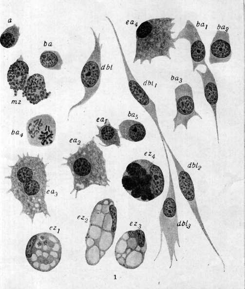
Figure 1. Cellular elements of the blood and connective tissue of the mollusk Anodonta: a, ba-bc - non-granular basophilic amoebocytes (ba - mitosis); dbl, dbl1 - dbl3 - their transformation into desmoblasts (connective tissue cells); e1-e4 - eosinophilic (granular) amoebocytes; ez1-ez4 - excretory cells; mg - intermuscular granular tissue cells (cellules spheruleuses of Kollmann). (According to Zavarzin.)
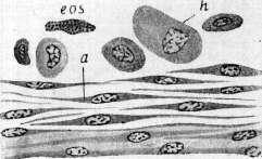
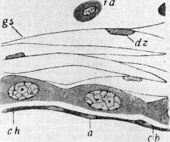
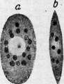
Figure 2a. Cellular forms of the blood of the rhinoceros beetle larva: a - small undifferentiated form from the hematopoietic organ; b, c - non-granular amoebocytes; d, e - "round cells" (granular amoebocytes); f - eosinophils. Figure 2b-d. Formation of a connective tissue capsule around a foreign body in the rhinoceros beetle (three successive stages): a, db, dz - non-granular amoebocytes transforming into ground substance (os); h, mg - "round cells"; eos - eosinophil; ch - chitin. (According to Lazarenko.) Figure 3. a, b - erythrocytes (b - in profile) of the annelid Pista cristata in a fresh state (dark droplets - fat); c, d - non-granular amoebocytes of the same annelid; e, f - granular amoebocytes of Perinereis cultrifera. Figure 3a. Eleocyte of Amphitrite rubra (light droplets - fat; dark large granules - excretory granules) (According to Romieu.) (See Figure 2a-d, 3 and 3a on the following insert table.)
2b
2d
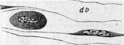
Sh § 3a 2c To the article Blood. BLOOD
Connective tissue. Under certain experimental conditions, and in particular upon the entry of a foreign body into the cavity of an animal or into the connective tissue, a connective tissue capsule forms around this foreign body. The formation of such a capsule proceeds entirely (Fig. 2b-a) at the expense of basophilic amoebocytes, which flatten out on the surface of the foreign body (the phenomenon of so-called thigmotaxis), fuse into a syncytium, and, through the differentiation of protoplasm, give rise to the formation of ground substance. The vascular wall in invertebrates sometimes has an endothelium and sometimes does not. There are indications that both the endothelium and the vascular wall itself are formed from the same amoebocytes (Danini). It is very characteristic that where there are no sedentary cells in the connective tissue (e.g., in insects), the vessels also lack an endothelium. In many invertebrates, the blood contains respiratory pigments. Most often, these are iron-containing hemoglobin, hemerythrin (red-yellow), chlorocruorin (green), and copper-containing hemocyanin (blue). The question of the colorless pigment achroglobin remains open and is decided rather negatively. In cases of colorless blood, the pigment is either absent altogether or is present in very weak concentrations of hemocyanin. Pigments are either dissolved in the blood plasma (many annelids, in particular the earthworm, some mollusks, and others) or are concentrated in special specialized cells, called erythrocytes by analogy with vertebrates (Fig. 3). Erythrocytes in invertebrates are found both in the vascular blood and in the cavity fluid. Sometimes they are present only in the coelomic lymph and are absent in the blood; more rarely, the opposite occurs. Erythrocytes are known in nemerteans, annelids, gephyreans, and mollusks. Their distribution is of a random nature. Sometimes, out of two closely related species [e.g., the mollusks Solen legumen (Griesbach) and Solen vagina, the annelids Polycirrus haematodes and Polycirrus caliendrum (Romieu)], one has erythrocytes, and the other does not. Erythrocytes in most cases are nucleated, have not particularly constant sizes, but a constant, usually round or oval shape. They are incapable of amoeboid movements. Their protoplasm is filled with pigment (hemoglobin or hemerythrin) and often contains urates and fat as inclusions. Thus, invertebrate erythrocytes can also combine reserve and excretory functions. Very rarely (the only case is the annelid Magelona), erythrocytes are also non-nucleated, just like those in mammals. These erythrocytes do not contain inclusions and are found only in the vascular blood (Romieu). The progenitor form for all the listed elements, up to and including erythrocytes, is a small non-granular basophilic amoebocyte, which, multiplying by mitosis, differentiates in various directions (Fig. 1 and 2a). Mitoses are found, although more rarely, also in granular amoebocytes. Specialized cells (erythrocytes, eleocytes, excretory cells, connective tissue cells, etc.) usually no longer multiply and gradually degenerate. Multiplication occurs in the tissue, in the blood, and in the coelomic lymph. The question of the existence of special hematopoietic organs in invertebrates remains to a large extent open and in most cases is decided negatively. Lymphoid organs have been described in cephalopods, insects, and decapod crustaceans. Spontaneous foci of hematopoiesis scattered in the connective tissue (crayfish, insects) have also been described. True lymphoid organs consist of a connective tissue reticular stroma, in the loops of which are located small basophilic non-granular amoebocytes multiplying by mitosis. The differentiation of the products of their multiplication always takes place outside the lymphoid organs and is likely heteroplastic. In many invertebrates (e.g., arthropods, mollusks, etc.), there exist special phagocytic organs, similar to lymphoid ones, but containing in their stroma only excretory-phagocytic cells (nephrophagocytes), which also originate from non-granular amoebocytes (see above). -In vertebrates, the blood is separated from the connective tissue both functionally and morphologically much more sharply than in invertebrates (including annelids). In connection with this, the cellular elements of both the blood and the connective tissue are more differentiated. While in invertebrates there are usually only two main types of amoebocytes, in most vertebrates there are at least three types of them (leukocytes); in this respect, only insects, which have three types of amoebocytes, approach the vertebrates. Besides erythrocytes, in the vascular blood of vertebrates, one distinguishes non-granular leukocytes (lymphocytes, monocytes) and granular leukocytes, or granulocytes (eosinophils, special leukocytes, basophilic leukocytes). -The situation is clearest with erythrocytes. In the blood plasma of vertebrates, hemoglobin is never present. Likewise, other respiratory pigments are not found. The erythrocytes of lower vertebrates have nuclei; in adult mammals, they are non-nucleated. In any given species, erythrocytes are distinguished by a much greater constancy of size than in invertebrates. The largest erythrocytes are found in amphibians and reptiles, the smallest in birds and mammals. The shape is oval in most nucleated erythrocytes (among non-nucleated, in the llama and camel) and round in the erythrocytes of most mammals and some fish. -Another non-motile specialized element of vertebrate blood are the thrombocytes of lower vertebrates and the blood platelets of mammals. Thrombocytes are small nucleated cells, somewhat similar to erythrocytes, but lacking hemoglobin; the morphological significance of non-nucleated blood platelets remains completely unexplained. -Vertebrate leukocytes are subdivided by most authors into two large groups: granular and non-granular. These names are valid mainly only for humans and mammals, and in some vertebrates, leukocytes belonging to the granular group do not contain specific granulation in the protoplasm (e.g., in many fish). Granular leukocytes are specialized forms incapable of reproduction. In animals with the most differentiated blood elements (birds, mammals), three types of granulocytes are distinguished: special, basophilic, and eosinophilic. In the overwhelming majority, special leukocytes are usually found. These leukocytes sometimes possess specific granulation (mammals, birds, reptiles, amphibians, some fish) and sometimes lack it (some fish). The granulation itself is extremely diverse in different species: it is sometimes neutrophilic and fine (dog, human, frog) and sometimes acidophilic and coarse (birds, reptiles, rabbit). Special leukocytes are particularly diverse in fish. Eosinophilic granulocytes represent a more constant form and are present in almost all vertebrates [with the exception of many fish, which, however, often have leukocytes with large protein inclusions resembling the protein cells of crayfish and other similar forms (see above)]. Eosinophils are usually few in number (2-5% of the total number of leukocytes), but in reptiles (in snakes), they are, on the contrary, very numerous. Eosinophils are distinguished by a more constant character of granulation; it is always sharply acidophilic and larger than in special leukocytes. --A less defined cellular form are the basophilic, or mast, leukocytes. In fish, they are apparently, with very rare exceptions, absent altogether. Furthermore, the basophils of mammals differ strongly from the basophils of birds, reptiles, and especially amphibians. While the basophilic leukocytes of mammals (constituting about 1% of the total number) differ quite sharply from the so-called mast cells of connective tissue, the basophils of birds, reptiles, and especially amphibians have a different character. The blood basophils of amphibians cannot be sharply delimited from the mast cells of connective tissue (Maximow), and the basophils of reptiles and birds may perhaps be immature myeloid elements (Kleineberger and Walter). In any case, under the name of basophils, non-identical forms are described in various vertebrates. The lymphocytes of lower vertebrates (fish, amphibians, perhaps birds and reptiles) are cells 7-10 μm in size with a narrow rim of basophilic non-granular protoplasm and a round nucleus. They are unconditionally undifferentiated elements capable of transforming into various specialized forms via a heteroplastic pathway. Recently (Maximow), they are even called hemocytoblasts (see below), and not lymphocytes. These lymphocytes can arise from reticular cells and themselves, by increasing the plasma rim, give rise to monocytes and macrophages. There are indications (Alfeeva, I. Kraft) that fibroblasts can also arise from these lymphocytes. Thus, the lymphocytes of lower vertebrates, in their potencies, are very similar to the basophilic non-granular amoebocytes of invertebrates. In the blood, they are always found in relatively significant quantities. In higher forms, and mainly in mammals, two forms can be distinguished among lymphocytes: one, concentrated mainly in hematopoietic organs, called the large lymphocyte (lymphoblast, hemocytoblast), and the other, formed in lymph nodes and present in significant numbers in the blood, called the small lymphocyte.
In the majority of mammals (man, dog, horse), there are fewer lymphocytes in the blood (about 20%) than granulocytes, but in some animals (rabbit, etc.) they are in the predominant (about 60%) majority. The question of monocytes is even more complex because, at the present time, it must be considered proven that a portion of monocytes may have a connective-tissue histiogenous origin and represent rounded-off reticular and endothelial cells and histiocytes of loose connective tissue that have entered the bloodstream. Thus, monocytes can have both a histiogenous origin and a hematogenous one (from a large lymphocyte). In any case, monocytes are forms that are little specialized, possessing significant nephrophagocytic capacity (macrophages). In lower vertebrates (mainly in fish, partly in amphibians), all blood elements are less specialized; furthermore, in these groups, there are elements absent in other vertebrates. Besides the aforementioned protein cells of fish, there are sometimes pigment leukocytes here. In addition, excretory inclusions are sometimes encountered in the leukocytes of fish. All this brings the blood of fish closer to the blood of invertebrates. Hematopoiesis has been studied mainly in mammals (Maximow) and amphibians (Drzewina, Maximow) and less well in birds (Danchakoff, Myasoyedov). With regard to hematopoiesis, vertebrates present a rather variegated picture. The main difference from invertebrates consists in the fact that while in the majority of the latter, the multiplication of cellular elements takes place in the blood (cavity) fluid itself, in vertebrates, hematopoiesis has been moved into the connective tissue, into the hematopoietic organs, and usually only mature forms enter the blood, with the exception perhaps of some fish and reptiles, in which multiplication and differentiation of cellular elements are possible even in the vascular blood.
A. Zavarzin. IV. Comparative data on the blood of vertebrates. Total amount of blood. The most accurate results are provided by the direct determination method of Welcker (1854) in the modifications of Müller and Abderhalden (Abderhalden, 1899, 1902; Müller, 1901, 1921). In view of the fact that this method, based on the determination of the amount of hemoglobin, yields its total amount not only in the blood but also in the muscles and other organs, it is not entirely accurate and produces errors of up to 8% (the amount of hemoglobin in the muscles is equal to about 5% of its total amount). Indirect methods by introducing isotonic solutions of salt, dextrin, dyes, antitoxins, etc., into the blood, as well as determination by the formation of hemoglobin-CO, yield more accurate but also not absolute results, especially since the figures obtained are subject to strong individual fluctuations. The following figures provide an idea of the total amount of blood in vertebrates. A dog weighing 10,350 g contains blood equal to 7.98–8.25% of its weight; for a dog weighing 5,780 g, the corresponding figures are 8.02–9.00%. The ratio of blood weight to body weight has the following values for some animals: lamprey – 5.2%, perch – 3.0%, frog – 5.7%, pigeon – 9.2%, mouse – 7.6%, rabbit – 5.5%, goat – 6.2%, cat – 6.6%, dog – 7.4%. For individual classes, the corresponding figures are as follows: fish – 2.0%, amphibians – 4.8%, reptiles – 5.8%, birds – 8.2%, mammals and humans – 6.8% (Welcker). All determinations of this kind have a very relative significance, and fluctuations within the limits of one species are very large. Thus, in the direct determination of the total amount of blood in 52 rabbits, the following figures were obtained. Average body weight 535 1,078 1,971. Percentage ratio of blood weight 5.65 5.20 4.49 4.35. Average body weight 2,250 2,494 3,147. Percentage ratio of blood weight 4.42 4.47 4.70. Attempts to derive general rules based on the surface area or volume of the animals studied (Dreyer; 1913) did not lead to definite results. The dependence of the differences on the biological properties of the objects studied—their warm- or cold-bloodedness, their herbivorous or carnivorous nature—is also not clear. The question is further complicated by the fact that it is not possible in all cases to account for the interrelations between blood and lymph. Chemical data. When investigating the chemical composition of blood serum alone, it turned out that the differences within the class of mammals are small, but that they are very large when investigating whole blood, which indicates the existence of corresponding differences in the composition of erythrocytes. Thus, according to the content of Na2O, P2O5, and K2O, groups I (human, pig, horse, rabbit), II (bull, goat, sheep), and III (cat and dog) relate as 1:5:10. The erythrocytes of the horse, pig, and rabbit generally do not contain Na, but are rich in K (4.489–5.250‰); in other mammals, the amount of Na is relatively large (bull 2.281‰), and the amount of K is relatively small (0.720‰). The differences in the amount of P between birds and mammals are very large (chicken – 7.510‰, human – 1.870‰). The ratio between the amount of erythrocytes and serum: human – 434.2:565.8; pig – 436.0:564.0; dog – 561.6:438.2; cat – 432.4:568.6; horse – 391.6:608.4; chicken – 391.5:608.5; at the same time, the blood of omnivores and carnivores is richer in erythrocytes (466.1‰) than the blood of herbivores (363.4‰); birds occupy an intermediate position in this respect (429.7‰). The amount of solid parts and water in the blood depends on the ratio of the amount of serum and erythrocytes. In mammals, the dry residue is equal to from 214.1‰ (omnivores and carnivores) to 194.4‰ (herbivores); in birds, it is equal to 204.1‰; in fish – 127.0‰; in frogs – 10%. The (relative) amount of hemoglobin in different classes: mammals – 100; birds – 83.4; reptiles – 46.2; amphibians – 41.5; fish – 38.1 (Korniloff; 1876). Quantitative data: per 1,000 weight parts of blood, there is iron, hemoglobin, and hematin: human: 0.545–129.7–6.18; dog: 0.550–139.8–6.24; cat: 0.457–122.3–5.18; pig: 0.525–134.1–5.96; horse: 0.492–126.4–5.58; rabbit: 0.430–123.5–4.88; chicken: 0.466–102.4–4.93; frog: 0.425–101.2–4.82 (Preyer; 1871). In general, the blood of mammals is richer in hemoglobin than the blood of birds, but at the same time, birds (122.1‰) occupy an intermediate position between omnivores and carnivores (152‰) on the one hand and herbivores (116‰) on the other. If we take the average hemoglobin content in the blood of mammals as 100, it turns out that for omnivores and carnivores it will be equal to 117.0, for herbivores – 89.6 (for non-ruminants – 94.7, for ruminants – 86.2), for birds – 94.3. The dependence of hemoglobin content on sex in animals is generally the same as in humans. Thus, in 1,000 parts of blood, hemoglobin is contained in males and females: in humans – 125.5–109.4; pigs – 166.8–154.0; dogs – 138.3–146.5; rabbits – 121.2–116.0; bulls – 112.0–104.0; chickens – 122.6–91.6. Chemical composition of plasma and serum. Since the amount of fibrin, or fibrinogen, in plasma is equal to 0.5–1% of the entire mass of plasma, it can be considered that the composition of serum and plasma is almost identical. The percentage content of water and solid parts in the blood in different classes of vertebrates is as follows: in mammals – 911 and 89, in birds – 934 and 66; in fish (with the exception of the eel) – also 934 and 66. Influence of sex on water content: men – 903, women – 906, bulls – 903, cows – 910; male cats – 909, female cats – 912. The content of mineral salts in serum is generally significantly higher in birds than in mammals, exceeding it by 12.5%, with the exception of Na, the amount of which in birds is lower than in mammals. It is possible that the higher Ca content in birds is related to the production of the calcareous shell. Protein is contained in plasma, or in serum, in an amount of up to 75% of the dry residue. Regarding the qualitative composition of proteins, significant differences are noted between different animals. Thus, the serum of warm-blooded animals contains all 3 types of serum albumins (α, β, γ with corresponding coagulation points: 70–73°, 76–80°, and 82–85°) with the exception of the horse, bull, and sheep, in which α-albumin is absent and in which coagulation occurs only at 76–80°. In cold-blooded animals, mostly only α-albumin is present, with the exception of the eel, which also has β-albumin, and the shark (Scyllium catulus), which has only β-albumin. Globulins. 1. Fibrinogen, which coagulates at 56°, is the same in all animals. The amount of fibrinogen in the blood per 1,000 parts of plasma is equal to: in humans – 4.2, in sheep – 6.0, in dogs – 4.6, in horses – 4.5, in pigs – 6.5 (Trendelenburg, Loewy; 1924). The average amount of fibrinogen per 1,000 parts of whole blood is equal to: in humans – 2.1, in pigs – 3.60, in dogs – 1.78, in cats – 2.03, in horses – 2.85, in rabbits – 3.75, in cattle – 6.03, in sheep – 3.80, in chickens – 5.85. In cold-blooded animals, the amount of fibrinogen is very small (Nasse; 1842). The amount of fibrinogen per 1,000 parts of plasma: in humans – 3.85, dogs – 3.09, cats – 3.59, pigs – 6.38, horses – 5.55, rabbits – 5.97, sheep – 5.13, bulls – 9.95, chickens – 6.61. The plasma of humans and carnivores is significantly poorer in fibrin than the plasma of herbivores and pigs. In the first group, max = 3.85% (human), in the second, min = 5.13% (goat). In males, the fibrin content is higher than in females. Content of urea and amino acids. Animal: Fibrin content in 1,000 parts of plasma. Cat: 4.39. 2. Serum globulin (paraglobulin) is contained in cold-blooded and warm-blooded animals in the same amount. The ratio of the percentage content of albumins and globulins in the serum of different animals is as follows: Protein. Warm-blooded animal. Cold-blooded animal. Difference. Total amount of protein: 6.51. Albumin: 3.61. Globulin: 2.90. 3.74. 1.12. 2.62. 2.77. 2.49. 0.28. Dependence of globulin and albumin content on physiological state: Rabbits. Normal. Starving (5 days). Protein in serum in %: 6.70. 7.70. (A) albumin: 4.80. 6.40. (G) globulin: 1.90. 1.30. 2.52. 4.92. Thus, in rabbits, the ratio between albumins and globulins doubled as a result of a 5-day fast; in the horse, a 24-hour fast was sufficient to obtain an effect 2.25 times greater. Content of non-coagulable N-substances. Content of residual N (average figures according to Bang). Animal. Residual N in blood. In plasma (in mg %). Pig: 27. 23. Horse: 35. 35. Sheep: 22. 19. Total content of residual N. (a) Urea. (b) Amino acids. Animals. Mammals (average): 30.6. 15.3. 1.0. Human: 6.0. 3.5. Pig: 0.9. Dog: 1.4. Cat: 0.9. Horse: 0.7. 1.4. 0.9. Rabbit: 0.9. Birds (average): 42.9. 8.5. 33.5. 0.25. Chicken: 33.0. 0.4. Pigeon: 0.1. Fish: Shark: 4.0. Carp: 0.4. According to the urea content in the blood, different animals are distributed as follows: mammals 0.00328%, birds 0.0182%, teleost fish 0.0238%, shark-like fish 1.8042%. Shark-like fish occupy exactly the same exceptional position with regard to extractive substances. Animals. Mammals (except humans). Human. Pig. Dog. Horse. Bull. Sheep. Birds: Chicken. Fish: Shark.
Bony marine [fish] Freshwater [fish] Creatinine Creatine 4.94 6.80 5.31 3.12 5.14 5.95 3.34 3.74 36.0 6.20 5.46 Uric acid 2.0 0 0 0 0 0 1.67 4.22 ? Thus, the blood of birds is incomparably richer in uric acid than the blood of mammals, which is connected with the fact that in the former, urine also contains mainly uric acid. Amino acid content in the blood of various animals. Animals Human Amino acid N 7.13 Pig 8.43-10.90 Dog 4.60-10.20 Cat 8.68 Ox 6.58 Sheep 7.0 Animals Rabbit Chicken Shark Bony fish Amino acid N 9.20 20.29 39.0 28.0 NH3 content in blood (in mg%). Mammals...............
3.94
Allantoin is absent in human blood; in the blood of the bull and pig there is 0.78 and 0.57 mg% respectively, which corresponds to the presence of this compound in the urine of the latter. Sugar and reducing substances are contained in different animals in the following amounts: in mammals 81 mg%, in birds 201 mg%, in amphibians 65 mg%, in fish 50 mg%. Within the same species, fluctuations reach up to 100%. Thus, for dogs, figures from 79 to 162 have been found, but it is necessary to take into account the phenomena of emotional hyperglycemia in laboratory animals, occurring under the influence of tying, fright, etc. (Cannon; 1911). Among mammals, the lowest sugar figures in the blood are found in cold-blooded animals and ruminants (50-57 mg%); in non-ruminants, this figure reaches up to 90 mg%. Fats, lipoids, cholesterol (content in blood in mg%). Animals: Neutral fats, Fatty acids, Cholesterol, Lecithin. Human: 62-78, 180. Pig: ... Dog: ... Cat: ... Horse: ... Rabbit: ... Neutral fat is contained only in plasma, but not in erythrocytes. Fatty acids and cholesterol are distributed between them approximately evenly. The lecithin content in erythrocytes is higher than in plasma. Pigments. The coloration of plasma or serum is more or less characteristic for different species. In the horse, it is lemon- or golden-yellow; in the bull, red-yellow; in the rabbit and pig, almost colorless. The color of the plasma is conditioned by the presence in it of pigments: lipochromes or luteins and bile pigments, especially bilirubin. Animal lipochromes—carotenoids—are close to plant lipochromes: xanthophyll (C40H56O2), soluble in alcohol, and carotene (C40H56), soluble in ether (Willstatter, Escher; 1910). A complete or almost complete absence of lipochromes characterizes the pig, dog, goat, rabbit, and guinea pig. A high content of lipochromes is characteristic of humans, bulls, chickens, and pigeons. Usually, either one or the other pigment is present; but if both are present, one usually strongly predominates. Thus, in humans and bulls, carotene predominates; in the chicken, xanthophyll. Carotene is also found in the horse; but it is completely or almost completely absent in the sheep or goat. The amount of lipochromes fluctuates within individual species quite widely and depends on the content of lipochromes in the food; but it has been clarified that each given animal species is characterized by the ability to pass only a quite specific lipochrome from the intestine into the blood. If bulls are fed food that does not contain the carotene typical for them, but is rich in xanthophyll, the latter still does not penetrate into the blood. Reverse relationships are found in chickens (Hyman, Miiller; 1916). Bilirubin. There is a great deal of data on the comparative content of bilirubin in different animals. Dog serum does not contain it. In the serum of the pig, ram, goat, rabbit, and guinea pig, only traces of it have been found. In the bull, it is sometimes contained in a fairly large amount. In the horse, there is mostly 0.02‰. Chemistry of erythrocytes. Average water content 62.5%. The content of mineral salts in erythrocytes in various animals is very different. Thus, for example, in the pig, horse, and rabbit, sodium is completely absent, while in the bull, goat, and sheep there is a lot of it; in humans, there is little. The reverse relationship is observed in the case of potassium. The content of P2O5 is very characteristic for different classes: 1.31‰ for mammals (1.902 for non-ruminants, 0.727 for ruminants), 6.685 for birds. The amount of iron is generally the same everywhere, averaging 0.154% Fe2O3. An exception is ducks (0.229% Fe2O3). It is possible that this is connected with their diving way of life, requiring large reserves of O2 in the blood. Hemoglobin. In general, the Hb content in birds (23.4%), if one does not count ducks, is lower than in mammals (31.8%). Percentage content: Mammals, Birds (including ducks), Ducks only. Hematine: 0.111, 0.115, 0.160. Hemoglobin: 1.26, 1.30, 1.82. Hb: 31.8, 27.1, 38.2. Thus, there is no parallelism between the figures relating to the content of iron and hematine on the one hand and Hb on the other. It is possible that different hemoglobins contain unequal amounts of iron or that they are not identical in colorimetric relation. Different hemoglobins can differ from one another in their ability to crystallize, in the shape of crystals, in light absorption, and in resistance. Crystallization. Oxyhemoglobins of the squirrel, rat, horse, and dog crystallize easily and are difficult to dissolve. Decomposition time of Hb (in minutes). Animals: 1/4 NaOH, 1/4 CH3COOH. Human: 0.73, 1,440.0. Pig: 3.3, 1.1. Dog: more than 1,440.0, 1,290.0. Cat: 80.0, 21.0. Bull: 137.0, 11.5. Horse: 21.5, 40.5. Rabbit: 40.0, 132.0. Guinea pig: 26.0, 49.0. Pigeon: 330.0, 153.0. Eel: 191.0, 41.0. Tench: 64.0, 141.0. Oxyhemoglobin of the cat crystallizes with difficulty. Oxyhemoglobin of humans, monkeys, bulls, sheep, pigs, rabbits, pigeons, and fish (exception: herring) dissolves with great difficulty and crystallizes easily (Preyer; 1871); the shape of crystals in the majority of mammals is rhombic, in the squirrel it is hexagonal, in the pigeon it is tetragonal, in the hamster it is monoclinic. Reduced hemoglobins dissolve more easily and crystallize with more difficulty. During the crystallization of putrefying blood, the character of the crystals can be different; thus, for example, in the horse, instead of normal prismatic crystals of the rhombic system, hexagonal ones can also be obtained. Light absorption also definitely points to the difference between different hemoglobins. The study of their resistance in relation to NaOH and CH3COOH leads to the same results (see table on p. 638 below). Data of elementary analysis of erythrocytes. Elements: Animals, C, H, N, S, Fe, O. Horse: 54.68, 7.07, 17.40, 0.66, 0.46, 19.74. Cat: 54.57, 7.22, 16.38, 0.57, 0.34, 20.23. Guinea pig: 54.60, 7.25, 16.52, 0.62, 0.35, 20.66. Chicken: 54.12, 7.36, 16.78, 0.58, 0.48, 20.68. [Unspecified]: 59.47, 7.19, 16.45, 0.86, 0.34, 22.50. Thus, for 1 atom of iron in the horse, there are 2 atoms of sulfur, and in the dog 1 atom; in the chicken, for 2 atoms of iron, there are 9 atoms of sulfur. Formed elements. Erythrocytes. For the function of respiration, the absolute amount of Hb in the erythrocyte is important on the one hand, and on the other, the area of its contact with the surrounding environment, since the binding of O2 occurs only on its surface. Therefore, it is assumed that, in general, small but numerous erythrocytes are characteristic mainly of animals with very intense metabolism, in contrast to animals with more sluggish metabolism, which are characterized by larger but less numerous erythrocytes. This relationship, however, is not a general rule. For humans and domestic mammals, the amount of Hb is a constant value, as a result of which all fluctuations in the amount of Hb correspond to specific changes in the number of erythrocytes. On 1 µ2 of the surface of an erythrocyte of a human and domestic animals, there are 32·10-14 g of Hb. In a comparative study of animals of one species, the difference between the average numbers relating to individual specimens does not exceed a few percent, while the difference between individual erythrocytes of one specimen can reach 40%. The amount of Hb in one erythrocyte fluctuates for all animals around the average figures of 30.3-31.7·10-14 g. However, it is necessary to note that for carnivorous and omnivorous animals, generally higher figures were obtained than for herbivorous ones. Number of erythrocytes. Erythrocytes are present in all vertebrates with the exception of the amphioxus (Amphioxus). The precise determination of their number became possible only recently, when it was clarified that previous authors, working with the Thoma-Zeiss chamber, fell into an inevitable error, since they did not take into account the circumstance that in different animals the speed of erythrocyte sedimentation is not the same. Therefore, the counts of the number of erythrocytes in lower vertebrates turned out to be especially inaccurate, since the higher the content of Hb in erythrocytes, the faster their sedimentation occurs. The shortcomings of the Thoma-Zeiss chamber are avoided when using the Bürker chamber. Animals: Number of erythrocytes in 1 mm3 (in millions), Bürker, Thoma, Difference in %, % Hb in blood, Hb in one erythrocyte (in 10-12 g), Sedimentation speed in chamber at 0.1 mm (in sec), Sedimentation speed in 1 minute (in mm). Frog: 0.452, 0.452, 1.068, 16-18, 14.6, 15.2, 14-16.6, 16.9, 12.1, 14.3, 11.6, 11.7, 8.5, 322, 54, 26-35, 25, 21, 19, 19, 18, 7, 50, 56-79, 9.3, 99, 103, 148, 0.375, 0.120, 0.076-0.107, 0.088, 0.065, 0.061, 0.058, 0.041. Pigeon: ... Dog: ... Rabbit: ... Pig: ... Distribution of Hb on the surface of erythrocytes. Animals: Number of erythrocytes in 1 mm3 (in millions). Dog: ... Pig: ... Rabbit: ... Bull: ... Horse: ... Sheep: ... Goat: ...
Human. Hb content in one erythrocyte, expressed in 10-12 g: 6.59, 7.44, 5.86, 5.72, 6.94, 10.70, 13.94, 5.00, 24, 22, 20, 19, 18, 11, 8, 30. Diameter (in µ), Surface (in µ2): 7.26, 82.7; 6.60, 68.4; 6.60, 68.4; 5.94, 55.4; 5.94, 55.4; 4.62, 33.6; 4.00, 25.1; 7.92, 98.4. On the other hand, errors also arose from the fact that it was not taken into account that the fluids used for observations were not applicable to all erythrocytes to the same extent; for example, Hayem's fluid, widely used in the study of human blood, is not applicable for the study of goat and bull blood, as it causes agglutination and shrinkage of erythrocytes in them, which cannot but affect the accuracy of the study (Behrens; 1923). In general, the dimensions of erythrocytes are always inversely proportional to their number; however, there are exceptions to this rule. Thus, in the llama, erythrocytes are larger than in humans; but their number normally reaches up to 13 million against 5 million in humans; however, the thickness of erythrocytes in the llama is significantly less than in humans, as a result of which the total volume of its erythrocytes is correspondingly lower, approaching the human norm. Among all vertebrates, mammals have the most numerous and at the same time the smallest erythrocytes. Hb content per 1 µ3, expressed in 10-14 g: 29, 32, 29, 34, 33, 33, 32, 31. Animals: Mammals: Monkey, Human, Dog, Cat, Mouse (black, white), Rat, Rabbit, Guinea pig, Horse, Bull, Goat, Pig, Elephant, Sheep. Number of erythrocytes (in millions); Diameters (in µ). Birds: Pigeon, Raven, Chicken, Duck, Ostrich, Finch. Reptiles: Turtle, Lizard. Amphibians: Frog, Triton, Proteus. Fish: Barbel (bony), Eel, Skate (cross-mouthed). The figures given are averages. Thus, among mammals, the elephant, having the largest erythrocytes, has the smallest number of them; among birds, the ostrich occupies the same position, and among reptiles and amphibians, the proteus. Of the fish, cartilaginous ones have the smallest number of erythrocytes compared to bony ones. The comparative volume of erythrocytes and the relative content of Hb in them are visible from the following table. Dependence of the number of erythrocytes on age. The number of erythrocytes increases with age, with the exception of the bull, in which the number of erythrocytes is higher at an early age than in adult animals. Thus, in a guinea pig weighing 490 g and 23 cm long, the number of erythrocytes is 5.3 million; in a fetus weighing 3.5 g and 3.2 cm long - 1.9 million. A fetus weighing 25.6 g and 11 cm long has 3.52 million erythrocytes, and a newborn guinea pig (72 g, 12 cm) - 4.9 million erythrocytes (Bethe; 1891). In an adult bull, the number of erythrocytes is 6.503 million, and the ratio of the number of leukocytes to erythrocytes is 1:729; in a calf, the corresponding figures are 8.523 and 1:548. Dependence of the number of erythrocytes on sex. In men, the number of erythrocytes is higher than in women (5.5:5.0). In sexually mature males, their number, as well as the Hb content, is higher than in females. In castrated animals, these numbers are lower than in normal ones. Very strong fluctuations in the number of erythrocytes are observed in amphibians depending on the sexual cycle, and in accordance with this, depending on the spawning period. But it is quite natural that the influence of the environment (temperature, humidity, nutrition) also has an effect at the same time. The dependence of the number of erythrocytes on environmental conditions is especially noticeable in aquatic animals; thus, when keeping amphibians on land, a gradual thickening of the blood occurs in them and a corresponding relative increase in the number of erythrocytes; in a frog that has lost 13.1% of its initial weight, there are 1,284,000 erythrocytes in 1 mm3; after a loss of 34.8% of weight, their number is already 2,588,000 (Durig; 1901). The sizes of erythrocytes vary greatly depending on the species. Structure of erythrocytes. Mammals have round erythrocytes with the exception of camelids (Tylopoda), in which erythrocytes have an elliptical shape. Birds, reptiles, amphibians, and fish also have elliptical erythrocytes. In lampreys, erythrocytes are round. For the structure of erythrocytes, the questions of the existence of a membrane and stroma in them are extremely important. The question of the membrane is apparently decided in the sense that at the boundary with the environment, i.e., plasma, the outer layer of erythrocytes has the character of a two-phase colloidal system (Berczeller; 1922). But besides this, there is undoubtedly a preformed stroma in erythrocytes, which determines their specific shape. In close connection with the stroma are apparently the so-called marginal hoops, best expressed in the erythrocytes of many amphibians and fish (Meves; 1901). These hoops have a fibrillar structure and undoubtedly determine the characteristic shape of elliptical erythrocytes, including the erythrocytes of the llama (Jolly; 1920). Some authors describe them in humans as well (Romieu; 1922). In any case, it seems very likely that the so-called Cabot rings are also similar in their origin to the indicated fibrillar formations. Nucleus. In mature erythrocytes, nuclei are generally present only in birds, reptiles, amphibians, and fish; but individual specimens are also found without nuclei, which in all probability is associated with phenomena of degeneration, karyolysis, etc. But although in the indicated forms the presence of a nucleus is inherent in both adult and embryonic erythrocytes, the latter are still distinguished (especially in amphibians) by their large size. In ontogenetic development, the erythrocytes of all vertebrates first pass through the stage of an erythroblast—a cell with a large nucleus and narrow protoplasm. The next ontogenetic stage, characterized by a relatively somewhat smaller nucleus, is the normal form of erythrocytes for fish and amphibians; a further relative decrease in the nucleus leads to a type typical for reptiles and birds, and finally, the disappearance of the nucleus gives the erythrocyte of mammals. Thus, in the lower classes of vertebrates, the phylogenetic type of erythrocytes is determined by the arrest of its development at an earlier ontogenetic stage (Minot; 1903). Blood platelets. In mammals, blood platelets have the character of peculiar small elements containing azurophilic granulation in the protoplasm. In all lower vertebrates, starting from birds, platelets or thrombocytes are represented by spindle-shaped cells resembling an erythrocyte in their structure. They have a large elliptical nucleus, mostly with a longitudinal fold on the membrane. The protoplasm does not contain hemoglobin. In serological and immunological respects, the thrombocytes of lower vertebrates fully correspond to the platelets of mammals. The size of the spindle-shaped cells is approximately 1/2-1/3 of the corresponding erythrocytes. Apparently, these cells also have phagocytic functions. G. Epstein. V. Total mass of blood. Mass of blood. The widespread idea that the total amount of blood in a human is approximately 1/13 of his weight is based on data obtained by Welcker and Bischoff in 1856 from two decapitated people using very imperfect methods. Subsequently, many methods were devised for determining the mass of blood in vivo; at present, mainly two methods are used for this purpose: CO inhalation (inhalation method) and the introduction of colloidal dyes (dye method). The inhalation method was proposed as early as 1882 by Grehant and Quinquaud, but came into use only after it was improved and simplified by Haldane, Smith, Plesch, and Whipple. The subject (human or animal) is given a certain (harmless) amount of CO to inhale, and after a few minutes, the CO content is determined by the Van Slyke and Salvesen method in a portion of blood taken from a vein; in another portion of the same blood, the ratio of the volume of erythrocytes to plasma is determined using a hematocrit (see), on the basis of which the mass of erythrocytes (CO is absorbed only by them) and the total mass of blood are then calculated.
Example: 50 cm3 of CO inhaled; 0.2 cm3 of CO found in 10 cm3 of blood; the volume of erythrocytes in the hematocrit is equal to 45%. The mass of erythrocytes is equal to: 50 * 10 * 0.45 = 2,250 cm3; the mass of blood is equal to: 2,250 : 0.45 = 5,000 cm3. The dye method was proposed in 1906 by Kottmann, but the dyes he used (indigo, methylene blue) were not suitable for this purpose; only after Keith, Rowntree, and Geraghty in 1915, and other authors following them, demonstrated the harmlessness and suitability of acidic colloidal dyes (Vitalrot, Trypanrot, Trypanblau, Kongorot), did this method begin to be used on a large scale. Its principle consists in injecting a certain amount of dye solution into the vein of the subject under study and, after 4 minutes (the time required for the uniform distribution of the dye in the blood), extracting a certain amount of blood from a vein in the other arm and comparing the stained plasma in a colorimeter with a standard of a certain concentration. Since the dye is not adsorbed by erythrocytes, the amount of plasma in the organism can be calculated from the degree of its concentration in the plasma, and the total mass of blood is then calculated on the basis of hematocrit data. There are numerous variations of this method. The method of injecting colloidal dyes yields less accurate data in nephrosis and especially amyloidosis (due to the greater permeability of the dyes through vessels and, mainly, due to the elective adsorption of the dye Congo red by amyloid substance) (Benhold, Griesbach); however, in the first minutes after the injection of the dye, when the plasma is colorimetered, these interfering factors have not yet had time to manifest sufficiently. Also, in cases of splenomegaly, excessively large amounts of blood were obtained by the dye method, and this was explained by the absorption of the dye by the phagocytic apparatus of the spleen (Greppi, Hartwich, May); however, Volsky and Sheveleva did not find such an increase in blood mass in malarial splenomegaly. Rather, in splenomegaly, one can expect greater lability of the circulating blood due to an increase in the splenic reservoir (see below). Depending on the basic research methodology used and one or another of its modifications, the average "normal" figures for blood mass obtained by different researchers diverge sharply and fluctuate between 48 and 92 cm3 per 1 kg of weight, not to mention the extreme figures (33 cm3 and 104 cm3 per 1 kg). Especially low average figures were obtained using the inhalation method (Haldane and Smith—48 cm3, Plesch—53 cm3); this is explained by the fact that this method reveals only those erythrocytes that are circulating in the blood at a given moment, while the CO does not reach those that are in the spleen and other erythrocyte depots; when Haldane and Douglas lengthened the interval of time between the inhalation of CO and the extraction of blood (mixing time) and the CO was thus able to penetrate into the erythrocyte reservoirs, the figures for blood mass turned out to be much more significant—78 cm3 per 1 kg. Of the researchers who worked with the dye method, Griesbach, Greppi, and Gordon found average blood figures between 66 and 75 cm3 per 1 kg of weight; the majority, however, and among them Keith, Rowntree, and Geraghty, Brown and Rowntree, Seyderhelm and Lampe, found from 83 to 92 cm3 per 1 kg; according to Chernyak's data, the average norm = 83 cm3, including 44 cm3 of plasma and 39 cm3 of erythrocytes per 1 kg. Since the average figures diverge even among authors using the same methodology, one must admit that here (just as in relation to individual differences found by each researcher), constitutional and social characteristics play a role, entailing differences in the degree of corpulence, development of musculature, and adipose tissue. All these questions still await their resolution. Partly, this difference in the normal average values of blood mass among various authors is explained by the fact that until recently, no precise distinction was made between the mass of circulating blood, which is in a state of rapid movement in the bloodstream, and the mass of blood in blood reservoirs or depots, where the blood, as it were, stagnates, being excluded from active circulation by special mechanisms (primarily in the spleen by the contraction of the muscular elements of the Schweigger-Seidel capillaries, in the liver thanks to the contraction of the sluice apparatus of the hepatic veins of Arey and Simonds, in the subcutaneous subpapillary plexus, etc.). Experiments by J. Barcroft and his school showed that a decrease in the size of the spleen (during an increase in ambient temperature, a drop in atmospheric pressure, physical exertion, fits of anger, digestion, pregnancy, and some other states) is accompanied by the entry of this reservoir blood into the general circulation and can thereby increase the total blood mass, as well as change the ratio between plasma and cellular mass, since the blood of the splenic depot, ejected upon the contraction of its muscular trabeculae into the general circulation (sang de chasse of Binet), is much richer in erythrocytes, leukocytes, and platelets than the circulating blood (partly due to their sedimentation by gravity during blood stasis). However, this separate accounting of the volume of circulating and reservoir blood mass has still hardly been used in clinical practice, except in the question of different types of cardiovascular decompensation (see Circulation). In clinical practice, the determination of blood mass has found fairly wide application in the study of various kinds of anemic states and diseases in which so-called "blood thickening" and "plethora" were assumed. These vague terms, and especially the latter with its subdivisions, contributed to the emergence of a number of incorrect pathogenetic concepts. The study of blood mass made it possible to replace them with completely rational concepts proposed by Brown and Rowntree, which deserve the widest dissemination: Normovolaemia, Hypervolaemia, Hypovolaemia. In the presence of a normal blood mass, they speak of normovolaemia, with an increase in blood mass—of hypervolaemia, with a decrease—of hypovolaemia. Depending on the ratio between the mass of erythrocytes and plasma (by hematocrit), they then divide each of these categories into simple (normal ratio), polycythemic (relative increase in erythrocyte mass), and oligocythemic (relative decrease in this mass). Thus, 9 categories are obtained, into which all changes in blood mass fit and which give a clear idea of these changes: I. simplex, polycythaemica, oligocythaemica. Blood mass in anemias. After acute hemorrhages in humans and experimental bloodletting in dogs, Smith, Plesch, Whipple, and others found that the loss of blood is very quickly replaced by a corresponding increase in plasma mass, as a result of which the blood mass may even exceed the initial one, especially with the rapid onset of erythrocyte regeneration. In chronic hemorrhages, however, a noticeable decrease in blood mass may occur (Plesch, Griesbach, Mendershausen, Chernyak), but almost exclusively at the expense of erythrocyte mass (hypovolaemia oligocythaemica), while the plasma mass remains almost unchanged or even increases. In so-called secondary anemias of various origins, Keith, Rowntree, and Geraghty, and Griesbach found the blood mass to be normal; Gordon, Hartwich, and May found an increase in relative blood mass (evidently in connection with the emaciation of the patients), but some decrease in the absolute mass; similar data were obtained by Cipriani and Giorgio; in Chernyak's cases, both the absolute and relative blood mass turned out to be within the norm (on average—82.6 cm3 per 1 kg). At the same time, however, the ratio between erythrocytes and plasma turns out to be sharply disturbed: the mass of the former is sharply lowered (23.7 cm3 instead of 39), the mass of the latter is increased (58.9 cm3 instead of 44). Thus, for "secondary anemias," the type normovolaemia oligocythaemica can be considered characteristic. In pernicious anemia, the blood mass fluctuates depending on the stage of the disease: during remission, it is normal; during relapses, it decreases, with the plasma mass increasing but unable to compensate for the loss of erythrocytes; in the pre-death period ("coma"), Mendershausen found a new increase in blood mass due to the dilution (Verwässerung) of the plasma. According to Rusznyak's data, the blood mass in pernicious anemia is normal, and during remission, it is increased. In leukemia, the determination of blood mass is complicated by splenomegaly (see above); Keith and Greppi obtained very large figures; however, Mendershausen found oligocythemic hypovolaemia or normovolaemia in three cases. He obtained the same data in three cases of lymphogranulomatosis. In chlorosis, the authors' data diverge: Smith and Plesch found significant hypervolaemia, Griesbach—both hyper- and hypovolaemia, Hartwich and May—hypovolaemia with normal plasma mass. Blood mass in polyglobulias. Of great interest are the data obtained regarding the true polycythemias of Vaquez and Gaisböck. In all such cases, a significant increase in total blood mass was found, reaching in one case of Brown and Griffin up to 224 cm3 per 1 kg.
Seyderhelm and Lampe drew attention to the fact that in polycythaemia rubra Vaquez, an increase in the relative mass of erythrocytes is always observed, sometimes reaching 80% of the blood mass (polycythemic hypervolemia), while in Gaisböck's polycythemia, the ratio between the mass of erythrocytes and plasma remains more or less normal (simple hypervolemia or, according to Seyderhelm and Lampe, polyemia). Among symptomatic polyglobulias, one should note the polyglobulia observed in pulmonary tuberculosis. The contradiction between the external pallor of these patients and the composition of their blood was explained at one time by Grawitz as a consequence of "blood thickening," masking the actual anemia. Studies of blood mass, conducted by Brieger on 23 tuberculosis patients, showed, however, that neither the total blood mass nor the plasma mass in them is reduced, and in cases of significant emaciation, they may even be relatively increased. Similar data can be found in Mendershausen. Chernyak's studies yielded an average blood mass in tuberculosis patients of 86 cm3 per 1 kg, and plasma of 48 cm3. Grawitz's hypothesis should therefore be rejected, and the polyglobulia of tuberculosis patients recognized as quite real. In congenital heart defects accompanied by polyglobulia, Plesch and Griesbach found a sharp increase in blood mass due to erythrocytes with a normal or even slightly reduced amount of plasma (polycythemic hypervolemia). For changes in circulating blood mass during states of collapse and cardiac decompensation, see Circulation, pathology. Regarding kidney diseases, the opinion has long been established that the anemia, edema, and hypertension accompanying them are caused by hydremia (see) or "hydremic plethora" (see Plethora). Numerous studies of blood mass show, however, that an increase in it is observed in only about half of nephrotics, while in the other half and in nephritics there is normovolemia or hypovolemia, and in anemic ones—oligocythemic hypovolemia. The anemia of Bright's disease patients is thus a true anemia (Seyderhelm and Lampe, Brown and Rowntree) of the hyporegenerative type (Tareyev). The formation of edema and its reduction are accompanied in kidney patients by significant fluctuations in blood mass; however, no constant relationship between "blood edema" and tissue edema is observed (Brown and Rowntree, Tareyev). There is also no definite dependence between blood mass and nephritic hypertension (Tareyev). Regarding other types of hypertension, there are observations about a parallelism between fluctuations in blood pressure and blood mass (Griesbach, Plesch); however, all authors agree that the level of pressure is not determined by blood mass. Among other pathological states, one can note a decrease in the relative mass of blood and plasma in obesity. After weight loss, both an increase and a decrease in blood mass are observed (Brown and Keith). In connection with this, one should point out the observations of Thompson and Chernyak, who saw an increase in blood mass in myxedematous patients under the influence of thyreoidin. Conversely, under the influence of insulin, blood mass, according to Villa, noticeably decreases. Changes in blood mass are observed under the influence of pilocarpine, atropine, morphine, and ether anesthesia (Griesbach). Among physiological states affecting blood mass, one should note pregnancy, in which most authors find hypervolemia, whereas Neubauer found a normal blood mass. In newborns, relative hypervolemia was found when calculated per 1 kg of weight, and a decrease in blood mass when calculated per body surface area (Seyderhelm and Lampe).
Ya. Chernyak. VI. The ratio between the formed elements and blood plasma. Blood represents a suspension of formed elements (erythrocytes, leukocytes, and platelets) in liquid plasma. Left to itself, blood outside the organism soon clots completely; the ratio of serum, which separates subsequently due to the retraction of the blood clot, to this clot does not repeat the initial ratio of the liquid part and the formed elements of the blood due to the precipitation of fibrin and the varying compactness of the blood clot. A more or less accurate idea of the richness of blood in plasma can be formed if the blood is defibrinated and left to independent sedimentation. To avoid defibrination and better preserve erythrocytes, Marcano diluted blood with a formol solution of Na2SO4 with the addition of NaCl, left the blood to stand in a small graduated conical vessel, and calculated the percentage content of formed elements in the blood based on the volume of the sediment. Biernacki, instead of diluting the blood, added minimal amounts of oxalate (2 mg per 1.0), and Grawitz constructed a special device based on the same principle—the Blutvoluminimeter. However, during independent sedimentation, intercellular spaces filled with plasma remain between the blood corpuscles; therefore, more accurate results are obtained by mechanical centrifugation. Such an apparatus, the hematocrit (see), modeled after the lactocrits of the dairy industry, was constructed by Hedin and widely used by Daland. The hematocrit consists of a pair of graduated capillary tubes fixed in a special metal stand with a device for centrifugation. Hedin added oxalate to the blood at 1:1,000 and diluted the blood before centrifugation with an equal volume of 0.9% NaCl solution. It is very difficult to obtain truly accurate data with a hematocrit, since oxalate and citrate in substantia cause significant changes in the volume of erythrocytes, as well as hemolysis; their isotonic solutions are often not isotonic for pathological blood. Griesbach preliminarily defibrinates the blood (the loss of erythrocytes in this process is so small that it is not revealed by comparative counting). It is better to add hirudin (or heparin) to the blood. Koeppe and Capps, with rapid manipulation, using a powerful centrifuge at 5,000 revolutions per minute, manage without any anticoagulant or are content with paraffin-coating the capillaries; the separation of blood into layers occurs so quickly that clotting is prevented, and at the same time so completely that all intercellular fluid is removed; this changes the usual conditions of light refraction, and the column of erythrocytes appears transparent, lacquered (in the absence of hemolysis). In the absence of such a centrifuge, it is preferable to work with a large amount of blood in accurately calibrated centrifuge tubes of 15 cm3, which gives greater accuracy of results. The percentage content of plasma in blood can also be determined indirectly by the content of some substance in whole blood and in plasma (the rule of mixtures). Thus, Bunge used for this purpose the fact that in some animal species Na is found exclusively in the plasma and is absent in the erythrocytes. Hoppe-Seyler, for the same purpose, determines the fibrin content in whole blood and in plasma. It is also possible to determine the distribution of an added indifferent substance that does not penetrate the erythrocytes, or certain physicochemical properties of blood and plasma that are sharply different for the formed elements and the liquid part (e.g., electrical conductivity—Höber), specific gravity (Grawitz), viscosity, and especially refractometry (Alder). As a result of all the cited methods, it has been found that the formed elements normally constitute slightly less than half of the entire blood, on average 44%. Froehlich cites figures of 45.13% for men and 41.38% for women. Figures from other authors often fluctuate within significantly wider limits. Venous blood contains a larger volume of erythrocytes due to their swelling from CO2. Muscular work causes an increase in the total volume of erythrocytes due to an increase in their number caused by the contraction of the spleen (Barcroft). Large fluctuations in the normal volume of erythrocytes have been described in various mammals [27.8 in the goat and 39.4 in the pig (Goetze)]. In human pathology, depending on anemias or erythremias, the fluctuations range from 9% to 80%. The determination of erythrocyte volume is important for studying fluctuations in the size of an individual red blood corpuscle and its Hb saturation. The so-called Volume index of Capps, which expresses the ratio of the percentage change in the mass of erythrocytes to the percentage change in their quantity in a given blood sample, is a constant value in normal conditions and gives characteristic deviations in various forms of anemia: a decrease in secondary anemia and an increase in pernicious anemia; in the latter, this increase in the volume index is an earlier sign than the increase in the color index (Haden). The saturation index, on the contrary, remains below normal even in pernicious anemia, i.e., the increase in the color index occurs only at the expense of the size of the corpuscle. During spontaneous sedimentation or centrifugation of blood, lighter nucleated erythrocytes, reticulocytes, and erythrocytes with malarial parasites accumulate predominantly in the upper layers, which can be used to enrich smears with the corresponding elements. Above the column of erythrocytes in the hematocrit, a layer of leukocytes accumulates (about 1% in normal conditions and significantly more in leukemias). Platelets are located in the upper layers; by repeated centrifugation, it is possible to isolate platelets in an almost pure form, especially from horse blood. It must be remembered that in circulating blood, significant mutual movements of water, salts, protein, etc., occur between erythrocytes and plasma under the influence of organ work, metabolism, etc., in accordance with which the known percentage ratio of plasma and formed elements is not a CONSTANT value.
Tareev. VII. Morphological composition of blood and the methodology of its investigation. Morphological composition of blood. In healthy people, blood in a fasting state reveals a fairly constant morphological composition, fluctuating within relatively narrow limits, especially for the same individual. In different people, fluctuations in the morphological composition of blood can be caused by sex, age, climatic, and racial factors, as well as various other genotypic and paratypic factors. Changes in the morphological composition of blood, especially during repeated examinations in one individual, have great diagnostic significance and are widely used in the clinic. Usually, for diagnostic purposes, the determination of the quantity of formed elements of the blood (erythrocytes, leukocytes, and platelets) by counting them (see Counting chambers) is used, as well as the study of their qualitative composition (see Leukocyte formula, Hemogram, Erythrocytes, Bizzozero platelets) and the determination of the amount of Hb. The number of red blood corpuscles in an adult male usually fluctuates normally between 4.5-5 million per 1 mm3, in a woman—between 4-4.5 million per 1 mm3. The number of platelets normally fluctuates from 130 to 750 thousand (usually 250-300 thousand). The number of white blood corpuscles fluctuates within the range of 6-8 thousand. Any shifts and deviations from the cited quantities are considered pathology. The qualitative composition of blood is studied in dry preparations, resp. in a thick drop, after their preliminary staining (see below—Methodology of investigations). In this way, the so-called leukocyte formula is calculated, and the character, size, pigment saturation, peculiarities of shape, pathological inclusions in erythrocytes, granulation, structure of the leukocyte nucleus, as well as the quantity and structural peculiarities of platelets are determined. The amount of Hb is determined with the help of hemometers (see). For more details, as well as about all deviations from the norm of various blood elements, see Anemia, Anisocytosis, Infant, Hemogram, Hemoglobin, Leukemia, Leukocytes, Leukocytosis, Leukopenia, Leukocyte formula of blood, Erythrocytes, and corresponding articles about individual diseases. Methodology of investigation. By morphological investigation of blood, the quantitative and qualitative composition of its formed elements is established. This investigation is either purely scientific in nature or pursues a practical goal as an auxiliary method for establishing the diagnosis of a disease and its course. For such an investigation, a small amount of blood is required, which in humans is easily obtained by pricking the tip of the finger or the earlobe. The skin is preliminarily cleaned and degreased—washed with warm water and soap, then with ether and alcohol. Naegeli recommends heating the hand when taking blood from the finger in

Fig. 1.
hot water to obtain active hyperemia and so that it is not necessary to squeeze the blood. It is best to take blood in the morning on an empty stomach. The puncture is usually performed with a Frank needle (Fig. 1) or a steel pen, from which half of the tip must be broken off. The first drop is wiped away, and then a fresh one is taken each time. In animals (rabbit, guinea pig, etc.), blood is taken by puncturing an ear vein with a syringe needle; in mice and rats, the tip of the tail is cut; in fish, a fin is incised; in birds, blood is obtained by pricking the comb or from a wing vein. First, blood is drawn into a pipette to determine the amount of hemoglobin (see Hemometers), and then into mixers for red and white blood cells for counting them in chambers (erythrocyte count—see Counting chambers). For dilution, either a 3% NaCl solution or Hayem's fluid (see Hayem's solution) is used. It is necessary to ensure that there are no air bubbles and that no blood clots form. By shaking gently for 2–3 minutes, a uniform distribution of the suspended elements is achieved in the mixer bulb, in which a glass bead is placed for this purpose.
To fill the chamber, a small drop of the suspension is applied to its center and then quickly covered with a coverslip; the latter is first placed on one edge at an acute angle, then quickly lowered; the first drops from the mixer are preliminarily discharged onto filter paper. The chamber must be filled with the suspension, there should be no air bubbles, and the liquid must not go beyond the limits of the groove surrounding the chamber. On both sides of the groove, the surface outside is slightly moistened so that the coverslip adheres better and Newton's rings are formed. For the counting technique, see Counting chambers. For a human, the average norm is 4,500,000–5,000,000 erythrocytes per 1 mm3. Leukocyte count. Into a Thoma mixer, behind the bulb of which there is a mark 11 (dilution by 10 times), blood is drawn up to the 1.0 mark (in cases of leukocytosis, up to 0.5). In leukemias with high leukocyte counts, a mixer for erythrocytes is used. The diluting fluid is diluted acetic acid with an admixture of dye to reveal the nuclei of leukocytes: Acid. acet. glac.—3.0, Gentiana violet (1% aqueous solution)—3.0, Aq. dest.—300.0. For the counting technique, see Counting chambers. The average norm for the number of white blood cells for a human is 6,000–8,000 per 1 mm3.
For counting blood platelets, Bizzozero, according to the generally accepted Fonio method (Fonio), first applies a drop of 14% Magnesium sulfuricum to the tip of the finger, makes a puncture through it, then the protruding drop of blood is mixed with the drop of solution, and a smear is made on a glass slide (a drop of Magn. sulfur. solution can also be applied after the puncture). The smear is best stained according to Giemsa, then a window proposed by Schilling is inserted into the eyepiece for the convenience of counting platelets and erythrocytes. Having counted how many blood platelets there are per thousand erythrocytes, one can calculate their content in 1 mm3 of blood by first determining the number of erythrocytes in 1 mm3. Besides the Fonio method, there are many other methods.
Determination of the color index (Farbeindex). From the amount of hemoglobin obtained in percentages and the number of erythrocytes in 1 mm3, the so-called color index is calculated, i.e., the index of hemoglobin content in each erythrocyte. This calculation is based on the premise that in the norm there is maximum saturation of the erythrocyte with hemoglobin. The norm corresponds to 100% hemoglobin at 5,000,000 erythrocytes per 1 mm3. The color index is calculated by the formula: Color index = (found amount of Hb) / (found amount of erythrocytes) * (100 / 5,000,000).
found, quantity. B. x 5,000,000 - 100 x found, quantity of erythr. Normal, color index = 1; in hyperchromia the color index is above unity, in hypochromia—below (see Hyperchromasia). Further examination of blood corpuscles is conducted in three directions. I. Examination in a fresh state. A drop of B. is placed on a microscope slide and covered with a coverslip (the slides must always be well washed and degreased). The preparation is sealed at the edges with paraffin. The B. of warm-blooded animals is examined during prolonged observation under a microscope on a heating stage—most convenient is an electric stage with easily adjustable t°. Erythrocytes stack into characteristic coin-like rolls; free erythrocytes reveal in places a biscuit-shaped, biconcave form, some of them have the appearance of a bell. Here it is also established whether there is poikilocytosis (see) or anisocytosis (see) of erythrocytes. In path. cases, vacuoles can be discovered in erythrocytes. Of the granulocytes, eosinophils stand out especially sharply due to their brilliant, coarse granulation. After some time, fibrin threads appear with clusters of blood platelets. Granulocytes and mononuclear cells reveal amoeboid movement; lymphocytes move extremely slowly. In malaria, pigment is sometimes discovered in mononuclear cells and in neutrophils. This same preparation, when examined in a dark field (Dunkelfeld), reveals more sharply the granulation of neutrophils, moving in accordance with the protoplasmic currents present in the cell; upon the death of the cell, the granulation begins to reveal Brownian movement. Granulation is also revealed in mononuclear cells (Naegeli). Hemoconia—lipoid particles—are also clearly visible in the dark field. Parasites of B.—spirochetes and trypanosomes—are perfectly visible in the dark field. II. Examination of formed elements in a stained state. A cell of B., like any other cell of the organism, obeys certain laws in relation to coloring substances, conditioned by the properties of its membrane and structure. In view of the fact that both the permeability of cell membranes and the stainability of cellular structures are different in a living and a dead cell, the staining of blood cells can be supravital and postmortem; as an intermediate link, this includes the so-called pre-death, or post-vital staining of dying cells. 1. Supravital staining is divided according to the law of permeability of the living membrane into two groups: 1) staining with basic and 2) acidic dyes. 1) Supravital staining with the help of basic dyes. Of the basic dyes in hematological technique, Neutralrot and Janusgrun are used primarily for supravital staining. However, many authors consider this staining to be atonal as well. Basic dyes, as soluble in lipoid mixtures that are part of the cell membrane, easily penetrate the cell, staining preformed structures here, primarily by adsorbing on their surfaces. Like many other organic coloring substances, Neutralrot is an indicator of reaction; it changes its color depending on the reaction of the medium in which it is located. The change in color of formations stained with Neutralrot, up to their complete decolorization, is thus a very subtle reagent for revealing the variability of protoplasm. Penetrating into the protoplasm of the cell, Neutralrot stains special formations—granules; Janusgrun is considered a specific dye for mitochondria. Staining of B. with Neutralrot-Janusgrun according to the Sabin method (Sabin). For this staining, two solutions are prepared: 1) Neutralrot 0.1 g per 10 cm3 of absolute alcohol and 2) Janusgrun—an alcoholic solution of the same concentration. On the day of the examination (or the day before), 20 drops of the first solution and 15 drops of the second are added to 5 cm3 of absolute alcohol, shaken, and applied to a microscope slide, carefully washed so that the dye flows freely from the slide placed vertically on filter paper. After the slide covered with the dye is completely dry, a drop of the blood under investigation is placed on it, covered with a coverslip, and placed for 1/2 hour in a moist chamber. Staining is considered complete after orange Neutralrot-granules and mitochondrial granulation (stained green by Janusgrun) are revealed in the protoplasm of lymphocytes and mononuclear cells; Neutralrot-granules are also present in neutrophils. The nuclei of leukocytes remain completely colorless. Sabin considers the rosette-like arrangement of Neutralrot-granules corresponding to the centrosphere to be characteristic of normal mononuclear cells. However, deviations are also often encountered. An arrangement of Neutralrot-granules in a rosette is also present in myeloblasts. Janusgrun-mitochondria and Neutralrot-granules are also discovered in erythroblasts. Both Janusgrun-mitochondria and Neutralrot-granules are preserved for some time in young erythrocytes. In normal conditions, they are discovered in insignificant quantity (1-5 in the preparation); with increased regeneration, their number increases to many in each field of view. Upon the death of erythrocytes, the granules and mitochondria begin to reveal Brownian movement and then disappear, after which, after some time, the granular-filamentous substance (see below) begins to be stained by Neutralrot. On the basis of this, Freifeld considers Neutralrot-Janusgrun staining to be supravital, whereas other authors consider it pre-death. 2) Supravital staining with acidic coloring substances. The method of penetration of acidic coloring substances, insoluble in lipoids, into the cell is completely different from that of basic ones: these substances enter the cell thanks to the physiol. variability of its membrane (physiol. permeability according to Hober); in other words, they enter the cell by way of phagocytosis. Therefore, the greater the phagocytic capacity a given cell possesses, the more easily it is stained by acidic dyes. Supravital staining with acidic dyes is used by injecting them into animals. Injection of Lithioncarmin according to Ribbert (Ribbert). A solution of the dye, as concentrated as possible, is taken and carefully filtered. 5-10 cm3 or more is injected into a vein, depending on the size of the animal. The peripheral B. and the organs of the animal are examined. Indigocarmin according to Heidenhain (Heidenhain) is introduced into animals intravenously in the form of a 0.4% aqueous solution, quantitatively depending on the weight of the animal; thus, rabbits receive 35-60 cm3, dogs—150-1,500 cm3. After various intervals of time, the animal is killed, the organs are washed through the arteries with absolute alcohol and fixed in absolute alcohol. Injections of Pyrrol I or Isaminblau according to Goldmann (Goldmann). A 1% aqueous solution is taken at 1 cm3 per 20 g of the animal's weight (massage the injection site); injections are made repeatedly every 6-7 days. The organs are fixed in formalin and cut on a freezing microtome. The phagocytosed dye is discovered in some mononuclear cells and in the reticulo-endothelium of the organs. 2. Pre-death (supravital) staining. The most usable are Brillantkresylblau, Nilblausulfat, Methylviolett, Neutralrot, Methylen-azur. For this examination, it is more convenient to use not a drop of B. covered with a coverslip, but smears of B. A smear can be made on a microscope slide or on a coverslip, as can be seen in Fig. 2. For the smear,
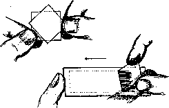
Figure 2.
The second or third drop of blood, appearing at the site of the puncture under light pressure, is taken. The object slide is quickly applied to the convexity of the drop, approximately a finger's width from the edge of the long side of the slide, then the object slide is fixed with the left hand, and the coverslip is brought close to the drop with the right hand; the drop of blood is distributed along its edge and follows it as it is moved. Thus, a uniform smear is obtained that does not reach the edges of the object slide. A smear on a coverslip is made as follows: the coverslip is quickly applied to the drop of blood; a film from steam forms around the resulting drop on the glass, which quickly disappears; then, when another coverslip is applied, the drop is distributed between both coverslips due to capillarity, which are then quickly separated, as shown in Fig. 2. Smears must be made carefully, as they to a significant extent determine the possibility of obtaining a good preparation. - Staining with Brillantkresylblau. A saturated solution of the dye in absolute alcohol is prepared (approx. 1 g of Brillantkresylblau per 80 cm3 of absolute alcohol). A little of the dye solution is applied to a warmed object or coverslip with a glass rod and quickly spread with another object slide (similar to a blood smear); a barely noticeable bluish-lilac film is obtained on the glass. Slides prepared in this way are suitable for use for a long period. A blood smear is then made on the glass covered with the dye in the usual way, which is immediately (while wet) transferred for 3-5 minutes into a moist chamber; the latter is a Petri dish or a Drigalski dish, in which a roll of moist filter paper is placed around the circumference (there should be no water on the bottom of the chamber). In the chamber, Brillantkresylblau stains the granulo-filamentous substance of the erythrocytes; then the preparation is removed from the chamber and left to dry in the air, after which it is examined under immersion (see separate table, Fig. 2). On average, the blood of a healthy person contains from 1‰ to 8‰ reticulocytes, i.e., young erythrocytes with substantia granulo-filamentosa. - Regarding the origin of the granulo-filamentous substance, the opinions of authors differ. Thus, Schilling considers it a derivative of the basophilic stroma of polychromatophilic erythrocytes, whereas Sabin and other authors believe that the granulo-filamentous substance originates from the mitochondrial apparatus. Schilling combines Brillantkresylblau staining with Giemsa staining in such a way that the vitally stained smear is fixed for 3 minutes with methyl alcohol and then stained with the usual Giemsa mixture. The preparations turn out very beautiful; however, part of the vital granulation is often lost; therefore, such preparations are less suitable for counting. - The technique of staining with Nilblausulfat is completely analogous. Schilling recommends it for detecting erythrocytes in erythrocytes in pernicious anemia and hemolytic anemias. Staining with methyl violet of Heinz bodies. In cases of poisoning with blood poisons (aniline, nitrobenzene, potassium chlorate, hydrogen arsenide, etc.), leading to the formation of methemoglobin in the blood, one or several bodies appear in the erythrocytes, so-called Heinz bodies, which do not always stain well with the above-described Brillantkresylblau method and stand out very well when stained with methyl violet (1 g of methyl violet is dissolved in 100 cm3 of a 0.6% aqueous solution of NaCl). A drop of the indicated solution is added to a drop of blood on an object slide, covered with a coverslip, and kept in a moist chamber. Heinz bodies appear as dark violet grains (see separate table, Figs. 4 and 5). 3. Staining of fixed blood smears. This method of blood examination has the greatest application in hematology. Staining of the preparation requires its preliminary fixation, for which various fixing liquids (or vapors) are used, as well as high temperature. The effect of subsequent staining depends to a large extent on the speed and quality of the fixative. Solutions of some dyes simultaneously fix and stain the preparation. P. Ehrlich, staining white blood cells with Triacid, believed that it was possible to perform a chemical analysis of the cell with the help of dyes in the sense that its alkaline components would be stained by acidic dyes, and acidic ones by alkaline dyes. Accordingly, leukocytes were subdivided into neutrophils, eosinophils, and basophils depending on the stainability of their granulation. However, the incorrectness of the basic tenets of this theory was soon proven by Ehrlich's student Michaelis, who showed the entire complexity of the processes occurring during staining and established that the role of chemical processes here is usually small, and physical-chemical processes come to the fore (see Histological technique). Staining of fuchsinophilic granulation of lymphocytes according to Schridde and mitochondria according to the Schridde-Altmann method. 1) A thin fresh blood smear on an object slide is immediately fixed in Formol-Müller (1:9) for 1-2 hours. 2) Rinse for a few minutes in running, and then in distilled water. 3) 1/2 hour in a 1% solution of osmic acid (in the dark). 4) Quickly rinse in water. 5) Staining in the following solution: 100 cm3 of a cold-saturated filtered solution of aniline in distilled water + 20 g of acid fuchsin. Filter. The dye is poured in a high layer onto the object slide, heated 5-6 times over a flame, each time until small vapors appear, then completely cooled. 6) The dried dye is wiped from the edges of the object slide with filter paper, then the preparation is differentiated in a solution of one part of a saturated alcoholic solution of picric acid and seven parts of 20% alcohol until it becomes yellowish. 7) Rinse in absolute alcohol. 8) Xylene. Canada balsam. - By this method, Schridde revealed fuchsinophilic granulation in lymphocytes, considering it specific for this type of cell, since they are not revealed by this staining in either myeloblasts or erythroblasts. Freifeld modified the Schridde-Altmann staining, fixing the preparations for up to an hour in 1% osmic acid and applying weak heating of the blood smears made on a coverslip over a small flame of a spirit lamp for 15-20 minutes. The preparation is repeatedly heated; vapors should not appear. With such a technique, fuchsinophilic granulation of the character of mitochondria is detected in lymphocytes, mononuclears, myeloblasts, erythroblasts, and also in very young erythrocytes (see separate table, Fig. 3). Klein worked with a similar methodology and arrived at similar results. - Staining with Ehrlich's Triacid. Triacid contains in solution Methylgreen, in which three basic groups are saturated with two acidic dyes: Orange G and acid fuchsin. 1) A blood smear (preferably on a coverslip) 1-2 days old is fixed by heat on a heated copper plate in a place where a drop of water placed on it does not boil, but rolls off in the form of a ball (Leidenfrost phenomenon); the fixation time is 10-20 seconds; the coverslip is placed with the smear facing down. 2) The dye is poured onto the preparation with a pipette and kept for 5-10 minutes. 3) Rinsing with water until the dye stops coming off. 4) Dry with filter paper. Canada balsam. The granulation of neutrophils is clearly stained a reddish-violet color, the granules of eosinophils are a bright copper-red color (see separate table, Fig. 6). In the remaining blood cells, granulation is not revealed. The nucleus is stained diffusely, structurelessly in a bluish-greenish color, in places with the precipitation of dark green grains. Staining with Eosin-Methylenblau (Jenner, May, and Grünwald). The preparation is stained immediately after preparation. The dye is dissolved in methyl alcohol and therefore preliminary fixation is omitted. 1) Undiluted dye is poured onto the preparation for 2-3 minutes. 2) As much distilled water is added as there was dye poured before. Stain for 5-15 minutes. 3) Thorough rinsing in distilled water until the preparation becomes pink. 4) Dry. Canada balsam. With this staining, the granulation of basophils, eosinophils, and neutrophils is beautifully revealed (see separate table, Fig. 7); the remaining white blood cells are without granulation in the protoplasm. A similar picture is given by Leishman's staining. - Staining with Methylenblau. Various methylene blues are used, for example, Methylenblau medicinale purissimum from foreign firms (Höchst, Grübler), as well as those of Russian production; usually a 1/2-1% solution or the so-called Löffler's alkaline methylene blue, or Manson's Boraxmethylenblau is used. Preparations fixed with alcohol and heat are stained for up to one minute with the appropriate solution. A finer staining is given by a strongly diluted methylene blue with prolonged staining; for example, a 1% aqueous solution of Methylenblau med. pur. (5 drops per 20 cm3 of tap water) when stained for one hour completely reveals the basophilic granulation of erythrocytes, polychromasia, as well as the basophilia of the protoplasm and pathological granulation of neutrophils.
Schmidt and Schwarz stain the basophilic granulation of erythrocytes according to Manson; Koch uses Azure II; Teleky uses polychrome methylene blue. Dyes containing Methylene Azure. After Romanovsky established that under the influence of alkali on methylene blue, Methylene Azure is formed from it, which contributes to obtaining an excellent panoptic staining of blood preparations, many methods for preparing azure-containing dyes were proposed. The best combination is solutions of Azure II-eosin. This staining has become especially widespread in the form of the Giemsa solution. Giemsa staining: 1) Fixation in absolute methyl alcohol for 3 minutes. 2) Staining with a diluted Giemsa solution: 15-16 drops of the commercial solution per 10 cm3 of distilled water. 3) Rinse with tap water. 4) Dry with filter paper. Very bright preparations are obtained if, according to Pappenheim, the preparation is first stained according to May-Grünwald, and then with the Giemsa solution (fixation with methyl alcohol is omitted, as it is contained in the May-Grünwald solution). Results of staining (see separate table, Figs. 9 and 10): nuclei are cherry-violet. Figure 1-14. Red and white blood cells under various stains: 1. Neutral red-Janus green; 2. Brilliant cresyl blue; 3. Modified staining according to Schridde-Altmann; 4. Methyl violet, Heinz bodies; 5. Heinz bodies when stained with Brilliant cresyl blue; 6. Ehrlich's Triacid; 7. May-Grünwald; 8. Methyl green-pyronin Pappenheim-Unna; 9. Azure II-Eosin (Giemsa); 10. Basophilic-granular erythrocytes and Jolly bodies (below) when stained with azure-eosin; 11. Carbol-fuchsin-methylene blue (normal); 12. Carbol-fuchsin-methylene blue (neutrophil toxicity); 13. Oxidase reaction; 14. Peroxidase reaction. er-erythrocyte, nb-normoblast, mb-myeloblast, n-neutrophil, e-eosinophil, b-basophil, m-mononuclear, l-lymphocyte. Figure 15. Elements of bone marrow. 20-hour culture of hemocytoblasts from the blood of a patient with acute myeloid leukemia. Hematopoiesis in vitro: H-hemocytoblasts; M-mitoses of neutrophilic myelocytes; S-segmented neutrophil; e-erythroblasts; D-degenerating cells (Zenker-formol, paraffin, section, azure-eosin). Figure 16. Plasma of normal blood. Figure 17. Serum of normal blood. Figure 18. Blood serum in cardiac congestion. Figure 19. Blood serum in pernicious anemia. Figure 20. Blood serum in mechanical jaundice. (To the illustrations for the articles Hematopoiesis, Blood.)
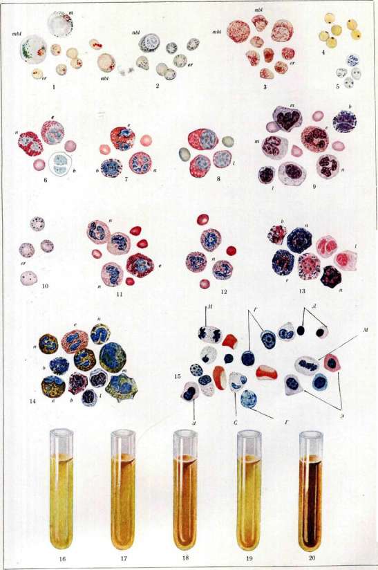
To the articles Hematopoiesis, Blood.
colors, pyknotic nuclei are often blue. The structure of the nucleus is well differentiated. The granulation of neutrophils is violet; the granulation of eosinophils, depending on pH fluctuations, is either bright red or somewhat brownish or bluish. The azurophilic granulation of mononuclears and the azurophilic granules of lymphocytes stain well (for the essence of the staining, see Giemsa), which, thanks to eosin, become bright red in color (the so-called Romanowsky effect). The chromatin of malaria plasmodia and trypanosomes stains the same azuro-red color. III. Examination in a thick drop according to Ross. To detect a small quantity of malaria plasmodia, it is necessary to use a thick drop, in which parasites can be easily seen after hemolysis of erythrocytes. Preparation of the smear: two drops of blood are placed on a microscope slide, which are spread with a platinum needle in the form of a circle approximately 1 cm in diameter; then the preparation is dried well (for speed, it can be placed in an incubator). A solution of Giemsa stain prepared in the usual way (or the Azur II-Eosin solution described below) is poured onto the unfixed preparation; after 3 minutes, the stain is carefully drained by slightly tilting the preparation; at the same time, a fresh solution of stain is added, which is left for 25 minutes. Then the preparation is carefully rinsed in a horizontal position with distilled water and dried in a vertical position (under no circumstances should it be dried with filter paper). Schilling uses a thick drop for an approximate count of the number of polychromatophils; the latter reveal a basophilic stroma that has precipitated in the form of a network. In a thick drop, eosinophils are also determined in cases where there are very few of them in blood smears. Nocht's method. It is possible to obtain azur-eosin solutions fully equivalent to the patented Giemsa stain according to Nocht's method. Two solutions are prepared: 1) 1.0 Azur II (preferably Grübler's or Merck's) is diluted in 1,000 cm3 of distilled water; 2) 1.0 eosin is dissolved in 1,000 cm3 of distilled water. Then each solution is poured separately into dropping bottles. To establish the correct mixture for staining blood elements, several test smears are prepared, each of which is stained with a slightly different mixture, for example, for 3 cm3 of distilled water, 4 drops of Azur II solution and 4 drops of eosin solution are taken (ratio 4:4) or 4:5, 4:6, etc. A preparation with cherry-red nuclei and revealed neutrophil granulation establishes the correct mixture of stains. Determination of changes in neutrophils occurring during various intoxications using azur-eosin staining. In various diseases (pneumonia, purulent processes, typhoid fever, tuberculosis, etc.), the granulation of neutrophils becomes larger and polymorphic, and bluish clumps (Döhle bodies) appear in the base of the protoplasm along with the disappearance of granulation and vacuolization of the protoplasm. Sharp changes in neutrophils can be seen on any preparation well stained with azur-eosin; for weaker changes, one can only use a stain with a specific concentration of hydrogen ions. For this purpose, Holborn (Grübler firm) prepares Giemsa with pH=5.4; the water used for dilution must be neutral. The reaction of the water can be determined with the help of hematoxylin: add (with clean tweezers) a few grains of hematoxylin to 10 cm3 of distilled water; not earlier than 1 minute and not later than 5 minutes, the water should slightly turn violet. If after 5 minutes the water does not acquire this color, one can add dropwise a 1% solution of Natrii carbonici until the correct color reaction appears and thus correct the reaction of the water. Nägeli, Gloor, and others use a titrated Giemsa solution, whereas Mommsen proposed determining pathological granulation with a buffer solution at pH=5.4. With such staining, only pathological granulation is revealed, while normal granulation is not stained. Buffer solution: 0.1 N sodium hydroxide (Merck) - 21.6 cm3, glacial acetic acid (Merck) - 27.0 cm3, distilled water up to 1,000.0 cm3 [can be obtained ready-made from Grübler (Hollborn)]. Stain solution: Giemsa solution (Grübler) - 10 cm3, distilled water - 40 cm3, buffer solution - up to 100 cm3. Stain for 1 hour; old preparations - up to 2 hours. Rinse with buffer solution. Dry. Pathological granulation of neutrophils when stained with carbol-fuchsin-methylene blue according to Freifeld. When stained with carbol-fuchsin-methylene blue, the protoplasm of normal neutrophils and neutrophilic myelocytes is devoid of any granulation (see separate table, Fig. 11), whereas pathological granulation in neutrophils (see separate table, Fig. 12) and in myelocytes is revealed in the form of violet-blue granules and clumps of various sizes and shapes - from dust-like granulation to fairly large clumping. Staining method. Two solutions are prepared: 1) 1.0 Fuchsin für Bac. (Grübler) (stock) is dissolved in 15 g of 96° alcohol while heating; after the solution cools, 100 cm3 of a 5% solution of carbolic acid (Acid. carb. cryst.) is added to it; 2) 1% aqueous solution of Methylenblau med. pur. (Grübler). A composition prepared ex tempore is poured in a high layer onto a preparation fixed for 3 minutes with methyl alcohol: to 20 cm3 of tap water, 7 drops of the first solution are added, shaken, and then 5 drops of the second solution are added and shaken again. Stain for one hour, after which the preparation is rinsed with water and dried with filter paper. For successful staining, it is necessary that the glassware be clean, without alkali or acid, and also that the drops from the dropping bottles are always of the same size. The drops of methylene blue must be identical to each other, just as the drops of carbol-fuchsin must be. In case of unsuccessful staining, if the leukocyte nuclei turn out red, the preparation can be counterstained for two minutes with methylene blue (5 drops of 1% methylene blue per 20 cm3 of tap water). Preparations stained according to Giemsa stain well with carbol-fuchsin-methylene blue (without preliminary destaining), and vice versa. Staining of pathological granulation of neutrophils with carbol-fuchsin-methylene blue is infallible in terms of diagnosis; pathological granulation of myelocytes can be established only by this method. Carbolpyronin-Methylgrün staining. The stain solution (Pappenheim-Unna) can be obtained ready-made from Grübler (Leipzig) or the stain is prepared from its components as follows: Methyl-grün 00 cryst. gelbl. - 0.15 g, Pyronin - 0.25, Alcohol 96% - 2.5, Glycerin - 20.0, Acid. carb. - 0.5, Aq. dest. - 100.0. Both pyronin and methyl green are alkaline stains, with the former staining primarily the basophilic substance, and the latter the chromatin of the nucleus. The protoplasm of lymphocytes and plasma cells stains particularly intensely in bright red (see separate table, Fig. 8); a cell whose protoplasm does not stain red is definitely not a lymphocyte, but not vice versa, since any cell with basophilic protoplasm stains in the same bright red color, e.g., an erythroblast. The basophilic granulation of erythrocytes is also bright red. The nuclei of lymphocytes are greenish-blue, the nuclei of leukocytes are violet. Staining method: 1) fixation by heat or any fixing liquid; 2) staining for 5-10 minutes; 3) rinsing with tap water; then, as usual. Winkler-Schultze oxidase reaction. In the protoplasm of leukocytes, unlike lymphocytes, substances are found, in all probability of the nature of the oxidative enzyme oxidase, which, upon exposure to the reagent described below, lead to the formation of indophenol blue. 1) Fixing liquid: 1 part 40% formaldehyde + 9 parts 95% alcohol, fix for a few seconds. 2) The following is poured onto the fixed preparation: a) for 3 minutes, a slightly diluted 1% aqueous alkaline solution of a-Naphthol. The solution is prepared as follows: a-Naphthol, when heated in distilled water, rises to the top and floats in liquid form; at this moment, a crystal of caustic alkali is added; then a-Naphthol dissolves in water; b) without rinsing or drying the preparation, a 1% aqueous solution of Dimethylparaphenylendiamin is poured onto it. After a few minutes, the granulation of the leukocytes becomes dark blue. The preparation is counterstained with a strongly diluted solution of Ziehl's fuchsin. The majority of neutrophils give a sharply positive reaction, and only individual neutrophils contain few oxidase granules; eosinophils all have a positive reaction, whereas basophils give a positive reaction only in the maturation stage - basophilic myelocytes. A portion of mononuclears also gives a positive reaction (see separate table, Fig. 13). As for myeloblasts, they give a positive oxidase reaction only at the stage of greatest maturation (transition to promyelocytes).
The so-called labile oxidase reaction (Nadi reaction according to Graeff), which is obtained without preliminary fixation after the action of an alkali-free solution of a-Naphthol in a 1% solution of Dimethylparaphenylendiamine, is performed on sections. Lymphocytes sometimes give a positive labile oxidase reaction. The peroxidase reaction was proposed by Kreibich and Fischel; it is obtained very well by the method of Graham: fixation with a liquid consisting of 1 part 40% formalin + 9 parts 95% alcohol; then the preparation is slightly rinsed with water and a benzidine solution is poured onto it for 5 minutes. The latter is prepared as follows: to 10 cm3 of 40% alcohol, several crystals of benzidine and 0.02 cm3 of hydrogen peroxide (pipette from a Sahli hemometer) are added; then rinse. Usually, a positive reaction is revealed in the form of golden-brown grains, less often bluish (the first stage of the action of benzidine). One can counterstain with thionine, methylene blue, or azure-eosin. Only fresh preparations are stained; stained preparations are preserved for a long time. The results of staining in general for the oxidase and peroxidase reactions are the same (see separate table, fig. 14). Various modifications of the peroxidase reaction have been proposed, e.g., the Naphthol-peroxidase reaction (Loele); the technique proposed by Epstein also yields good results. - Epstein's method: 1) fixation of a blood smear (not older than 48 hours) with a mixture of Alcohol (95°) - 90.0, Formol (40°) - 10.0; 2) rinse with distilled water (15-20 sec.); 3) pour on for 3 min. a solution of: Alcohol (40°) 100.0 + a-Naphthol - 1.0 + 3% H2O2 - 0.2 (according to Graham); 4) rinse with distilled water; 5) stain for 15 min. (up to 14 hours) in the following solution: Toluidinblau - 1.0, Lith. citricum - 1.0, Aq. dest. - 100.0; 6) quickly rinse with distilled water (1 sec.); 7) rinse in a 1% tannin solution (1 sec.); 8) dry with filter paper. The granulation of neutrophils, eosinophils, and mononuclears that gives the peroxidase reaction stains bright green. Erythrocytes are greenish; otherwise, the characteristic staining of the azure-containing mixture. Detection of lipoids in the granulation of leukocytes. Sehrt proposed a special method of staining blood smears with Sudan; with the help of this stain, it was clarified that the granulation of both neutrophilic and eosinophilic leukocytes, as well as mononuclears, is sudanophilic, i.e., that it gives a characteristic lipoid stain. A modification of this stain by Goldman yields very good results; in this, Goldman established that the granulation of leukocytes, which stains with Sudan, always gives an oxidase reaction. - Staining method: 1) Fixation of a fresh blood smear for 3 min. with a solution of 1 part Formol (40%) and 4 parts Spirit. vini rectif.; 2) rinse with distilled water; 3) rinse with a weak alcohol solution (30-40°); 4) stain for 15 min. with the following solution: Spiritus 70° - 100.0 + H2O - 20.0 + Sudan III (in excess) + a-Naphthol 1.2 + H2O2 (3%) - 0.3 (the alcohol solution of Sudan and a-Naphthol is boiled for 5 minutes; hydrogen peroxide is added to the cooled solution); 5) rinse with distilled water; then the author stains the nuclei for 1-3 min. with Böhmer's hematoxylin (5 drops of hematoxylin + 2-3 drops of Liq. ferri sesquichlorati). Better staining of nuclei is given by Carozzi's hematoxylin [0.25 g cryst. hematoxylin + 0.025 potassium iodate (KIO3) + 1.0 potassium alum + 25.0 glycerin + 100.0 distilled water]. Staining results: granulation of neutrophils, eosinophils, and mononuclears is bright orange-red. - Histogenetic study of the formed elements of blood is performed mainly on embryological material and experimentally. Stains for sections from tissues and organs are the same as those used for smears, adapting them accordingly to histological technique. Recently, the development of blood cells has also been studied by the method of tissue cultures. E. Freifeld. VIII. Physico-chemical properties of blood. Blood is a sticky, opaque liquid of red color, with a salty taste. The specific gravity of blood is 1.050-1.060; according to Bamberger, for men - on average 1.057.5, for women - 1.053; according to Schmaltz - 1.059, resp. 1.055. The specific gravity depends mainly on the number of erythrocytes or Hb, and less on the properties of the serum. The specific gravity of erythrocytes is 1.090, of serum - 1.028-1.032, of plasma - 1.029-1.034. The specific gravity of serum is determined mainly by the protein content, since their percentage content significantly exceeds the content of other solid substances of the serum (urea, sugar, NaCl). Fluctuations in the content of sugar, urea, bile pigments, and fat, even under pathological conditions, cannot significantly change the specific gravity of the serum. - Under physiological conditions, an increase in the specific gravity of blood is observed during strong physical exertion, under the influence of heat, and during the withdrawal of fluids. These changes, however, are soon leveled out. In the morning, the specific gravity of blood is somewhat higher than in the second half of the day. In pathology, thickening of the blood with a corresponding increase in specific gravity is described in cancer of the esophagus and pylorus (up to 1.070), in long-lasting diarrhea (in cholera, dysentery - up to 1.080), and in severe burns (up to 1.073). In cholera, the determination of the specific gravity of blood has, according to Rogers, significance in that an increase to 1.063 represents an indication for intravenous infusions. Less significant increases in specific gravity accompany erythrocytosis in congenital defects and decompensation of heart defects. A decrease in the specific gravity of blood is characteristic of the depletion of blood of formed elements, consequently for anemias, in which the specific gravity falls to 1.040 and lower, approaching the normal specific gravity of serum; a decrease in the specific gravity of serum is observed when it is depleted of proteins due to renal and cachectic edema. In general, in modern clinical practice, in most cases, the determination of the specific gravity of blood and serum is replaced by a count of erythrocytes, determination of Hb, and refractometry of serum, which give a more direct answer to those main changes in the composition of blood that predominantly influence the fluctuations of specific gravity. Technique for determining specific gravity. With the pycnometric method of Schmaltz, blood (about 0.1) is drawn into a chemically pure special capillary, weighed, and then the weight of exactly the same volume of distilled water is determined in this same capillary. The areometric method of Hammerschlag consists in allowing blood to fall drop by drop into a liquid whose specific gravity approximately corresponds to blood (a mixture of chloroform with a specific gravity of 1.485 and benzene with a specific gravity of 0.88), observing whether the drop falls or sinks in this mixture, and by adding one or the other ingredient accordingly, a mixture is obtained in which a new drop of blood remains suspended; the specific gravity of this mixture is determined with an areometer, which also indicates the specific gravity of the blood under investigation. Rogers accelerates the determination by preparing a series of mixtures of glycerin with water with specific gravities from 1.040 to 1.070. By placing drops of blood into separate mixtures, the specific gravity of the blood is easily selected. Barbour and Hamilton determine the specific gravity of blood, as well as other liquids, by the time of the fall of a drop of the test liquid through a certain layer of a mixture of xylene and bromobenzene compared to the time of the fall of standard solutions of K2SO4. E. Tareev. The viscosity of blood depends on 1) the viscosity of the plasma, 2) the number of erythrocytes and their volume, 3) the Hb content, 4) the amount of CO2 in the blood, 5) the salt composition of the plasma, and other conditions. A particularly close dependence is noted between the volume of erythrocytes and the viscosity of blood. According to Frey, the volume of erythrocytes in volume percent = 2.4 n2, where n is the viscosity value (see Viscosity, Viscosimetry). - The surface tension of blood, both whole and defibrinated, is less than the surface tension of water. The difference between the surface tension of blood, plasma, and serum is very insignificant. With a decrease in t°, the surface tension increases. When determining the surface tension of serum, values higher than those in subsequent determinations are obtained at first. Approximately half an hour after the first determination, the lowest value is obtained. The highest value is called dynamic surface tension, the lowest - static. For normal human serum, the value of static surface tension = 58-57 dyn/cm at 16-18° and 47 dyn/cm at 37°. The surface tension of blood under pathological conditions undergoes significant changes: it decreases in eclampsia, uremia, in cardiovascular diseases, when bile salts are contained in the blood, when the CO2 content in the blood increases, etc. An increase in surface tension is encountered significantly less often. It has been noted that the surface tension of serum giving a positive RW is increased. The cited data on the change in the surface tension of blood cannot, however, be considered firmly established and require verification. The osmotic pressure of serum and defibrinated blood is the same and represents a very constant value in the normal state.
The maximum fluctuations under physiological conditions correspond to Δ = 0.54–0.59°, on average 0.56°, in gram-molecules of osmotic concentration of serum = 0.3 gram-molecules. In different parts of the circulatory system, Δ of blood can have a different value; for example, in the portal vein Δ = 0.692–0.602°; in the hepatic veins Δ = 0.722–0.633°; CO2 increases Δ. The main place in the osmotic concentration of blood belongs to crystalloids, primarily NaCl (about 60%). Serum proteins account for only 0.01° of the Δ value. During pregnancy, Δ decreases, averaging 0.54°, while in various forms of nephritis it increases to 0.60–0.71°. Of the constituent parts of serum, NaCl has the main significance for electrical conductivity, just as it does for osmotic pressure, and its content in the serum accounts for about 63% of the total electrical conductivity value; of the remaining 37%, the greater part is accounted for by bicarbonates. The electrical conductivity of serum is quite constant and changes significantly only in pathological cases. A change in the CO2 content in the blood is not reflected in the electrical conductivity value of the serum, which is explained by complex processes of ion exchange between erythrocytes and plasma when the CO2 content in the blood changes (see below). Little is known about physiological influences on the electrical conductivity of serum: muscular work apparently increases the electrical conductivity of serum, while the influence of nutrition remains unclear. Pregnancy does not change electrical conductivity. The electrical conductivity of plasma and serum is the same. The electrical conductivity of blood is significantly lower than that of serum, since the ability of formed elements to conduct current is negligible. Animals Δ·10-4 Plasma Δ·10-4 Blood Δ·10-4 Formed elements pulp 105.3 103.7 112.9 107.4 63.4 62.8 36.9 43.3 1.63 1.67 1.70 2.17. The opinion that existed for a time that the electrical conductivity of blood corpuscles could be taken as equal to 0 turned out to be erroneous: by an indirect method, Höber managed to show that the electrical conductivity of the endoglobular content of erythrocytes corresponds to a NaCl content in solution of 0.2%. The determination of electrical conductivity gives an idea of the total sum of ions. At present, methods have also been proposed for determining the concentration of individual types of ions. The method for determining the ion concentration of alkali metals is based on the general principle of concentration cells (see Gas chain), and its peculiarity lies only in the preparation of the corresponding electrodes. Michaelis uses electrodes made of alkali metal amalgam, which is kept without access to air in a special vessel that makes it possible to renew the surface of the electrode, which oxidizes in the air, as needed. Preparation of the electrode: about 200 cm3 of pure and dry mercury is placed in vessel A (Fig. 3); after cutting several pieces (about 5 g) of Na or K with a cork borer, they are quickly transferred into tube a. Vessel A is evacuated several times, filled with hydrogen, the free end of tube a is sealed, it is slightly heated, allowing the molten metal to flow into the mercury, and the capillary K is sealed. After shaking the contents of vessel A, tubes b and d are connected with rubber, vessel B is evacuated through passage E, tap H is opened, and the amalgam is allowed to flow into reservoir B; the remaining free part of reservoir B is filled with hydrogen through tap H. By turning tap Hs, tube I, which serves as the electrode, is filled with amalgam. When determining, after immersing the electrode in the liquid, the latter is connected by means of a tube filled with agar and potassium chloride to a saturated KCl solution, into which the tip of a calomel half-element is lowered. (Fig.—see p. 675–676). The method of determining the concentration of hydrogen ions in blood or plasma has become widely used. Three different methods have been proposed: 1) electrometric (see Gas chain, Active reaction), 2) colorimetric (see Indicator method), 3) gas-analytical. 1. The only correct method for the electrometric determination of the H-ion concentration of blood, according to Bigwood, is the study of plasma separated from formed elements at the same pressure of O2 and CO2 that was in the blood. For precise work, the electrodes must be re-platinized before each determination. 2. Colorimetric method for determining the active reaction of plasma according to Cullen. Necessary reagents: 1) Standard phosphate solutions, prepared according to the table. pH Na2HPO4 cm3 KH2PO4 cm3 7.00 61.1 38.9 7.05 63.9 36.1 7.10 66.6 33.4 7.15 69.2 30.8 7.20 72.0 28.0 7.25 74.4 25.6 7.30 76.8 23.2 7.35 78.9 21.1 7.40 80.8 19.2 7.45 82.5 17.5 7.50 84.1 15.9 7.55 85.7 14.3 7.60 87.0 13.0 7.65 88.2 11.8 7.70 89.4 10.6 7.75 90.5 9.5 7.80 91.5 8.5. 2) Indicator solution: in a measuring flask with a capacity of 100 cm3, 1.05 cm3 of a 0.04% phenol red solution and a few drops of 0.02 n NaOH are added to 95 cm3 of a 0.9% NaCl solution to pH = 7.4–7.6; the flask is filled to the mark with NaCl solution. Procedure: paraffin oil is added to 5–20 cm3 of solution No. 2 in flat-bottomed test tubes, under the layer of which 0.4–1.0 cm3 of plasma is released. After stirring the contents of the test tube, the resulting color is compared with the color of the standard solution + indicator in a Walpole comparator; the pH for t° 38° is calculated by the formula: pH38 = pH found at the temperature of the experiment + 0.01 (t° - 20°) - 0.23; the temperature at which the determination was performed is measured by immersing a thermometer in a test tube with diluted plasma. 3. The gas-analytical method for determining the pH of blood, proposed by Hasselbalch, is based on the following principles. The active reaction of blood possesses significant constancy, which is caused by 1) the peculiarities of its chemical composition (see Buffer properties of blood) and 2) a number of physiological processes (respiration, work of the kidneys, glands of the digestive tract, etc.). Of the mineral constituents of blood, bicarbonates (NaHCO3 + CO2) and phosphates (Na2HPO4 + NaH2PO4) possess buffer action; the significance of the latter, however, is only secondary, since their concentration in blood is very small (about 3 millimoles). For CO2 in the presence of bicarbonates in solution, the equation is valid: [H'] = K · [CO2] / [Bic] · α, where [H'] is the concentration of H ions, [Bic] is the concentration of the salt NaHCO3; [CO2] is the concentration of carbonic acid, α is the degree of dissociation of bicarbonate at a given t°; K is the first dissociation constant of carbonic acid. Introducing the notation 10-pK instead of K and taking the logarithm, we get: -pH + log [Bic] + log α = -pK, where pH = -log [H']; from here: pH = pK + log [Bic] / [CO2] · α.
(1), where pH = pK1 + log a. Normally, deviations of pK1 from the mean values (6.10 at 38°; 6.18 at 20°) do not exceed ±0.03. It is therefore sufficient to find the concentration of CO2 present in the plasma in a dissolved state [CO2] and the concentration of bicarbonates [Bic], i.e., the value of the fraction [Bic]/[CO2], to possess all the data for calculating the pH. The gas-analytical method for determining pH consists precisely in determining the value of the fraction [Bic]/[CO2]. The amount of free CO2 in the plasma depends 1) on the tension of carbonic acid in the blood and 2) on the solubility coefficient (see Blood gases). In volume percent, the amount of dissolved CO2 is equal to 0.1316 pa... (2), where p is the pressure of CO2; a is the solubility coefficient. The concentration of bicarbonates, expressing it in volume % of CO2, is found by subtracting from the total amount of carbonic acid found that part of it which is in a dissolved state: [Bic] = CO2 - 0.1316 pa... (3). The value of the product 0.1316 a at different temperatures can be judged from the table. Temp. According to Bohr According to Van Slyke and McLean 15° 20° 25° 30° 38° 0.1307 0.1128 0.0975 0.0855 0.0712 0.1225 0.1057 0.0913 0.0800 0.0668. When working not with plasma but with blood, a correction depending on the amount of O2 in the blood must be introduced into the value of a for serum: a for blood = (1 - 0.00677h) a for serum, where h is the amount of O2 in volume % (v%). Substituting expressions (2) and (3) into equation (1), we obtain: Thus, to calculate the pH, it is sufficient to know the pressure of CO2 in the blood and the total amount of CO2 in the plasma, expressed in v%. The pressure of CO2 in the blood can be determined by several methods. 1. The direct method was proposed by Krogh and consists in the fact that a small vessel with a capacity of about 2 cm3 (microtonometer; Fig. 4) is filled to the top with blood through a bent end (hirudin, heparin, Na2C2O4, NaF are used as substances preventing blood clotting) and is closed from above by a thermometer entering the rubber, which has a small cup at the end. The cup carries with it a bubble of air, which is shaken with the blood at a temperature of 38° to equalize the gas pressure, and then transferred to the Krogh micro-apparatus (Fig. 5), previously filled with acidified physiological solution. An essential part of the gas analysis apparatus is a graduated narrow glass capillary, ending at the bottom in a small funnel and having a side passage closed by a screw. Leaving the bubble p near the bent end after shaking, the latter is introduced into the funnel (1) of the micro-apparatus filled with physiological solution. By pushing the thermometer deep into the tonometer, the bubble p is driven out of the tonometer and transferred into the funnel (1). Subsequently, by turning the screw S, the bubble p is sucked into the capillary, where its volume is measured at a certain temperature; after this, having placed the apparatus in an inclined position, the physiological solution is sucked out of the funnel and replaced with a 10% solution of caustic alkali. The apparatus is again placed in a vertical position, and by turning the screw S, the bubble is carefully brought into contact with the alkali; taking care that the alkali does not flow into the capillary, the screw h is rotated and the bubble is moved up and down for better absorption of CO2. Leaving the bubble in the lower part of the capillary, the alkali is sucked out, replaced with physiological solution, and, by again sucking the bubble into the capillary, its volume is measured. The difference between the 1st and 2nd reading after correction for temperature gives the amount of CO2 contained in the bubble. Knowing the initial volume of the bubble (1st reading), the percentage content of CO2 in it is calculated, and knowing the atmospheric pressure, the partial pressure of CO2 is calculated. The accuracy of the analysis depends mainly on the cleanliness of the capillary. The tension of CO2 in the blood can be determined indirectly as follows: in a sample of blood taken with the necessary precautions for gas analysis, the total amount of CO2 is determined either by pumping out (see Blood gases), or in the Barcroft apparatus (see Barcroft apparatus), or by the Van Slyke methods (see Van Slyke methods). Then several samples of blood (usually three) are saturated with gas mixtures containing different amounts of CO2. In the blood saturated with a gas mixture of one or another composition, the total amount of CO2 is determined by the same method as before, and the data obtained in v% are plotted graphically, plotting the found values on the ordinate axis. The pressure of CO2 in the gas mixtures with which saturation was performed is also determined (expressing the CO2 pressure in mm of mercury), and the data obtained are plotted on the abscissa. By connecting the points located in the coordinate system, a curve of CO2 binding by blood at various pressures of the latter is obtained. Example of arterial blood analysis: total amount of CO2 in blood without preliminary saturation - 50 v%; total amount of CO2 after saturation: at a partial pressure of CO2 of 31 mm - 45.5 v%, at a partial pressure of CO2 of 38.5 mm - 49.0 v%, at a partial pressure of CO2 of 45.0 mm - 52.8 v%. The graphical representation of the results obtained gives the curve ab (Fig. 6). If a perpendicular is dropped onto the abscissa from the point of the curve corresponding to the total amount of CO2 found without preliminary saturation, the intersection point of these two straight lines will indicate the CO2 pressure that was in the blood (40 mm). Some difficulties arise when determining the CO2 pressure in venous blood by this method. Blood containing reduced hemoglobin binds more CO2 than oxidized blood (by 5-6 v%); the CO2 binding curve of the former lies higher than that of the latter (Fig. 7). To move to the CO2 binding curve for oxygen-saturated blood and the curve characteristic of venous blood (to introduce a correction for venousness), for every volume percent of O2 missing until full saturation, the CO2 binding curve should be raised, according to some researchers by 0.34 v%, according to others by 0.27 v%, on average by 0.3 v%. Having introduced the appropriate correction, the CO2 tension in venous blood can be determined from the CO2 binding curves and the total amount of CO2. A more complex method is saturation with a gas mixture containing oxygen under a pressure corresponding to the tension of this gas in the venous blood under study. The CO2 pressure in the blood can also be judged by analyzing alveolar air. It is believed that the data from the analysis of alveolar air by the Haldane method correspond to the CO2 tension in arterial blood, and by the Plesch method - in venous blood. Analyses of alveolar air began to be used with confidence to judge the CO2 pressure in the blood after verifying the data obtained by direct determination according to Krogh. Having determined the CO2 tension in the blood by some method and using formula (2), the amount of CO2 dissolved in the plasma is calculated in v%. In the given example, v% CO2 = 0.1316 a * 40. To find the amount of CO2 in a bound state, i.e., the term [Bic], one should subtract the amount of CO2 dissolved in the plasma from the total amount of CO2 found in the plasma without preliminary saturation. It is also recommended to determine the CO2 binding curve not for whole blood, but for plasma; for clarifying the CO2 tension, this is indifferent, only in one case a point corresponding to the total amount of CO2 in the blood is found on the binding curve, and in the other, in the plasma. When determining CO2 binding curves for plasma, it is necessary to saturate whole blood with a gas mixture, then, having protected the blood from contact with air using a layer of paraffin oil, it is centrifuged and the plasma is taken for analysis (true plasma). If the amount of CO2 in a dissolved state in the blood is calculated for various CO2 pressures and the values obtained are plotted graphically, a straight line is obtained starting at the origin of the coordinate system and going at a small angle to the abscissa axis (the angle doc shaded in Figs. 6 and 7 represents the amount of dissolved CO2 at various pressures). The distance from the points of the curve ab (Fig. 6) to the points of the straight line od located correspondingly in relation to the abscissa (e.g., h) gives the expression (in v%) of the amount of CO2 in a bound state. The value h, corresponding to a CO2 pressure equal to 40 mm, is called the reserve alkalinity of the blood (see). If the concentration of hydrogen ions in the blood is determined at a CO2 pressure equal to 40 mm, the resulting hydrogen index is called the reduced pH; the hydrogen index calculated according to the CO2 pressure that was found in the blood is called the regulated pH. The simplest method for determining the CO2 binding curve for blood or plasma was proposed by Fridericia.
A centrifuge tube a (Fig. 8), containing 7-10 cm3 of blood, protected from coagulation by the addition of Na2C2O4 + NaF, is connected by a rubber tube, equipped with a screw clamp C, to a cylinder B (capacity about 500 cm3), closed with a stopper with a capillary tube with a stopcock D passing through it; clamp C is screwed shut. The cylinder is placed in a thermostat and evacuated with a water-jet pump until the mercury rises in the manometer tube E to a certain predetermined level (e.g., 660 mm). Then, by turning the three-way stopcock F, cylinder B is connected to a gasometer filled with pure CO2, or to a Straub apparatus, and CO2 is slowly admitted into B, controlling the amount of the latter by the drop of mercury in tube E. When the pressure has dropped by the desired amount (e.g., to 630 mm), by turning stopcock F, the gasometer and cylinder B are disconnected; by turning stopcock H, B is connected to the atmospheric air to equalize the pressure. After closing stopcock D, the bulb B is freed from the system of tubes; after removing the rubber tubing, screw clamp C is unscrewed; the bulb is turned into a horizontal position and the blood is allowed to spread along the walls of the cylinder. By rotating the bulb for 9 minutes, the blood is saturated with the gas mixture, then the bulb is moved to a vertical position, allowing the blood to drain into the test tube. Clamp C is screwed shut, and depending on whether blood or plasma is being analyzed, one either cuts the rubber below the screw clamp and immediately draws the blood for analysis (e.g., in a Barcroft apparatus) or cuts the rubber above clamp C and unscrews the clamp, having previously poured paraffin oil into the segment of rubber above C. The test tube is centrifuged, and the plasma that has collected above the formed elements and under the oil is taken for examination. When analyzing blood, it is sufficient to take 2-2.5 cm3 of blood into test tube a. By introducing different amounts of CO2 into cylinder B, the saturation is repeated with new portions of blood several times, and according to the above, several points of the carbon dioxide binding curve for blood or plasma are obtained. The method for determining the CO2 binding curve proposed by Straub is designed for subsequent blood analysis in a Barcroft apparatus. In a tonometer O (with a capacity of 250-300 cm3), with stopcock B closed (Fig. 9), 2-3 cm3 of blood is placed, after which the upper opening of the tonometer is closed with a stopper. Under pressure from a special burette A, the required amount of CO2 is admitted into the tonometer, stopcock B is closed, and the tonometer is placed in a thermostat (t° 37-38°), where the blood is saturated with the gas mixture by shaking. After 10 minutes, having connected the tonometer through stopcock B to a mercury manometer, one determines how much the pressure inside the tonometer is higher than atmospheric. Then the blood is allowed to collect in part A, and by turning stopcock B to connect with pipette E, the latter is allowed to fill with blood. By turning stopcock B to connect with d, the blood is released under an ammonia solution into the vessel of the Barcroft apparatus. To analyze the gas mixture, the tonometer is connected to a Haldane apparatus for gas analysis, into which the necessary portion is drawn and analyzed. Example of calculating the CO2 pressure in the tonometer: the pressure in the tonometer is increased by 60 mm; the corrected barometer reading = 760 mm. The total pressure in the tonometer is 760 + 60 = 820; minus the water vapor pressure (47 mm) = 773 mm. The CO2 content found in the gas mixture = 773 * 0.3 = 0.3%; whence the CO2 pressure = 2.32 mmHg. The Straub method has no advantage over the Fredericia method, since with the latter, for greater accuracy of work, one can determine the pressure of the gas mixture in the tonometer and secondarily check the CO2 pressure by analyzing the gas mixture in a Haldane apparatus. When determining the active reaction of blood, a number of precautions should be observed when taking blood and storing it until analysis: blood taken without any clamping of the vessel into a syringe under paraffin oil is transferred either through a needle or (after removing the needle) through a rubber tube into a test tube containing 3-5 cm3 of paraffin oil, along the walls of which a mixture of Na2C2O4 + NaF has been previously rubbed. The amount of the mixture of sodium oxalate and sodium fluoride (1:1), finely ground in an agate mortar, should be 0.2-0.3% of the amount of blood that is intended to be collected. After mixing the blood with a fused glass rod, the test tube is placed in snow. If some time must pass from the moment of taking the blood until the analysis, it is desirable to close the test tube in which the blood is located under a layer of oil with a stopper with a hole for the oil to escape. This hole is closed with a glass rod when the stopper is pushed in to the proper depth. In this form, blood can be stored for 5-6 hours. When examining plasma, the test tube closed as described is centrifuged, and after carefully opening the stopper, the plasma is collected from under the oil. Returning to the question of the stability of the active reaction of blood, it should be pointed out that solutions of alkaline bicarbonate salts in the presence of free CO2 change their inherent hydrogen ion concentration more easily than plasma, and the latter more easily than blood. The suspension of red blood cells possesses the most pronounced buffer properties. The objects studied: CO2 pressure 20 mm, 40 mm; NaHCO3... 7.64, 7.46; Serum... 7.37, 7.27; Suspension of blood formed elements... 7.15, 7.04; Difference... 0.30, 0.27, 0.19, 0.11. The table data indicate that bicarbonates are not the only buffer system of blood. Hemoglobin is of significantly greater importance. The significance of hemoglobin as a buffer becomes clear when comparing the CO2 binding curves: 1) by a NaHCO3 solution, 2) by serum separated from formed elements at a CO2 pressure = 44.55 mm Hg, 3) by plasma saturated with the gas mixture together with erythrocytes (true plasma), and 4) by blood (Fig. 10). It turns out that serum separated from erythrocytes possesses a completely different ability to bind CO2 than true plasma and blood. Curve III intersects curve II only in one place, namely, corresponding to the CO2 pressure at which the serum was separated from the erythrocytes. Curve III runs analogously to IV. This is explained by the fact that with an increase in CO2 pressure in the blood, there is a movement of anions (Cl) into the formed elements, due to which the ability of the plasma to bind CO2 increases. This process of ion movement corresponds to the Donnan equilibrium rule (see Donnan equilibrium), with hemoglobin playing the role of an ion incapable of penetrating the membrane. At the present time, it can be considered proven that serum proteins also possess a buffer effect: the ability of the serum ultrafiltrate to bind CO2 turned out to be significantly less than that of the part of the serum that did not pass through the filter, i.e., enriched with proteins (Fig. 11). The constancy of the active reaction of blood is maintained by a number of physiological processes, among which respiration must be placed in the first place. The slightest increase in CO2 tension in the blood, leading to a shift of the active reaction of blood to the acid side, causes an increase in pulmonary ventilation, consequently a decrease in CO2 pressure, and a return of the blood pH to the initial level. Under physiological conditions, changes in the active reaction of blood are insignificant and fluctuate in different individuals within narrow limits: 7.30-7.44 at 38°. In pathological states, the hydrogen ion concentration can change quite significantly. A change in pH to the acid side has received the name acidosis (see), and to the alkaline side—alkalosis (see). These terms are unsatisfactory, since, firstly, the active reaction of blood does not sufficiently reflect the state of the general acid-base economy of the organism; secondly, an excessive accumulation of products of an acid reaction in the blood (e.g., lactic acid) is repeatedly observed, accompanied by a shift of pH to the alkaline side. It should be kept in mind that the active reaction of blood depends on the most diverse factors: the qualitative and quantitative composition of blood electrolytes, the amount of plasma protein, the number of formed elements, the concentration of hemoglobin, the pressure of O2 and CO2, and other conditions. A more or less correct assessment of the acid-base economy of the blood can be made only by determining the blood pH, the ability to bind CO2, the hemoglobin concentration, and the total amount of plasma bases. Van Slyke considers it sufficient to determine the total amount of blood CO2 and the active reaction. Van Slyke depicts the various changes in the acid-base economy of the blood with the scheme shown in Figs. 12 and 13. Legend for Fig. 12: I—uncompensated alkali excess; II and III—uncompensated CO2 deficit; IV—compensated alkali or CO2 excess; V—normal acid-base equilibrium; VI—compensated alkali or CO2 deficit; VII and VIII—uncompensated CO2 excess; IX—uncompensated alkali deficit. To assess shifts in the active reaction of blood, Henderson proposes the following terms: pH does not go beyond the limits of normal fluctuations—euhydria; pH is lowered—hyperhydria; raised—hypohydria. Blood gases—see Blood gases and Respiration (gas exchange in the lungs).
Severin. IX. Chemical composition of Blood. The chemical composition of Blood, despite the continuous influx and efflux of various gaseous, mineral, and organic substances, represents a comparatively constant value. The organism maintains the constancy of certain chemical ingredients of Blood with exceptional tenacity—the so-called chemical constants of Blood. Such constants include: the active reaction of Blood (its pH), its freezing point (Δ, resp. the number of molecules dissolved in it), the ratio between individual cations, and a number of others. Under physiological conditions, insignificant fluctuations in the chemical composition of blood are extremely quickly equalized by the organism through the most diverse means (respiratory and excretory organs, etc.). The following table gives an idea of the narrow limits within which the normal chemical composition of blood fluctuates. [Graph/Table content: CO2 pressure in mm Hg, absorption pH]
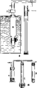
S3
for bringing into equilibrium with atmospheric air to the gasometer
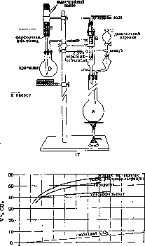
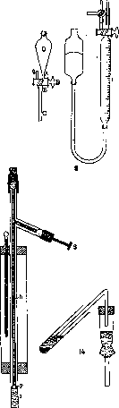

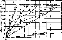
Normal composition of human blood (according to data from various authors). Volume and constituent parts
- 100-200 150-190 58-101 44-79 3.1-8.6 13.8-19 38.9-75 » » » » ,8 » » ,8 » » * Of this amount, about 60% is in the form of esters. Proteins. Blood plasma proteins constitute about 6-8% of the plasma or about 3/4 of its dry residue; almost the same ratios apply to serum; in relation to whole blood, plasma proteins constitute 3-4%. The main proteins of blood plasma are globulins and albumins. In these main groups, one can distinguish more or less distinct subgroups. To the number of blood plasma globulins belong: 1. Fibrinogen, distinguished by a relatively low degree of dispersion, low salting-out limits [precipitates from five-fold diluted plasma at 13-28% saturation with (NH4)2SO4], low coagulation temperature (56°), and the ability to convert into fibrin (during blood clotting). Fibrinogen constitutes 0.1-0.4% of plasma. In pathological cases, especially in diseases proceeding with leukocytosis (with the exception of leukemia), its content may increase (hyperinosis) up to 1.5%; in malaria, pernicious anemia, leukemia, and some other diseases, a decrease in fibrinogen content is observed (hypinosis), reaching even (very rare cases) its complete disappearance. In its relation to blood clotting, fibrinogen has very important biological significance. 2. Fibrin-globulin, also possessing low salting-out limits and a low coagulation temperature, but remaining in the serum after the separation of fibrin; its quantity is small; some authors believe that it is preformed in the plasma, others that it is formed at the moment of fibrinogen clotting. 3. Paraglobulin (2-3% of plasma), representing a mixture of globulins, which the Hofmeister school divided by means of fractional precipitation with salts into two main fractions: a) euglobulin, salting out at 28-36% saturation with (NH4)2SO4 and existing in the plasma in solution due to the action of protective colloids, and b) pseudoglobulin, salting out at 33-46% saturation with (NH4)2SO4. The overlapping of the salting-out limits casts doubt on the individuality of these fractions. Serum albumin (4-6% of plasma) also apparently does not represent a homogeneous substance, as evidenced by the inconstant coagulation temperature, different salting-out limits, and different forms of crystals. There have been attempts to divide the group of albumins into different fractions, for example, soluble and insoluble in water (pseudo- and eualbumins). The coagulation temperature of serum albumin (70-85°) strongly depends on the salt content. Serum albumin can be obtained in crystalline form (see Proteins). Further, in blood plasma, there are nucleoprotein and seromucoid, contained in small quantities (0.1-0.25 g per 1 l) and very little studied. Nucleoprotein falls into the globulin fraction upon precipitation. Seromucoid belongs to glycoproteins; it does not coagulate upon boiling, as a result of which it can be mistaken for albumoses. It remains unclear how many groups and which groups of globulins and albumins are in blood serum. There are all kinds of gradual transitions from one fraction to another. It is possible that the matter lies not so much in chemical differences as in differences in the physicochemical state of one substance. It should be noted that the division into fractions is revealed not only in the above-listed properties but also concerns the serological, immune, and enzymatic properties of globulins. Not only the total amount of plasma proteins is important, but also the mutual ratio of the fractions, which changes strongly in many pathological processes. The ratio of globulins to albumins, the protein coefficient, is denoted by a fraction, equating the amount of globulins to 1; in normal conditions, it fluctuates from 1:1.5 to 1:2.3. Furthermore, the expression "blood protein picture" (Bluteiweissbild) is accepted, by which is understood the ratio of both fractions, expressed as a percentage of their total amount; in normal conditions, this ratio = 33/67; an increase in the relative amount of globulin is denoted as a shift to the left of the blood protein picture. Finally, one can simply indicate the amount of globulin as a percentage of the total amount of protein, for example, 33%. There are indications that the amount of globulins increases during various immunization processes, but not in proportion to the degree of the immunization titer. In some pathological processes, especially in diseases accompanied by leukocytosis, the content of globulins in blood can become higher than the content of albumins. In nephritis, the protein coefficient can reach 1:11.3. Little is known yet about the origin of blood proteins. Apparently, blood-forming organs (bone marrow) play a known role in this process. Many observations point to the liver as an important organ for the formation of fibrinogen. A number of authors believe that not any specific organs, but all organs participate in the formation of blood protein, and that it is a product of the destruction of the protein of the latter. The question of the genetic connection between globulin and albumin, important for clarifying the mechanism of changes in the protein coefficient and which occupied a number of authors, has not yet been clarified. Quantitative determination of proteins can be performed by coagulating all proteins by boiling (desired pH 5.5), weighing the washed and dried precipitate, and determining the nitrogen in it according to Kjeldahl (see Kjeldahl method); by multiplying the amount of nitrogen by the coefficient 6.25, one can calculate the amount of protein with a high degree of approximation. Using the Kjeldahl method, one can also determine the nitrogen falling separately to the share of fibrinogen, globulin, and albumin (distribution of blood nitrogen). For this, nitrogen is determined in plasma, serum, the filtrate after precipitation of globulins, and in the filtrate after precipitation of all proteins. Recently, a number of authors (Rona, Rausch-Nyack) have proposed nephelometric determination (see Nephelometry). The refractometry method is very convenient. For blood, one can speak of the refraction of plasma and serum. For investigating the refraction of blood plasma or serum, the Abbe or Pulfrich refractometers are most suitable; the first gives directly the refractive index, the second the so-called Pulfrich units—P.U. (1 P.U. corresponds approximately to Δn=0.00036-0.00038), from which, with the help of tables, one can find the refractive index. The refraction of blood plasma is composed of the refraction of water (n for 17.5° = 1.33320), the refraction falling to the share of inorganic and organic non-protein components [in normal conditions Δn (increase of the refractive index compared to the refractive index of water) = 0.00200-0.00277], and the refraction of proteins (in normal conditions Δn is about 0.01800); for plasma, the refraction of fibrinogen (Δn = 0.00080) is added here. As can be seen, proteins have the greatest influence on refraction, with more dispersed albumins having a lower refraction than less dispersed globulins (refraction of albumins in a 1% solution = 0.00177, refraction of globulins in a 1% solution = 0.00229; Robertson). The refraction of human serum under normal conditions = 1.34873-1.35168. Under normal conditions and in the majority of pathological states, the content of water and crystalloids in serum fluctuates within such narrow limits that fluctuations in refraction can be attributed to an increase or decrease in the amount of proteins (Reiss). The exceptions are only uremia and hyperglycemia, in which the amount of non-protein substances refracting light in the serum is strongly increased and must be taken into account when determining refraction. For refractometric investigations, one can use capillary, venous, or arterial blood, but it is essential that the vessels are not compressed during blood collection to avoid the mixing of tissue lymph with the blood being investigated. When investigating plasma, the use of anticoagulant substances is absolutely impermissible, as they change the refraction in a way that is difficult to account for. It is possible only to add a small amount of a good preparation of hirudin or heparin. It is best to perform determinations in the so-called natural native plasma, i.e., plasma obtained by rapid centrifugation in ice and in paraffined test tubes of whole blood without the preliminary addition of anticoagulant substances. When investigating serum, it should always be obtained in the same way, since depending on whether the serum was obtained by centrifugation of whole clotted blood or defibrinated blood or from natural plasma, the values of its refraction are different. Hemolysis, wherever it may be, is impermissible. Since refraction depends on almost all components of blood, it is hardly the case that its value has independent interest as a constant of one or another blood; rather, investigations of refraction are interesting because they make it possible to methodically simply determine quantitatively proteins in a small amount of blood and indirectly judge the content of water in it. For this purpose, 3 methods have been proposed. I. The Reiss method.
Having accepted the non-protein part of An as a constant value of 0.00277, and the An of serum protein (a mixture of albumins and globulins) in a 1% solution as 0.00172, Reiss compiled a table with the help of which, having the refractometer readings in R.E. at 17.5°, one can directly find the percentage of protein. Subsequently, the refraction figures used as the basis for the Reiss table turned out to be incorrect, and Robertson proposed using the constants: 0.00208 and 0.00195 (this latter number corresponds to a protein mixture consisting of 2/3 albumin and 1/3 globulin, as is usually the case in serum). Despite this correction, the Reiss method yields only approximate results, since its basic premises are incorrect; in reality: 1) the refraction of the non-protein part varies in different cases, 2) the An of a 1% serum protein solution also does not appear to be a constant value and changes depending on the ratio of albumin and globulin fractions in the protein of a given serum.
G. Derviz. II. Robertson's method. In contrast to Reiss, Robertson pointed out that the refraction of protein bodies is composed additively from the refractions of individual types of proteins (globulins, albumins). The Robertson method consists of three parts: the determination of the refraction of 1) the non-protein part of the serum, 2) albumins, and 3) globulins. 1) The refraction of the non-protein part of the serum is determined by boiling with an equal volume of 1/25 acetic acid and subsequent refractometric measurement of the filtrate. The refraction of 1/25 acetic acid is subtracted from the obtained number. The difference, multiplied by 2, gives the refraction of the non-protein part of the serum. In view of the fact that the non-protein part consists predominantly of NaCl, and other substances have a refractive index similar to NaCl, the obtained number is divided by 0.00160 (the refraction of a 1% NaCl solution). Thus, the percentage content of the non-protein part, expressed as NaCl, is obtained. 2) Determination of albumins. An equal volume of a saturated solution of ammonium sulfate is added to the serum; globulins are precipitated, and albumins remain in the solution. It is centrifuged, the clear liquid is aspirated, diluted with an equal volume of water, shaken, and refractometered. The refraction of 1/2 of the saturated (NH4)2SO4 solution is subtracted from the result. The difference, multiplied by 4, gives the refraction of the albumins and the non-protein part of the serum. By subtracting the number obtained in the first determination from this difference, and dividing the result by 0.00177 (the refraction of a 1% albumin solution), the percentage content of albumins is obtained. 3) Determination of globulins. The refraction of the unaltered serum is determined, the refractions of distilled water at the same temperature, the non-protein part, and the albumins are subtracted from it, and thus the refraction of the globulins is obtained. By dividing the obtained number by 0.00229 (the refraction of a 1% globulin solution), the percentage content of globulins is obtained. Berger based the determination of pseudoglobulin and euglobulin on this same principle. The Robertson method has several sources of error: 1) the refraction of a saturated (NH4)2SO4 solution is subject to fluctuations; 2) the methods of protein precipitation by boiling with acetic acid and salting out globulins by half-saturation with (NH4)2SO4 cannot be considered flawless; 3) the refractions for 1% solutions of albumin and globulin cannot be considered constant, since serum contains various types of albumins and globulins, and certain types may predominate in a given serum. Therefore, it is impossible to provide a constant refraction value for 1% solutions of globulin and albumin.
Yu. Gefter, G. Derviz. III. Rohrer's method is based on the fact that albumin and globulin solutions differ strongly in viscosity. Rohrer proposed a table in a coordinate system, by which, knowing two physical quantities for a given solution—its refraction and viscosity—one can find the percentage content of albumin and globulin in it. In the table, refraction in R.E. is plotted on the ordinate, and the viscosity of the serum, determined with the help of a Hess viscometer (see Viscosimetry), is plotted on the abscissa. The results obtained by the Rohrer and Robertson methods often do not coincide, especially in pathological cases, but for clinical purposes they are sufficiently accurate. The Reiss method is particularly quick to perform [in cases with a changing amount of non-protein components (e.g., nephritis), this method is not sufficiently reliable]. - Physiol. refraction figures of blood proteins. In a normal state, the serum refraction for a given individual fluctuates within relatively small limits (1-8 units in the 4th decimal place). Normal average refraction figures while fasting in a calm state are as follows. I Newborn . ! 1.34575-4798 48-54
6.6-8.2 For fluctuations in the magnitude of refraction, the following moments are especially important: 1) the exchange of water between the serum and the formed elements and between the serum and the tissues; 2) an increase or decrease in the non-protein part of the serum; 3) the formation or consumption of proteins. - The magnitude of refraction changes extremely insignificantly and randomly during the day, which is reflected only in tenths of a percent of protein. Drinking has a short-term effect on the magnitude of refraction, as water is quickly eliminated into the tissues or by the kidneys. Heavy sweating leads to an increase in refraction; during balneological heat procedures, there is a slight increase in refraction (up to 1 unit in the 3rd decimal place). Eating food has no effect on refraction, but with prolonged malnutrition, the amount of protein falls. Moderate work and movement have little effect on refraction; with more strenuous work, stronger changes occur. Upon overheating of the organism, a sharp left (see above) shift of the blood protein pattern occurs. - During pregnancy, an increase in the globulin fraction is observed. Changes in refraction (i.e., protein concentration) in pathological cases can depend on a primary change in the amount of protein (hypoproteinaemia, hyperproteinaemia) or on a primary change in the amount of fluid in the blood (hydraemia). During immunization processes, the protein content in the plasma increases, with the globulin fraction increasing. - In acute infectious diseases (influenza, pneumonia, scarlet fever, suppuration), there is first a certain decrease in protein, then an increase, which can last for quite a long time; the increase involves protein fractions in the following order: fibrinogen, globulins, and albumins. - In chronic diseases (e.g., tbc), an increase in protein content occurs, concerning mainly globulins. The protein coefficient decreases, and the blood protein pattern undergoes a left shift. In tbc, the increase in the globulin fraction proceeds in proportion to the severity of the disease, which makes it possible to use refraction as a prognostic sign. - In neoplasms, a decrease in protein content is more often observed, but an increase also occurs, which depends on the location of the tumor; during light therapy, the protein content usually increases, since decay products stimulate an increase in the globulin fraction. Changes in refraction and protein quantity occurring as a result of a primary change in the amount of fluid are observed mainly in diseases of the kidneys, the gastrointestinal tract, and disturbances of circulation and metabolism. - Renal insufficiency leads to a decrease in refraction, which can be detected in the serum before edema occurs (refraction reaches 1.3518-1.3543). In cases of stenosis of the esophagus and pylorus, when food intake is difficult, refraction sometimes rises up to 4 units in the 2nd decimal place. In cases of diarrhea, refraction changes differently. In compensated heart diseases, refraction does not change; in the onset of decompensation and an increase in CO2 in the blood, it falls due to the influx of water from the formed elements and tissues into the plasma. In diabetes and acidosis, a decrease in refraction also occurs (acidification, as for example during the injection of acidic buffer solutions, leads to significant hydraemia).
G. Derviz. Carbohydrates of blood. In blood are found glucose, β-fructose and very small amounts of glycogen (2-5 mg %); the content in blood of maltose, isomaltose and pentose has not been proven with certainty; upon administration per os of sucrose and lactose, these carbohydrates may temporarily appear in the blood. Some authors have expressed the hypothesis that the glucose of normal blood is not the usual equilibrium mixture of 2 stereoisomeric forms: α- and β-glucose, but represents an unstable γ-glucose, easily subject to cleavage, appearing under the influence of the combined action of the liver and pancreas; in diabetes mellitus, in the opinion of these authors, there is no γ-form in the blood, which is the only one that can be destroyed by cells, unlike the α- and β-forms. This hypothesis is insufficiently substantiated, as is the opinion of some authors that a significant part of the sugar is found in the plasma in a bound (to proteins) form; if such sugar exists in the blood, it is only in a very small amount compared to free glucose. That blood sugar is glucose was proven in 1846 by Magendie. Michaelis, Rona, and others showed that, contrary to the previous opinion, erythrocytes also contain sugar, the amount of which varies significantly among different animal species. In individual animal species, the amount of glucose in the plasma lies within the range of 0.05% to 0.2%; in healthy humans in the blood - from 0.08% to 0.116%; in plasma: 0.098-0.114%. Arterial blood contains 0.01-0.02% more glucose than venous blood. After fermentation of blood sugar by yeast, a reducing capacity of the blood remains, which is called residual reduction and may be due to the content in the blood of glucuronic acids and other both non-nitrogenous and nitrogen-containing substances. The magnitude of residual reduction under normal conditions is insignificant (0.005%). The amount of sugar in the blood of animals and humans can change under various conditions. Determination of the amount of sugar in the blood, along with urine examination, is of great importance for the clinic, in view of the fact that by determining sugar in the blood, it is possible to detect latent cases of carbohydrate metabolism disorders in the absence of glycosuria. There is no definite ratio between the amount of sugar in the blood and in the urine; the permeability of the kidneys for sugar fluctuates. Compared with a single determination of sugar in the blood, which is performed on an empty stomach, a better idea of the degree of carbohydrate metabolism disorder is given by plotting sugar curves, i.e., determining sugar in the blood at known intervals of time after a glucose load. An increase in blood sugar content, hyperglycemia, occurs when feeding with large doses; this phenomenon is of a transient nature and is called alimentary hyperglycemia; usually, within a few hours, the blood sugar amount returns to normal. Temporary hyperglycemia develops after Bernard's sugar puncture (see) and can occur under the influence of strong emotions, anesthesia, bloodletting, administration of caffeine, diuretin, adrenaline, etc., during poisoning with carbon monoxide, arsenic, mercury, morphine, amyl nitrite, etc. Hyperglycemia is encountered in febrile states, in organic brain diseases, in concussion of the brain, in mental diseases, and in skin diseases. The most severe and constant hyperglycemia (up to 0.4%, rarely higher) and the greatest deviations of sugar curves from the norm are observed in diabetes mellitus (see). Besides lesions of the pancreas causing diabetes, dysfunction of other endocrine glands can also lead to hyperglycemia, which is observed, for example, in Basedow's disease, pituitary lesions, especially in diseases with acromegalic symptoms; repeated injections of pituitary extract, electrical stimulation of the pituitary gland cause hyperglycemia. In nephritis, an increased sugar content in the blood is often observed, and glycosuria is often absent, which is explained by the fact that when the kidneys are damaged, their permeability for sugar decreases. Hypoglycemia is encountered significantly less often than hyperglycemia. Hypoglycemia is observed during prolonged fasting, in dropsy, in Addison's disease, after removal of the liver, in myxedema, and cretinism. For the clinic, the hypoglycemia that develops as a result of insulin injections (see) is of the greatest importance. During muscular work, the amount of sugar in the blood increases, but if this work leads to exhaustion, then, on the contrary, hypoglycemia may occur due to increased use of glycogen stores, e.g., after a marathon run (see Metabolism, carbohydrate). Yu. Gefter, S. Severin. For the determination of the amount of sugar in the blood, several methods have been proposed; of these, at the present time, the most common in clinical and laboratory practice is the Hagedorn-Jensen method, the principle of which consists of the following: a titrated solution of potassium ferricyanide K3Fe(CN)6 is added to the protein-free blood filtrate; the potassium ferrocyanide K4Fe(CN)6 formed after the oxidation of glucose precipitates in the presence of zinc sulfate; the excess K3Fe(CN)6 that did not react with glucose is determined iodometrically according to the following equation: 2K3Fe(CN)6+2HJ = 2K4Fe(CN)6+J2. For the determination of sugar, 0.1 cm3 of blood is sufficient. Necessary reagents (not containing impurities of iron salts): 1) 0.45% solution of zinc sulfate; 2) n/10 solution of NaOH; 3) 1.65 g of K3Fe(CN)6+10.6 g of anhydrous Na2CO3 dissolved in 1 l of distilled water; 4) 50.0 g of ZnSO4+250.0 g of NaCl in 1 l of distilled water; when using this reagent, 2.5% KJ is added to it; 5) 3% acetic acid (not containing iron); 6) soluble starch; 7) n/200 solution of hyposulfite (Na2S2O3). Procedure of determination: to precipitate blood proteins, 1 cm3 of n/10 NaOH solution (2) and 5 cm3 of 0.45% ZnSO4 solution (1) are introduced into a test tube. A white flocculent precipitate of zinc hydroxide forms. After introducing 0.1 cm3 of blood, the pipette is rinsed twice with the mixture in the test tube and left for three minutes in a boiling water bath. The protein coagulates. To the quantitatively collected protein-free filtrate, 2 cm3 of solution No. 3 is added and heated in a boiling water bath for 15 minutes. After cooling, 3 cm3 of solution No. 4 is added, then 2 cm3 of acetic acid (No. 5) and a few drops of starch solution (No. 6). The liberated free iodine is titrated with n/200 hyposulfite solution (No. 7). The titer of the hyposulfite is checked against an accurate n/200 solution of acid potassium iodate (KJO3+HJO3). In parallel, it is necessary to set up a control (blind) experiment, i.e., to conduct the determination as indicated, without adding blood. The calculation is performed according to the attached table. The table is used in such a way that in the left column (extreme) one finds the number of whole and tenths of a cm3 of n/200 Na2S2O3 solution spent during titration, and in the top horizontal row - the number of hundredths. The value found at the intersection of the lines drawn from these numbers in the table indicates the amount of mg of sugar in 100 cm3 of the blood taken for analysis. Example.
When titrating the contents of the control test tube, 1.98 cm3 of n/20 Na2S2O3 solution was used, which corresponds to 0.003 mg of glucose. When titrating the contents of the test tube with blood, 1.43 cm3 was used, which corresponds to 0.101 mg of glucose. The amount of sugar in 0.1 cm3 of blood = 0.101 - 0.003 = 0.098 mg, or 98 mg %. Lipoids are investigated in an ether or alcohol-ether extract of blood. Normally, in the blood serum of a fasting human, the individual components of the ether extract are distributed as follows: total amount of fatty acids - 0.25-0.47%, on average - 0.38%; neutral fat - 0.04-0.2%, on average - 0.11%; lecithin (phosphatides) - 0.17-0.26%, on average - 0.2%; cholesterol - 0.18-0.31%, on average - 0.2%. The ratio lecithin/cholesterol, which is quite
but constant, equal to 0.82-0.96. The total amount of ether extract of plasma is 0.72 g per 100 cm3. The content of these substances in the blood depends little on age or sex, but fluctuates extremely in connection with the type of diet. When fats are introduced into the organism, all parts of the ether extract increase sharply (alimentary l i p e m i a), which can often be established already by the external appearance of the serum, which is noticeably opalescent in these cases. During starvation, the amount of lipoids in the blood first increases, then, with developing exhaustion, falls below the norm. With dietary regimens poor in fat, the content of substances in the blood extractable by ether remains at a low level (see also Lipemia). Cholesterol in the blood - see Hypercholesterolemia. Lecithin in the blood is in some respects antagonistic to cholesterol (e.g., causes hemolysis, increases the stability of the erythrocyte suspension, etc.). Methods for determining lipoids in the blood were proposed by Bang and subsequently modified by numerous researchers, but even to the present time they cannot be recognized as technically simple and sufficiently accurate. When determining blood lipoids, the purity of glassware and reagents is of particular importance. The determination of fatty acids and cholesterol, according to Bang, in the modification of Bloor, is based on the oxidation of fatty acids and cholesterol with a solution of chromic acid, the excess of which is determined by iodometric titration. The reducing capacity of palmitic acid is 3.51; oleic acid - 3.61; stearic - 3.66; cholesterol - 3.92; lecithin - 3.11; cephalin - 3.12. The numbers denote the amount of cm3 of n/10 solution of bichromate which is reduced by one mg of the substance. Reagents: 1) n/10 solution of Na2S2O3; 2) n solution of potassium dichromate K2Cr2O7 (4.904 g to 1,000 cm3 of water); 3) starch solution; 4) 10% solution of KI; 5) solution of silver dichromate in sulfuric acid: to 25 cm3 of a 25% solution of AgNO3 in a large centrifuge tube, add 50 cm3 of a 10% solution of K2Cr2O7; the Ag2Cr2O7 precipitate is washed several times with water by centrifugation and subsequent suction of the liquid collecting above the precipitate, and then dissolved in 500 cm3 of concentrated H2SO4; 6) petroleum ether (boiling point 60-70°); 7) ether-alcohol mixture: 3 parts of 95% alcohol are mixed with 1 part of ether (both reagents distilled twice); 8) sodium ethylate, approximately n solution: 2-3 g of metallic Na are dissolved (cooling with ice) in 100 cm3 of absolute alcohol (the solution is kept in the dark). Distribution of cholesterol in the blood, according to Hueck, Wacker. Animal species: Rabbit, Horse, Dog. Whole blood: Free cholesterol, Cholesterol esters, Total amount of cholesterol. Serum: Free cholesterol, Cholesterol esters, Total amount of cholesterol. Ratio of free cholesterol to cholesterol ester: 0.07, 0.0618, 0.0997, 0.0228, 0.0360, 0.0469, 0.0928, 0.0978, 0.1466, 0.02, 0.018, 0.0365, 6.8, 8. Procedure of determination. Extraction: to 3 cm3 of plasma placed in a 50 cm3 volumetric flask, add gradually, with constant stirring, 40 cm3 of the ether-alcohol mixture (7), immerse in a boiling water bath until boiling begins, then cool and, after filling to the mark with the ether-alcohol mixture, filter through a degreased filter. - Determination of the total amount of lipoids. In an Erlenmeyer flask with a capacity of 100 cm3, add 2 cm3 of sodium ethylate solution (8) to 15-20 cm3 of the obtained filtrate and heat on a water bath until the smell of alcohol disappears. The residues of alcohol are removed by suction of air. To the viscous contents, add 1 cm3 of H2SO4 (1:3), heat on a water bath for 1 min, and extract the resulting solution for 2-3 min using 10 cm3 of boiling petroleum ether. After pouring the petroleum ether into a 25 cm3 volumetric flask, the extraction is repeated 3-4 times with 5 cm3 portions, which are poured into the same volumetric flask. Upon cooling, the liquid is brought to the mark with petroleum ether and closed with a stopper - extract No. 1 (E. 1). - Oxidation. 10 cm3 of the extract (E. 1) is evaporated in an Erlenmeyer flask equipped with a ground-glass stopper until the smell of petroleum ether disappears; the residues of it are removed by blowing air through the flask. To the residue, add as a catalyst 5 cm3 of the solution of silver dichromate in sulfuric acid (5), 3 cm3 of n solution of potassium dichromate (2) and place for 5 min in a drying oven at a temperature of 124°. - Perform a blank experiment under the same conditions with only the reagents as a control. Having taken out the flasks, stir their contents by rocking, tightly cover with ground-glass stoppers and place again in the oven for 10-15 min. Having taken out the flasks, pour into them, without cooling, 75 cm3 of water, 10 cm3 of 10% KI solution and quickly titrate with n/10 hyposulfite solution, avoiding excessive agitation at the beginning of titration to avoid loss of iodine. At the end, agitate energetically, add starch solution and titrate until the blue color disappears. The difference in the number of cm3 of n/10 hyposulfite solution spent on titration after processing only the reagents (blank experiment) and the analyzed mixture indicates the number of cm3 of n/10 bichromate solution used for the oxidation of fatty acids and cholesterol. - Example. During titration in the blank experiment, 33.0 cm3 were used, with the test sample - 23.45 cm3 of n/10 hyposulfite solution, which corresponds to 9.55 cm3 of n/10 bichromate solution or 9.55 / 3.60 = 2.65 mg of oleic acid. - To determine cholesterol, another 10 cm3 of the extract (E. 1) is evaporated as described above. To dissolve the residue, add chloroform in small portions, heat slightly and decant into a 10 cm3 graduated cylinder. Having repeated the extraction 3 times, add chloroform up to 5 cm3, add 1 cm3 of acetic anhydride and 0.1 cm3 of concentrated H2SO4. Simultaneously, process a standard solution containing 0.5 mg of cholesterol in 5 cm3 of chloroform in the same way. The solutions are mixed, left in a dark place for 15 m. and then colorimetered. - Calculation: the obtained amount of mg of cholesterol, multiplied by 3.92, is subtracted from the number of cm3 of bichromate used for the oxidation of the total amount of fat and cholesterol; the difference is divided by 3.60 (3.92 - the number of cm3 of n/10 chromic acid solution oxidizing 1 mg of cholesterol; 3.60 - the average of 3.59 and 3.61, i.e., the numbers characterizing the reducing capacity of palmitic and oleic acids). Determination of phosphatides (lecithin and cephalin, according to Bloor). Principle: lipoids isolated by precipitation of the alcoholic blood extract with acetone and magnesium chloride are oxidized with chromic acid. 1 mg of lecithin is oxidized by 3.11 cm3 of n/10 chromic acid solution, 1 mg of cephalin - 3.12 cm3; other lipoids possess a slightly different reducing capacity, which gives Bloor the right to adopt for all lipoids a factor equal to 3 cm3 of n/10 bichromate solution for 1 mg of phosphalipoid. - Into a 100 cm3 volumetric flask containing 75 cm3 of distilled 95% alcohol, pour 5 cm3 of blood with constant stirring; heat on a water bath until boiling, boil for 5 min; after cooling, bring to the mark with alcohol and filter through a degreased filter; 20 cm3 of the filtrate is evaporated to dryness, the residue is extracted by boiling with small portions of petroleum ether, which is poured into a graduated centrifuge tube with a capacity of 15 cm3. When the volume of the extract becomes equal to 10 cm3, it is centrifuged, the transparent centrifugate is poured into another graduated centrifuge tube, immersed in warm water and the petroleum ether is carefully evaporated, bringing the volume of the extract to 2 cm3. Add 7 cm3 of distilled acetone, 3 drops of a cold-saturated alcoholic solution of MgCl2; stir, centrifuge, pour off the transparent liquid and, after washing the precipitate with acetone, dissolve it in 5 cm3 of moist ether, free from peroxides. The transparent ether extract, quantitatively collected in a flask for oxidation, is evaporated, and the residues of ether are removed by suction of air. Adding 5 cm3 of silver solution (5) and 3 cm3 of n solution of potassium dichromate (2) to the flask, carry out oxidation and subsequent titration as described above (determination of fat and cholesterol).
The amount of phosphatides in the portion of extract taken for analysis is equal to (b-a)/3 mg, where a is the number of cm3 of N/10 hyposulfite solution used in the titration of the test sample, b is the number of cm3 of N/10 hyposulfite solution used in the titration of the blank sample, and 3 is the number of cm3 of N/10 bichromate solution required for the oxidation of 1 mg of phosphatides. For the determination of lipoid phosphorus, according to Bloor, 0.5 cm3 of a mixture of concentrated sulfuric and nitric acids (1:1) is added to the residue after evaporation of the alcohol-ether extract of blood, taken in a quantity of 5 cm3, and heated until a colorless, transparent solution is obtained. After diluting with water, neutralizing with alkali, and bringing the volume to 5 cm3 with water, phosphorus is determined colorimetrically in the resulting liquid (see below, blood phosphorus). Lecithin contains about 4% phosphorus. Determination of cholesterol, according to Szent-Györgyi. The method is based on the precipitation of free cholesterol with digitonin and the weighing of the resulting precipitate. In the blood extract (corresponding to 1 cm3), cholesterol is precipitated by the addition of 1 cm3 of digitonin solution (1 g of crystalline digitonin in 50 cm3 of 80% alcohol). The resulting mixture is evaporated on a water bath to half its volume, allowed to stand at room temperature, and after 1/4 hour, filtered under pressure through an asbestos filter. Using 7 cm3 of 80% acetone, the precipitate is washed into a centrifuge tube, immersed in a water bath (t° at 45-50°), the contents are stirred with a glass rod, and after 5 minutes, centrifuged. After suctioning off the liquid through an asbestos filter [Fig. 14 (p. 675-676)], the precipitate is washed first with 80% acetone, then with pure acetone. After suspending the precipitate in water (t° at 50°), it is completely transferred to the same filter, the water is suctioned off, and the precipitate on the filter is dried and weighed. To calculate the amount of cholesterol, the found weight of the precipitate is multiplied by 0.2431 (Windaus). Mineral substances of blood. Heubner provides the following table of the content of mineral substances in normal human blood plasma. Mineral substances Cl. HCO3 SO4. HPO4 Fluctuations (in mg%) | Concentration in average equivalents. limiting 320-400 3-15 355 160 22 10 Na........!
300 20 10 2.5 879.5 0.100 0.026 0.005 0.002 0.130 0.005 0.005 0.002 0.133 acid 0.142 bases. In various species of animals, as well as in different parts of the blood (plasma, serum, and blood cells), the content of mineral substances is not the same. These differences can be significant, as can be seen from the analyses of Abderhalden. 1,000 parts by weight of whole blood contain: Elements: Horse, Pig, Rabbit, Cow, Ox, Sheep, Goat. Na: 2.691, 2.406, 2.78, 3.635, 3.712, 3.638, 3.579. K: 2.738, 2.309, 2.108, 0.407, 0.407, 0.405, 0.396. Ca: 0.051, 0.068, 0.072, 0.069, 0.064, 0.07, 0.066. Mg: 0.064, 0.088, 0.057, 0.035, 0.036, 0.033, 0.04. Cl: 2.785, 2.69, 2.898, 3.079, 3.081, 3.08, 2.92. Inorg. P: 0.806, 0.749, 0.685, 0.171, 0.174, 0.19, 0.142. Water: 749.02, 790.56, 816.92, 808.9, 814.8, 821.67, 803.89. 1,000 parts by weight of serum contain: Elements: Horse, Pig, Rabbit, Cow, Ox, Sheep, Goat, Dog, Cat. Na: 4.34, 4.251, 4.442, 4.312, 4.316, 4.303, 4.326, 4.63, 4.43. K: 0.263, 0.27, 0.259, 0.255, 0.262, 0.256, 0.246, 0.226, 0.262. Ca: 0.111, 0.122, 0.116, 0.119, 0.111, 0.117, 0.121, 0.113, 0.04. Mg: 0.045, 0.041, 0.046, 0.044, 0.042, 0.041, 0.041, 0.04, 0.04. Cl: 3.726, 3.627, 3.883, 3.69, 3.688, 3.711, 3.691, 4.02, 4.17. Inorg. P: 0.071, 0.052, 0.064, 0.084, 0.062, 0.073, 0.07, 0.08, 0.07. Water: 902.05, 917.6, 925.6, 913.64, 913.38, 917.4, 907.69, 923.98, 926.93. 1,000 parts by weight of blood cells contain: Elements: Horse, Pig, Rabbit, Cow, Ox, Sheep, Goat, Dog, Cat. Na: 0.08, 0.15, 0.077, 2.23, 2.50, 2.13, 2.17, 2.82, 2.70. K: 1.94, 1.47, 1.236, 0.72, 0.69, 0.74, 0.67, 0.28, 0.25. Ca: 1.45, 1.65, 1.73, 0.017, 0.02, 0.016, 0.04, 0.07, 0.08. Mg: 613.15, 625.61, 633.53, 1.81, 1.87, 1.65, 1.48, 1.35, 1.04. Cl: 0.15, 0.35, 0.39, 0.45, 0.27, 1.29, 1.186. Inorg. P: 1.47, 591.85, 618.6, 604.7, 608.7, 644.2, 624.1. Water: 613.15, 625.61, 633.53, 591.85, 618.6, 604.7, 608.7, 644.2, 624.1. Mineral substances of the blood are for the most part in the state of a true solution, but in part they form compounds with colloids. Adsorption by colloids of both individual ions and salt molecules is also possible. When calculating the sum of acids and bases in the blood, a predominance of the latter is revealed (see above). The degree of ionization of individual salts depends on the ratio of ions present in the blood. The significance of electrolytes in the blood is determined by their following basic functions: 1) maintaining the osmotic concentration of the blood at a constant level; 2) maintaining the active reaction of the blood at a constant level; 3) influencing metabolism in cells; 4) influencing the state of colloids (see Ions, physiological action). For the life of the organism, not only the presence of one or another mineral substance in the blood is important, but also a specific quantitative ratio of their ions. Thus, for example, according to data by Gollwitzer-Meier, for the excitability of the respiratory center, the ratio in the blood of ions [K+]·[HPO4 + H2PO4-] / [Ca++]·[Mg++] is significant. At the present time, many methods have been proposed for determining the electrolyte content in whole blood, as well as in plasma and serum. When applying a method, it must be taken into account that: 1) in ash analyses, all compounds of the component under study are determined, both those bound to organic substances and those present in the blood in the form of mineral compounds; 2) in serum analyses, a portion of Ca, Mg, and HPO4 can precipitate with fibrin during coagulation; and 3) when studying the distribution of mineral substances between plasma and formed elements, attention should be paid to ensuring that the active reaction of the blood and its CO2 content do not change during the drawing of blood and its storage until analysis; otherwise, a change in the distribution of ions between plasma and blood cells may occur. The determination of the total amount of bases in blood serum is usually performed by the method of electrodialysis (see Dialysis). The determination of the dialyzable part of blood Na, Ca, Cl, etc., is done by the method of compensation dialysis (Rona). In a sample of serum, the total amount of the substance whose dialyzable part is to be established is determined. In a number of other samples, a larger amount of serum is dialyzed against the smallest possible amount of a salt solution of the electrolyte under study, and this external solution is made isotonic to the serum by adding other compounds not included in the study. The concentration of the salt of the substance under study varies in different parallel experiments, so that it is sometimes higher and sometimes lower than in the serum. After a certain time, for example, after 24 hours, equilibrium is reached between the external and internal fluid; dialysis ends, and the concentration of the substance under study in individual samples of the external fluid is determined. In one of the series of samples, the concentration does not change; consequently, it is equal to the concentration of the dialyzable part of the substance under study in the analyzed serum. Calcium is found in plasma partly in the form of free ions, partly in the form of the undissociated salt Ca(HCO3)2, and partly in combination with colloids. By the method of compensation dialysis, Rona and Takahashi established that the amount of free calcium ions constitutes 3 mg%, calcium of organic compounds constitutes 25-30% of the remaining amount of calcium, and up to 65-70% falls to the share of the undissociated salt. Different blood proteins possess different abilities (at the same pH) to bind calcium; 100 g of fibrin can bind 11.5 mg of calcium, globulin—37.0, and albumin—78.0. In total, plasma proteins can bind 1/3 of the total amount of calcium. The physiologically active part of calcium is considered to be its ionized form. For calcium ions, just as for hydrogen ions, a buffer system exists in the organism. According to Rona et al., a decrease in the concentration of hydrogen ions and an increase in the concentration of bicarbonate ions (HCO3-) and phosphate (HPO4--) leads to a decrease in the concentration of calcium ions (Ca++); reverse changes cause its increase. The calcium content in human blood is equal to 10-12 mg%. In blood cells, the calcium content is negligible (in the dog 2.5-3.0 mg% CaO) (Heubner). Correspondingly, in whole blood, there is less calcium than in plasma and serum. In humans, the Ca content in whole blood fluctuates from 5.3 to 8.9 mg%, in serum—from 9 to 13 mg%. The amount of calcium in the blood undergoes fluctuations during the year, with the lowest values observed in January and September, and the highest—in May and November. Venous blood, as a rule, contains slightly more Ca than arterial blood. Under normal conditions, the Ca content in the blood is fairly constant, and various influences on the calcium metabolism of a healthy organism change the Ca content in the blood only slightly and for a short time. The administration of Ca per os in small doses does not affect the Ca concentration in the blood; large doses (up to 20 g) produce a significant increase. Bloodletting of up to 1/5 of the total amount of blood can cause an increase in Ca in the blood during the first hour, and during the second and third hours, only insignificant fluctuations compared to the initial concentration.
S. Severin. On the specific action of the Ca ion—see Calcium. Tetany and spasmophilia have long been associated with disturbances of calcium metabolism, with hypocalcemia being observed both in infantile tetany (3.5–7 mg%) and in adult tetany, even in the latent period of tetany. Apparently, there is a dependency between a deficiency of calcium in the blood and an increase in the excitability of the neuromuscular system observed in tetany. Likewise, the removal of the parathyroid glands in animals leads to the development of tetany and a decrease in the amount of calcium in the blood (McCallum, Voegtlin). In contrast to rickets (see below), in tetany there are normal figures for the phosphorus content in the blood. Despite the fact that in rickets there is a strong depletion of calcium in the bones, the serum contains almost normal amounts of Ca or figures lying at the lower limit of the norm. With comparatively normal amounts of calcium, the amount of inorganic phosphorus in rickets is lowered (Howland, Kramer); the authors attach great importance to the ratio between Ca and PO4. The calcium content in the blood during tuberculosis was the subject of study by many authors; the majority believe that the amount of calcium in the blood remains within the norm, whereas Bergeim asserts that in tuberculosis there exists demineralization, which is also expressed in a decrease of calcium in the blood. In nephritis, the amount of calcium can decrease significantly, especially in cases of nephritis accompanied by signs of uremia, whereas in hypertension without renal symptoms, the amount of calcium was for the most part found to be normal. In eclampsia, the amount of calcium in the blood is for the most part lowered; some authors view hypocalcemia as an etiological factor causing the onset of eclampsia. In large bone fractures, a slight increase in calcium in the blood is observed with a simultaneous more significant increase in inorganic phosphorus (see also Metabolism, mineral). Methods of determination. Determination according to de Waard. In the case of examining whole blood, proteins are preliminarily precipitated or the blood is ashed; serum does not need to be burned. From the ash solutions after burning, or from the filtrate after precipitation of proteins, or from the serum, calcium is precipitated with a saturated solution of ammonium oxalate (C2O4 + Ca++ = CaC2O4). The precipitate of calcium oxalate washed with water is dissolved in strong HNO3, and the oxalic acid liberated during this is titrated with a solution of potassium permanganate: 5H2C2O4 + 2KMnO4 + 6HNO3 = 2KNO3 + 2Mn(NO3)2 + 8H2O + 10CO2. Determination in serum. 0.5 or 1 cm3 is introduced into a centrifuge tube and 0.5 cm3 of a saturated solution of ammonium oxalate is added. It is left to stand for 30 minutes, after which it is centrifuged. The liquid is carefully decanted as completely as possible. It is best to do this with the help of the device shown in Fig. 15 (pp. 675–676). Air is blown into the short tube, the liquid rises through the other tube, which serves as a siphon. The lower end of the long tube with a very narrow lumen is bent like a hook so that the precipitate is not sucked into the tube. After this, 2–3 cm3 of water are added, the precipitate is shaken, and it is centrifuged again. Having repeated the washing 2–3 times, 0.3 cm3 of strong HNO3 is added to the precipitate, it is heated on a water bath to 50–70°, and upon dissolution of the precipitate, it is titrated from a microburette with n/60 KMnO4 until the appearance of a faint pink color that persists for 2 minutes. Determination in whole blood. 0.5–1.5 cm3 of blood is placed in a platinum crucible and first dried over a low flame, then burned over a strong flame until complete ashing; the ash is dissolved in a few drops of diluted hydrochloric acid (no more than 1 cm3), and the solution is quantitatively transferred (by repeatedly rinsing the crucible with small portions of water) into a centrifuge tube. To the obtained CaCl2 solution, 0.5 cm3 of a saturated solution of (NH4)2C2O4 is added, it is placed in a boiling water bath, a few drops of concentrated NH3 are added, and it is carefully acidified with glacial acetic acid. The mixture is cooled and centrifuged after 15–30 minutes. The liquid is carefully decanted, and the determination is subsequently conducted as described for the analysis of Ca in serum. When determining calcium, distilled water must be double-distilled. The determination should be performed in test tubes made of Jena glass. Calculation. From the amount of cm3 of n/60 KMnO4 used for titration, 0.01 cm3 or 0.02 cm3 is subtracted—the amount necessary to obtain a noticeably pink color in a blank experiment. 1 cm3 of n/60 KMnO4 solution corresponds to 0.4 mg of Ca. The obtained result, relating to the taken amount of blood or serum, should be reduced to 100 cm3. Determination according to the Kramer-Tisdall method—see Calcium.
Potassium. The greater part of potassium, which passes entirely through an ultrafilter, is found in the blood in a dissociated state. Rona and Petow showed that potassium does not enter into combination with serum colloids. The potassium content in human serum in individual subjects is fairly constant and constitutes 16-24 mg%, in whole blood-150-200 mg%. According to Zondek, the potassium content in blood is connected with the activity of the vegetative nervous system. Thus, in vagotonics, lower numbers for blood were found than in sympathicotonics. However, numerous literary data on this question are far from uniform. An increase of potassium in blood is observed in renal insufficiency, in essential hypertension, asthma, and diseases of the circulatory apparatus with clearly expressed insufficiency. Byzlov notes a sharp increase of potassium in the blood of scarlet fever patients. The potassium content in blood to a significant degree depends on the number of erythrocytes; therefore, both in primary and secondary anemias, its quantity was found to be significantly lowered. In general, however, in various pathological conditions, comparatively few changes were observed in the potassium content. More significance was attached to the ratio between potassium and calcium. Kylin and Myhrman found the value of the coefficient K/Ca in essential hypertension to be equal to 2.08-2.97, whereas in the norm, fluctuations lie between 1.70 and 2.15. (See also Metabolism, mineral.) Determination according to Kramer and Tisdall. The method is based on the precipitation of potassium with cobalt nitrite in the form of the complex salt K3Na[Co(NO2)6]·6H2O and subsequent oxidimetric titration of the precipitate with a solution of potassium permanganate. Processing of blood. Blood or its serum is freed from proteins by the addition of trichloroacetic acid or by incineration. Into a volumetric flask with a capacity of 50 cm3, containing about 25 cm3 of double-distilled water, add a precisely measured quantity of blood (5-10 cm3) and 2-3 drops of octyl alcohol (to prevent foaming). To the hemolyzed blood, add slowly, with constant stirring, 12-13 cm3 of 12% trichloroacetic acid. After 10 minutes, bring to the mark and filter. Take a certain measured portion of the obtained filtrate, dry in a crucible to dryness, extract the residue with 0.1 N HCl solution and bring the volume to 10 cm3. The ash of two to three cm3 of serum or blood is repeatedly extracted with small portions of double-distilled water while stirring with a rod; the aqueous extracts are successively poured through a small ashless filter into a volumetric flask with a capacity of 5-10 cm3 and brought to the mark. Only alkaline salts pass into the aqueous extract. It is possible (less convenient) to conduct the determination directly in the serum or filtrate after the precipitation of proteins, without subjecting them to ashing. Subsequently, the determination of potassium is conducted in a specific portion of the flask's contents. Reagents. 1) 25 g of crystalline cobalt nitrate are dissolved in 50 cm3 of water and 12.5 cm3 of glacial acetic acid are added to the solution; 2) 120 g of NaNO2, not containing potassium, are dissolved in 180 cm3 of water; 3) to the entire quantity of solution (1) add 210 cm3 of solution (2) and draw air through the mixture until the removal of colored vapors possessing the characteristic odor of nitrogen oxides. The reagent is kept in the cold (no more than a month), and filtered before use. 4) Approximately 4N H2SO4 solution; 5) N/100 Na2C2O4 solution; 6) N/100 KMnO4 solution; 7) 50% NaNO2 solution. Course of determination. In a centrifuge tube, to 1 cm3 of liquid obtained after processing the blood or serum or plasma, add 0.5 cm3 of water and 0.5 cm3 of NaNO2 solution (7); after mixing, add first 2 cm3 of water, and then dropwise 2 cm3 of the cobalt solution (3), mix well, and after 45 minutes centrifuge. Decant the liquid carefully, pour 5 cm3 of water along the wall of the tube, mix with the residue of the liquid above the precipitate without shaking the latter, and centrifuge again. Washing with 5 cm3 of water and centrifuging is repeated 3-4 times. Then, to the precipitate, pour 2 cm3 of KMnO4 solution (6) and 1 cm3 of H2SO4 solution (4), mix with a rod, and heat in a boiling water bath for 1-1.5 minutes; at this point, the pink color should still be preserved (if a brown tint appears upon more prolonged heating, the analysis is spoiled). Into the warm solution, pour 2-3 cm3 of Na2C2O4 solution (5) and titrate its excess with KMnO4 solution (6) until the appearance of a faint pink color. Calculation. 1 cm3 of N/100 KMnO4 solution = 0.071 mg K. From the consumed quantity of N/100 KMnO4 solution, subtract 0.03 cm3, necessary for imparting a faint pink color to such a volume of pure water as was the volume of liquid during titration; multiply the difference by 2 (in order to convert the number of cm3 of N/100 solution into the number of cm3 of N/50 solution), subtract the number of spent cm3 of N/100 Na2C2O4 solution, and multiply the obtained difference by 0.071. The found result is converted to 100 cm3 of blood. Sodium. In the total concentration of blood cations, 90% falls to the share of sodium, the greater part of which is in combination with Cl. The NaCl content in human blood is from 560 to 600 mg%. Blood corpuscles contain 50-60% of the NaCl of plasma. Since the Cl content in human blood is about 350 mg%, the equivalent quantity of Na in blood should constitute about 230 mg%; however, the Na content in blood is on average equal to 330-340 mg%. Consequently, a small part of Na is found in the blood in other compounds, mainly in the form of NaHCO3 and partly, probably, in combination with proteins. Changes in the Na content in blood were observed comparatively little. A lowering of Na in blood is noted with a vegetable diet. Colorimetric determination of sodium according to Barrenscheen and Messiner. Sodium is precipitated in the form of a complex salt: uranyl zinc sodium acetate [(UO2)3ZnNa(CH3COO)9·9H2O]. For the colorimetric determination of sodium, the reaction of uranyl with potassium ferrocyanide is used: K4Fe(CN)6 + (UO2)(CH3COO)2 = (UO2)K2Fe(CN)6 + 2CH3COOK. A stable red-brown coloration is obtained. Simultaneously with the investigated solution, the same reaction is performed with a standard solution. Solutions: 1) 10 g of uranyl acetate and 6 g in 30% acetic acid are dissolved while heating in 50 cm3 of distilled water; 2) 30 g of zinc acetate and 3 g of 30% acetic acid are dissolved while heating in 50 cm3 of water; 3) both solutions are mixed warm and filtered after a day; 4) standard NaCl solution (0.254 g of chemically pure calcined NaCl is dissolved in water, 1 cm3 of chloroform is added, and brought with water in a volumetric flask to 1,000 cm3; 1 cm3 of solution = 0.1 mg Na); 5) saturated solution of zinc acetate in alcohol; 6) 20% solution of potassium ferrocyanide. Course of determination. Into a graduated centrifuge tube of Jena glass, introduce 0.9 cm3 of distilled water and pour in 0.1 cm3 of blood; the pipette is rinsed several times with the obtained solution. For the precipitation of protein and inorganic phosphorus, which interfere with the determination, add 3 cm3 of saturated alcoholic solution of zinc acetate (5), leave to stand for 1-2 minutes in a beaker with hot water. Upon cooling, if evaporation has occurred, add 96% alcohol to the initial volume and centrifuge or filter through a dry filter. Now, simultaneously process the obtained filtrate and the standard solution of sodium chloride; 2 cm3 of filtrate (= 0.05 cm3 of blood) and 1 cm3 of standard solution are introduced into two different centrifuge tubes of thick Jena glass. To the standard, add 3 cm3 of 96% alcohol. Into both tubes, add 2 cm3 of uranyl zinc acetate solution (3) and by the addition of 96% alcohol (approx. 1.2 cm3) bring the alcohol concentration in the test tube to 50%. Leave to stand for 30 minutes, centrifuge, aspirate the alcohol as completely as possible (as described above for the determination of Ca), and add 3 cm3 of 50% alcohol, carefully washing the walls of the tubes. Such washing is repeated 3 times. To the yellow precipitate, add 0.5 cm3 of diluted acetic acid and a little water.
The solutions are quantitatively transferred into 50 cm3 volumetric flasks and filled to the mark with water. 0.5 cm3 of a 20% solution of potassium ferrocyanide is added, stirred thoroughly, and colorimetrically measured after 3 minutes. The calculation is performed according to the formula: (H2 / H1) * 200 mg%, where H2 is the height of the column in the standard solution, and H1 is in the analyzed one. Magnesium. The magnesium content in human blood serum is 1.6–3.5 mg%. In contrast to Na and K, divalent magnesium is found in the blood only partially in the form of an easily dissociating salt. Cushny found only half of the total amount of Mg in the ultrafiltrate of cow blood serum. The non-filterable part is likely mainly in combination with proteins. The action of magnesium is generally similar to the action of calcium; but in contrast to calcium, it possesses the special property of acting depressively on the nervous apparatus of skeletal musculature. Determination according to Gadient. After removing calcium from the serum, magnesium is precipitated in the form of ammonium magnesium phosphate salt. Phosphorus is determined colorimetrically in the precipitate, and the amount of Mg is calculated from its quantity. Procedure of determination. To precipitate calcium, 2 cm3 of plasma or serum is introduced into a centrifuge tube, 3 cm3 of distilled water and 1 cm3 of a 3% solution of ammonium oxalate are added. After 30 minutes, it is centrifuged; 5 cm3 of the perfectly clear centrifugate (=1.6 cm3 of plasma) is introduced into a centrifuge tube, 1 cm3 of a 2% solution of ammonium phosphate and 2 cm3 of a 20% solution of ammonia are added, stirred thoroughly with a glass rod, placed in a water bath at 80° for 5 minutes, and left covered for 24 hours; it is centrifuged, the clear liquid is decanted as much as possible (as in the determination of Ca in blood), 2 cm3 of a 2% ammonia solution is added, it is centrifuged, and this washing is repeated 2 more times. The precipitate is dissolved in 0.5 cm3 of a normal solution of H2SO4 while heating. The resulting solution is poured into a 25 cm3 volumetric flask, brought to the mark with water, and the amount of phosphorus is determined colorimetrically. When calculating, the found amount of phosphorus should be multiplied by (24 / 30.6) = 0.784 in order to obtain the amount of Mg.
Chlorine (Cl) in plasma is found entirely in an ionized state; however, this does not exclude the possibility that a small part of the Cl ions is adsorbed by protein bodies. According to Hamburger and Nasse, chlorine ions, due to the complete permeability of blood corpuscles to them, can move from the plasma into the corpuscles and vice versa, thus changing the Cl content in the plasma. An increase in the tension of CO2 in the blood causes the transition of Cl ions from the plasma into the erythrocytes, and a decrease in the tension of CO2 causes the reverse process. Thus, chlorine ions to a certain extent regulate the alkalinity of the blood, releasing or binding the bases of the plasma. Quantitatively, Cl in the blood significantly exceeds all other anions. Snapper gives 360–380 mg% for serum and 130–160 mg% for erythrocytes, and he assumes that the chlorine content in serum and erythrocytes is almost the same if one takes into account only the endoglobular fluid without the stroma of the erythrocytes. Arterial plasma always contains more Cl than venous (according to Brunetti, by 12 mg%). Many researchers note fluctuations in the Cl content in plasma after food intake. During muscular work, changes in the Cl content in the blood are inconsistent. The determination of chlorides in the blood has no special clinical significance in view of the fact that their accumulation in the blood usually does not occur, since when chlorides are retained in the body, they pass into the tissues together with water. Most often, fluctuations in the Cl content in the blood should be attributed more to the thickening or dilution of the blood than to true hyper- or hypochloremia. Ambard proposed a coefficient indicating the ratio between the amount of chlorides in urine and in blood; but this coefficient has not gained practical significance. In nephrosis (with edema), the amount of sodium chloride in the blood sometimes decreases, whereas in interstitial glomerulonephritis it may increase. The amount of chlorides is decreased in diabetes (diabetes mellitus and diabetes insipidus), fever, pneumonia (however, before the crisis, the amount of chlorides increases); it is also decreased in anemia. At the height of digestion, a decrease in chlorides in the blood is observed; at the same time, an increase in the amount of bicarbonates in the blood is noted (see also Mineral metabolism). Determination of chlorides according to Prikladovitsky and Apollonov. Reagents: 1) n/100 NaCl solution (0.5846 g of chemically pure dry NaCl is dissolved in 1 liter of double-distilled water); 2) n/100 AgNO3 solution; the titer is established against the n/100 NaCl solution (1) with K2CrO4 indicator; 3) n/100 KJ + J2 solution: 0.83 g of KJ is dissolved in 20 cm3 of water, 0.2 g of iodine is added, and water is added up to 500 cm3; the titer is established before each titration against AgNO3 (2): 2 cm3 of n/100 AgNO3 solution, 0.05 cm3 of HNO3 (1:3), 3–5 cm3 of water, 3–4 drops of aqueous starch solution are introduced into a flask and titrated with iodine solution (3) until a faint blue coloration; 4) nitric acid in 1:3 dilution; 5) saturated KMnO4 solution; 6) glucose solution; 7) aqueous starch solution. Procedure of determination. 2–3 cm3 of water and 0.1 cm3 of blood or plasma are placed in an Erlenmeyer flask with a capacity of 25–35 cm3. The pipette is rinsed several times with the liquid in the flask. 2 cm3 (for blood) or 1 cm3 (for plasma) of HNO3 solution (4) and 2 cm3 of n/100 AgNO3 solution (2) are added, heated on a sand bath to boiling, 15–20 drops of saturated KMnO4 solution are added, and it is boiled for 10–12 minutes. After boiling, glucose solution is added dropwise while shaking slightly until decolorization. After cooling, the contents of the flask are filtered, rinsing the flask and washing the filter 3 times with water (3 cm3). 3–4 drops of starch solution are added to the perfectly clear filtrate, and the excess AgNO3 is titrated with iodine solution (3) until a faint blue coloration appears. Calculation: the amount of Cl in mg% = [A – (B – 0.01)] * 355, where A is the amount of cm3 of added AgNO3 solution, B is the amount of cm3 of iodine solution consumed for back-titration of the excess AgNO3, 0.01 cm3 is the correction for color.
Bicarbonate ion HCO3. The average content in plasma and venous blood is equal to 0.15% HCO3; in normal conditions, its amount is subject to significant fluctuations depending on the CO2 content in the blood (see Buffer properties of blood) and significantly decreases in acidosis. Phosphorus in the blood is contained both in the liquid part and in the formed elements. The formed elements are the richest in phosphorus. Abderhalden proposed distinguishing three fractions of phosphorus in the blood: 1) lipoid phosphorus, 2) protein phosphorus, and 3) inorganic phosphorus. Grünwald distinguishes also acid-soluble phosphorus, by which is understood the phosphorus of those compounds that pass into the solution upon precipitation of proteins. Normal P content in mg% in humans: Total amount, Acid-soluble, Lipoid, Residual. Male, Female, Male, Female, Plasma 7.7–13.8, 2.4–4.3, I.
9.8-13.0 | 3.0-4.5 | 2 Blood corpuscles 58.5-102.7 | 44.3-79.1 | 3 89.0-84.7 | 50.6-65.2 -3.8 -4.4 -8.6 3.1-8.3 5.1-7.4 6.0-9.2 13.7-20.0 14.9-19.8 0.1-1.3 0.5-1.3 38.9-75.8 42.3-59.5 with acids (e.g., trichloroacetic). This acid-soluble phosphorus includes 1) inorganic phosphorus and 2) phosphorus of organic esters of phosphoric acid of a nature not yet known (Heubner). Feigl designates this second part as residual phosphorus. In plasma and serum, inorganic phosphorus predominates; organically bound phosphorus (except for lipoid phosphorus) is found only in trace amounts (0.2-0.6 mg%, according to Feigl); in erythrocytes, the ratios are reversed. In rickets, the amount of inorganic phosphorus in the blood decreases, which is characteristic of rickets. Improvement in the clinical condition of rickets is preceded by an increase in inorganic phosphorus in the blood, so that during therapeutic measures, their effect can be observed by the curve of inorganic phosphates in the blood. In tetany, the amount of phosphates is normal; in rare cases of infantile tetany without concurrent rickets, even increased amounts of phosphorus have been found. In cases of parathyroid gland insufficiency, increased amounts of phosphorus in the blood have been observed. In cases of major bone fractures in adults, along with a slight increase in calcium in the blood, a rather strong increase in the amount of phosphorus in the blood is observed; it can persist for several days and up to 2-4 weeks. After union, the amount of phosphorus gradually falls. As a rule, in the absence of callus formation after a fracture, no increase in phosphorus in the blood is observed. In chronic nephritis with retention of nitrogenous products in the blood, there is also a significant increase in the amount of phosphorus in the blood. Along with the increase in phosphorus, acidosis usually increases (reserve alkalinity decreases). During heavy physical labor, the amount of phosphorus in the blood is usually increased; during mental labor, it increases by the end of the workday but returns to normal overnight (Gefter and Yudelovich) (for more details, see Metabolism, mineral; Fatigue). In winter, the P content in the blood is at its lowest, and in spring, it is at its highest (Hess). There are indications that these fluctuations depend on the action of ultraviolet rays. P compounds in the blood participate in carbohydrate metabolism and influence the intensity of glycolysis (see). Determination according to Fiske-Subbarow-Braunstein (Fiske, Subbarow). The method is based on the fact that blood phosphorus forms a complex compound with ammonium molybdate, which is reduced by aminonaphtholsulfonic acid (eiconogen) to form molybdenum blue. Simultaneously with the sample being tested, a standard solution of phosphorus of a certain concentration is processed. The color of the solutions is compared in a colorimeter. Reagents: 1) 25 g of ammonium molybdate dissolved in 1 l of water; 2) 5 N solution of H2SO4; 3) eiconogen solution: 15 g of NaHSO3 and 0.5 g of Na2SO3 are dissolved in approximately 50 cm3 of water; 0.25 g of eiconogen is added and the volume is brought to 100 cm3 with water; before use, a portion of this stock solution is diluted 4 times; 4) standard solution: 383.3 mg of KH2PO4 is dissolved in water (in a 1 l volumetric flask), 20 cm3 of 0.1 N H2SO4 solution is added, and the volume is brought to the mark with water (1 cm3 of this solution corresponds to 0.2 mg of P2O5); 5) 22% trichloroacetic acid solution; 6) 0.85% NaCl solution; 7) hydrogen peroxide (Perhydrol); 8) 0.1 N KMnO4; 9) chemically pure H2SO4 (specific gravity 1.84); 10) freshly prepared mixture of equal volumes of the 1st and 2nd solutions. Eiconogen is a photographic developer. Its solution must be colorless; otherwise, it is purified as follows: 1,000 cm3 of H2O is heated to 90° and 150 g of NaHSO3 and 10 g of crystalline Na2SO3 are dissolved in it. 15 g of eiconogen is added to the mixture, shaken, filtered after dissolution, cooled, and 10 cm3 of concentrated HCl is added. The precipitate is suction-filtered on a Buchner funnel; washed first with water, then with alcohol, until the draining alcohol becomes colorless. It is dried in the air in a dark place and stored in a dark bottle. Procedure for determination. 1. Inorganic phosphorus. 2 cm3 of NaCl solution (6) and 1 cm3 of freshly drawn blood (anticoagulants must not be used!) are introduced into a centrifuge tube; the pipette is rinsed several times by drawing the resulting solution in and out. 1 cm3 of trichloroacetic acid solution (5) is added, shaken, and after 10 minutes, it is filtered or centrifuged. The centrifugate (filtrate) is processed simultaneously with the standard solution. 2 cm3 of centrifugate, 2 cm3 of reagent (10), and 1 cm3 of eiconogen solution (3) are taken into a test tube. 2 cm3 of standard solution (4), 2.5 cm3 of trichloroacetic acid (5), and 10 cm3 of eiconogen solution (3) are introduced into a 25 cm3 volumetric flask and brought to the mark with water. The test tube and flask are placed simultaneously in a water bath at 37° for 5 minutes, cooled, and colorimetered. The calculation is performed for an Autenrieth colorimeter using a curve empirically established for the given wedge, cuvette, and reagents; for a Duboscq colorimeter, using the formula: P2O5 in 100 cm3 of blood = standard reading / test reading * 1.38 mg% (1 mg P2O5 = 0.437 mg P = 1.38 mg H3PO4).
2. Acid-soluble and total phosphorus. To determine total P, 0.5 cm3 of freshly drawn blood, diluted 10 times with water, is introduced into a Kjeldahl flask; for the analysis of acid-soluble P, 0.25-0.50 cm3 of the filtrate after protein precipitation with trichloroacetic acid is taken. The subsequent analysis proceeds identically. 2 cm3 of H2SO4 solution (2) is added to the contents of the flask, a glass bead is dropped in, the flask is covered with a small glass plate, and heated over a low flame until all the water has boiled off, after which it is heated for another 5 minutes over a strong flame. 2 drops of Perhydrol are added to the cooled liquid (directly to the bottom of the flask) and heated until complete decolorization, which persists for 5 minutes. If decolorization has not occurred, more Perhydrol is added until it does. After decolorization, the test and standard solutions are processed simultaneously. 5.5 cm3 of H2O is added to the Kjeldahl flask, rinsing the glass plate and walls, along with 0.5 cm3 of KMnO4 solution (8), 2 cm3 of molybdate solution (1), and 2 cm3 of eiconogen solution (3). 2 cm3 of standard solution (4), 5 cm3 of H2SO4 solution (2), 1.25 cm3 of KMnO4 solution (8), 5 cm3 of molybdate (1), and 5 cm3 of eiconogen solution (3) are introduced into a 25 cm3 volumetric flask and brought to the mark with water. Both flasks are placed in a water bath at 37° for 5 minutes, cooled, and colorimetered. Calculation.
standard reading. 320. for total... mg%. experience reading. Sulfur. Data on the normal and pathological concentration of sulfates in the blood are not uniform; there are few studies, and determinations were made mostly in ash, where part of the sulfates is formed during the combustion of protein sulfur. Heubner and Meyer-Bisch found in the serum dialysate free sulfuric and ethereal sulfuric acids of approx. 19 mg% in the normal state (including 6-8 mg% in the form of ethereal sulfuric acid) and up to 42 mg% in pathological cases. Loeper, Olivier, and Tonnet established the ratio between the amount of sulfuric acid sulfur and the total amount of sulfur: 80% S accounts for oxidized sulfur and 20% S for so-called neutral sulfur. This ratio changes in the direction of a decrease in oxidized sulfur in liver diseases, while neutral sulfur remains unchanged. Neutral sulfur increases in Addison's disease in connection with the formation in the body of pigments containing a large amount of sulfur. The total amount of sulfur increases in kidney damage accompanied by azotemia (see Metabolism, mineral). Determination of sulfates by the method of Hubbard, modified by Wakefield. Principle: sulfates are precipitated with a benzidine solution, the precipitate is dissolved in HCl, hydrogen peroxide and ferric chloride are added to the resulting solution, and then it is colorimetered, comparing the developed yellow color with the color of a similarly treated standard solution of benzidine chloride. Necessary reagents: 1) standard solution: a) 2.0071 g of benzidine chloride is dissolved in 500 cm3 of 0.2 n HCl solution; 1 cm3 of this solution is equivalent to 1.5 mg of sulfate; b) by diluting solution "a" with 0.2 n hydrochloric acid, standard solutions are prepared, 1 cm3 of which is equivalent to: 0.15; 0.03; 0.015 mg of sulfate (SO4); 2) 20% solution of sulfate-free trichloroacetic acid; for purification, a benzidine solution is added to the molten trichloroacetic acid (per 100 cm3 of acid - 5 cm3 of 0.5% benzidine solution) and distilled under reduced pressure; the distillate is free from sulfates and benzidine; 3) hydrogen peroxide (commercial Perhydrol), free from sulfates; before use, it is diluted 1:5 with distilled water; 4) 0.5% aqueous solution of FeCl3, not containing sulfates; 5) pure acetone (purification by distillation); 6) 0.5% solution of benzidine in acetone; the appearance of a yellow color is a sign of the unsuitability of the reagent. Procedure for determination of inorganic sulfates in blood serum. After clotting, the blood collected in a dry test tube is centrifuged, and 3 cm3 of serum is taken into a centrifuge tube with a capacity of 15 cm3, 3 cm3 of water and 3 cm3 of 20% trichloroacetic acid are added, and it is brought to 15 cm3 with water. It is stirred and centrifuged for 5 minutes; 5 cm3 of the clear centrifugate (C) is transferred to another centrifuge tube, 10 cm3 of solution (6) is added, and it is closed with a stopper to avoid evaporation of acetone. After not less than 1/3 of an hour, it is centrifuged for 30 minutes at a speed of 3,000 revolutions per minute. The liquid is drained, and the residue is removed by inverting the tube onto dry filter paper for 3 minutes. After wiping the edge of the tube, 15 cm3 of pure acetone is poured into it. After stirring the precipitate with a thin glass rod, the tube is closed with a stopper and centrifuged again for 15 minutes at the previous speed. After draining the liquid and again inverting the tube onto dry filter paper, the edges of the tube are wiped after 5 minutes, and 2 cm3 of n/10 HCl solution is added. If the precipitate does not dissolve upon shaking, the tube is slightly heated, without bringing the liquid to a boil. After cooling, 6 cm3 of water and 1 cm3 each of solutions (3) and (4) are added, thoroughly stirred, and after 5 minutes it is colorimetered, comparing the color with the standard solution. The concentration of HCl in the test and standard solutions must be the same, and the treatment of both solutions must be carried out simultaneously. The greatest color, which developed after 5 minutes, lasts for 10 minutes; the addition of 0.5 cm3 of concentrated HCl to both the standard and the test solution prevents rapid fading of the color. Calculation. The amount of mg SO4 in 100 cm3 of serum = (s/u) * w * 100, where s is the standard reading, u is the test solution reading, w is the amount of mg SO4 equivalent to the benzidine content in the standard. Serum containing large amounts of sulfates must be diluted more than 1:5 during protein precipitation. When the sample contains 0.06 mg SO4, the colorimetry conditions are best. Determination of the total amount of SO4 in serum. After protein precipitation, 2 drops of concentrated HCl are added to 5 cm3 of the centrifugate (C) (see above), and the centrifuge tube is immersed in a beaker with boiling water for 15 minutes (hydrolysis). After cooling, 10 cm3 of reagent (6) is added, and the procedure is the same as for the determination of inorganic sulfates. The content of inorganic SO4 in serum found by this method is 0.5-1.8 mg%, and the total amount of SO4 is 2.3-4.5 mg%. Determination by Denis. In the filtrate after precipitation of blood or plasma proteins, a BaSO4 precipitate is obtained, the amount of which is determined nephelometrically. Solutions: 1) a) 0.5437 g of K2SO4 is dissolved in 1 l of water; b) 10 cm3 of solution (a) is mixed with 90 cm3 of water (1 cm3 contains 0.01 mg S); 2) 5 g of HgCl2 is dissolved in 100 cm3 of water; 3) 1 g of BaCl2·2H2O is dissolved in 100 cm3 of water; 0.5 cm3 of HCl (specific gravity 1.125) is added to the obtained solutions 2 and 3; 4) 1% solution of (NH4)NO3. Procedure for determination. 5 cm3 of blood, serum, or plasma is mixed with 5 cm3 of n/10 HCl and 5 cm3 of corrosive sublimate solution (2) and 0.3 g of HgCl2 in powder form are added, stirred, and after an hour, during which the mixture is frequently and vigorously shaken, it is filtered through an ashless filter; 5 cm3 of the clear filtrate is mixed with 1 cm3 of (NH4)NO3 solution (4) and 4 cm3 of BaCl2 solution (3). For the standard, 10 cm3 of K2SO4 (1), 10 cm3 of corrosive sublimate solution (2), 4 cm3 of (NH4)NO3 solution (4), and 1 cm3 of BaCl2 solution (3) are taken. 10 minutes after turbidity, the solutions are compared in a nephelometer. Iodine is found in the blood in minimal quantities: from 0.002 mg% to 0.013 mg%. According to Veil, the iodine content in human blood is quite constant in the normal state; individual fluctuations are insignificant; up to 65% of I is found in the form of organic compounds and 35% in the form of inorganic ones. In summer, the iodine content is higher than in winter. The introduction of 0.5 g per os causes an increase to 1 mg% after 1.5 hours, gradually returning to normal within 24 hours. Injections of adrenaline and atropine increase the iodine content, while pilocarpine and choline decrease it. Hyperthyroidism proceeds with an increase in the concentration of iodine in the blood. After strumectomy, the amount of iodine in the blood decreases in parallel with a decrease in basal metabolism. During menstruation and in the last months of pregnancy, there is hyperiodemia. In blood diseases (pernicious anemia, leukemia), hyperiodemia is observed (Veil and Sturm) (see Metabolism, mineral). Determination of iodine in the blood. Fellenberg's method. Principle. After ashing the blood, the iodine found in the ash in the form of iodides is extracted with alcohol. The alcoholic extract is ashed again, and the iodine in the aqueous solution of the ash is either colorimetered after being released by a mixture of sulfuric acid and nitrite or titrated after being converted into iodate. By this method, amounts of iodine of 0.1-0.3 γ (1 γ = 10-6 g) can be determined. Bromine. The content in human blood is about 0.5-1.5 mg% (Gautier, Bernhardt, and Ucko). Aluminum, silicic acid, hydrogen thiocyanate, arsenic, lithium, zinc, copper, lead, silver, manganese were detected in the blood in trace amounts. Iron is found in the blood in the form of an organic compound in the hemoglobin molecule. Its content in the blood is 50-60 mg%. Fluctuations in iron content run parallel to changes in hemoglobin. Normally, there is no iron in the serum; according to Jolles and Erben, in severe hemolytic anemias, traces of iron are detected in the serum; this is not observed in chlorosis and leukemia. Determination of iron by Bergman. Ammonium thiocyanate and acetone are added to mineralized blood. Iron oxide solutions give a dark red color with an ammonium thiocyanate solution: FeCl3 + 3(NH4)CNS = 3NH4Cl + Fe(CNS)3. A standard solution is treated in parallel.
Reagents: 1) approximately 34% HBr solution; 2) n/10 KMnO4 solution, not containing iron; 3) chemically pure acetone; 4) standard iron solution [a) 4.3557 g of dry chemically pure FeCl3·6H2O is dissolved in water, bringing it to 1 l; b) 1 cm3 of solution a is diluted to 100 cm3 with water (in 1 cm3 - 0.009 mg Fe)]; 5) 5n (38% NH4CNS solution); 6) n/10 HCl solution. Procedure: 2 cm3 of H2O and 0.04 cm3 of blood are introduced into a test tube; the pipette is rinsed with the resulting solution. 0.2 cm3 of n/10 HCl and 2 cm3 of n/10 KMnO4 are added, and the test tube is placed in a boiling bath for 2 minutes. A brown precipitate forms. 2 drops of HBr (1) are added and it is again placed in the boiling bath for 2 minutes. The precipitate dissolves: if the liquid is not transparent, another drop of HBr is added and it is heated. The liquid is filtered through a dense ashless filter into a narrow measuring cylinder with a capacity of 20 cm3. The filter is carefully washed with H2O until the liquid level in the cylinder reaches 5 cm3. Into another cylinder of the same capacity, 2 cm3 of ferric chloride solution (4 b) is poured, the same number of drops of HBr as in the first, it is placed for 4 minutes in a boiling bath, after which it is brought to 5 cm3. Into both cylinders, if possible simultaneously, 5 cm3 of (NH4)CNS solution (5) is introduced; after mixing, 10 cm3 of acetone is added to each (up to 20 cm3); after mixing again, the cylinders are closed with stoppers and after 5 minutes they are colorimetered. The colorimetric reading must be performed quickly so that the acetone does not evaporate. Calculation. Blood taken 0.04 cm3. The amount of iron in the standard is 0.018 mg. The dilution of the blood and the standard is the same, the amount of Fe in the blood = (H1 / H2) * 0.018, where H2 is the reading of the standard, H1 is the reading of the blood.
Water. The water content in blood depends on many factors: exchange processes in tissues, excretory function of the kidneys, evaporation of water, protein concentration in blood, binding of water by plasma colloids, type of diet, species and age of the animal. Despite this, the water content in blood under physiological conditions is relatively constant. Regarding the water content in the blood of various animals, see the Abderhalden table provided above. For the determination of water content in blood, and fluctuations in its content, see Hydremia, as well as Metabolism, water. Extractive substances of blood are divided into two main groups—nitrogenous and non-nitrogenous. The former include urea, amino acids, uric acid, purine bases, creatine, creatinine, allantoin, carbamic acid, hippuric acid, indican, as well as ammonia. The nitrogen that is part of all the listed substances is often determined in total—in the filtrate obtained after the precipitation of blood proteins—and is combined under the general name of non-protein or residual nitrogen of blood (RN). The residual nitrogen of blood also includes, in small quantities, albumoses, peptones, seromucoid, and protein acids. Depending on the method used for the precipitation of blood proteins, the amount of residual nitrogen will be different; under normal conditions for human blood, the content of residual nitrogen is: according to Bang—19-39 mg%, according to Folin—28-42 mg%, according to Gettler and Baker—30-45 mg%, according to Hammett—35.6 mg%. Various substances, the nitrogen of which is part of the so-called residual nitrogen, are contained in normal blood in the following amounts: urea—19-32 mg% (its nitrogen constitutes 1/2 of the total residual nitrogen of blood); ammonia—0.02-0.03 mg%, amino acids—6-7 mg%; creatine—3-7 mg%; creatinine—1-2 mg%; uric acid—2-3 mg%; indican—0.025-0.082 mg%. To the total value of nitrogen, about 2 mg of nitrogen corresponding to phosphatides should be added. Some substances, e.g., urea and other components of residual nitrogen, which are also part of urine, are only end products of metabolism subject to excretion from the body; some amino acids serve as nutritional material for cells and tissues. Urea is distributed approximately equally between plasma and erythrocytes, while amino acids and NH3 are contained in greater quantities in the formed elements. In case of disturbance of intermediate metabolism, the fraction of amino acids in the blood increases. By the amount of residual nitrogen in the blood, one cannot judge the retention of residual nitrogen in tissues, since there is often no parallelism between the indicated values; muscle tissue, as well as the spleen, are the richest in residual nitrogen. Connective and especially adipose tissues contain a negligible amount of residual nitrogen. An increase in the amount of residual nitrogen is observed in various pathological processes, especially in kidney lesions: most of all in far-advanced nephrosclerosis, in acute glomerulonephritis, as well as in mercury poisoning, bilateral kidney cysts, reflex and mechanical anuria, and especially in uremia (see) of the azotemic type. Retention of nitrogenous products in the blood is also observed due to extrarenal causes. Thus, in lesions of the cardiovascular system, the content of residual nitrogen may be slightly elevated. In gastrointestinal disorders accompanied by vomiting and diarrhea, due to hemoconcentration, as well as in intestinal obstruction, an increase in residual nitrogen is observed. It is also encountered in febrile processes, pneumonia, lead poisoning, which is often accompanied by kidney damage, diabetes, acute yellow atrophy of the liver, and a number of other diseases accompanied by intensified processes of tissue decay (autolysis). The amount of residual nitrogen was found to be lowered in normal pregnancy (see also Metabolism, protein; Nephritis; Nephrosclerosis). Yu. Gefter, S. Severin. When determining the residual nitrogen of blood, it should be protected from coagulation with preparations of oxalate salts (Na2C2O4; K2C2O4; Li2C2O4) in an amount of about 20 mg per 10 cm3 of blood. Removal of proteins, which is necessary for various blood analyses, can be performed by various methods. 1. According to Folin. Reagents: 1) 10% solution of sodium tungstate (Na2WO4·2H2O). The preparation must dissolve easily in cold water and must not contain a large amount of carbonates. For 10 cm3 of the prepared reagent, no more than 0.4 cm3 of 0.1 N HCl solution should be used during titration (indicator phenolphthalein); 2) 2/3 N solution of H2SO4.—Precipitation: to a portion of blood (5-10 cm3), hemolyzed by the addition of a 7-fold volume of water, in a flask with a capacity of 100-150 cm3, 1 volume of reagent (1) is added, and 1 volume of reagent (2) is added dropwise with constant stirring. After thorough shaking, it is left to stand for 5 minutes. The color of the coagulated protein changes from dark red to dark brown. If this does not happen, the coagulation of the protein occurred unsatisfactorily (usually due to an excess of added oxalate). The analysis can sometimes be saved by the careful addition of 10% H2SO4 solution until foaming ceases and a brown color appears (shaking after the addition of each drop). The resulting mixture is filtered through a dry filter, pouring the first portions of the filtrate back onto the filter. 2. According to Benedict and Newton. Reagents: 1) Sodium molybdate solution: 25 g of molybdic acid (chemically pure) is boiled with 125 cm3 of 1 N NaOH solution until practically complete dissolution occurs. The resulting solution is filtered, the filter is washed with 100 cm3 of water, and the cooled filtrate is brought to 500 cm3 with water. 2) 0.4 N H2SO4 solution.—For the precipitation of proteins, a pre-prepared mixture of equal volumes of reagents 1 and 2 is used.—Precipitation: 1 volume of blood (5-10 cm3) is hemolyzed by the addition of 7 volumes of water; to the resulting solution, 2 cm3 of the mixture of reagents 1 and 2 are added, shaken, and filtered. 3. According to Schenk. Reagents: 1) 5% HgCl2 solution, 2) 2% HCl solution.—Precipitation: To 1 volume of blood, 1 volume of water and 2 volumes of solutions 1 and 2 are added, shaken thoroughly, and after a few hours (no more than 24) filtered. In the filtrate, mercury is precipitated with hydrogen sulfide, from which, after filtering off the HgS, one is freed by drawing air through it. The resulting filtrate can be concentrated by evaporation in a vacuum with a slightly acidic reaction. 4. According to Michaelis and Rona. Precipitation: Having diluted the blood with a 10-15-fold volume of water, a 5% solution of colloidal iron hydroxide is added (calculated at 4 cm3 of hydroxide solution per 1 cm3 of blood) and several cm3 of 10% MgSO4 solution (0.1-0.2 g of powdered MgSO4 can also be added). The mixture is shaken thoroughly (2-3 minutes) and, after letting it stand for 10 minutes, filtered. If the resulting filtrate is still colored (contains Hb), then the iron hydroxide solution should be added again for the final precipitation of traces of protein. The resulting clear filtrate, acidified with acetic acid, can be concentrated by evaporation under reduced pressure. To remove protein from serum or plasma, smaller amounts of iron hydroxide are needed: 1 cm3 of iron hydroxide solution per 1 cm3 of serum. 5. With trichloroacetic acid (10-20% solution). To hemolyzed blood, a solution of trichloroacetic acid is added with constant shaking in such an amount that the final concentration of trichloroacetic acid is not less than 2.5% (e.g., into a volumetric flask with a capacity of 50 cm3 containing 25 cm3 of water, 8 cm3 of blood is placed; with constant shaking, 12-13 cm3 of 12% trichloroacetic acid solution is added and brought to the mark with water). Foaming is prevented by the addition of octyl alcohol (1 drop). 6. According to Bang. Reagents: In a porcelain dish, 10 g of sodium phosphomolybdate, 10 g of Na2SO4, and 150 cm3 of water are placed; 15-20 drops of 25% NaOH solution are added and boiled for 15 minutes. After cooling, the contents of the dish are completely transferred to a two-liter volumetric flask, rinsing the dish several times with water, 30 g (16 cm3) of concentrated H2SO4 and 0.5 g of glucose are added, and brought to the mark with water.—Precipitation: Bang's paper (see Bang's micromethods), impregnated with 100-120 mg of blood, is allowed to dry in the air (about 5 minutes), then it is placed in a test tube, into which Bang's reagent is poured in such an amount that it completely covers the entire paper. After 1 hour, the contents of the test tube are filtered. Using Bang's reagent, one can, of course, precipitate blood proteins without collecting the latter on paper. 7. Proteins can also be removed from blood by ultrafiltration. The highest values for residual nitrogen of blood are obtained using trichloroacetic acid for protein precipitation, the lowest—by using phosphotungstic acid. The difference is about 2 mg per 100 cm3 of blood.
This amount of N is designated as peptide nitrogen. In a filtrate free of blood proteins, the nitrogen of organic compounds and ammonia is determined by the Kjeldahl method (see Kjeldahl method). Methods for determining residual nitrogen. 1. Bang's method. The formed NH3 is captured by a titrated solution of acid (see Kjeldahl method). The excess acid in the receiver is determined by iodometric or acidimetric titration (the former is more accurate). The course of the reaction: 3H2SO4 + 5KI + KIO3 = 3K2SO4 + 3H2O + 3I2 (a) 6I + 6Na2S2O3 = 6NaI + 3Na2S4O6
(b) The excess acid in the receiver, not bound by NH3, liberates, as can be seen from equation (a), an equivalent amount of free iodine, which is determined by titration with hyposulfite (b). The difference in the amount of cm3 of hyposulfite (1 equivalent of hyposulfite corresponds to 1 gram-atom of N) consumed during the titration of the acid in the receiver after the distillation of the blank experiment (control) on one side and the test liquid on the other, will indicate the amount of NH3 in the latter, and consequently of N (see Iodometry). 2. F o l i n ' s m e t h o d. After the precipitation of blood proteins according to Folin, 1 cm3 of the mineralization mixture is added to 5 cm3 of the filtrate placed in a test tube made of Jena glass with two marks (corresponding to 35 cm3 and 50 cm3). By heating over a small flame, the contents of the test tube are brought to a boil and boiled until completely decolorized. After cooling, the contents of the test tube are brought up to 35 cm3 with water, 15 cm3 of Nessler's reagent are added, centrifuged if necessary, and then colorimetrically measured, comparing with the color of a standard solution (see below). Preparation of the mixture for combustion (mineralization). To 50 cm3 of a 5% copper sulfate solution, 300 cm3 of 85% phosphoric acid are added. After mixing, 10 cm3 of concentrated chemically pure H2SO4 are added. For mineralization, a mixture diluted with water (1:1) is used. Preparation of the standard solution for colorimetry. 3 cm3 of (NH4)2SO4 solution [0.4716 g of chemically pure (NH4)2SO4 in 1 liter of ammonia-free water: 10 cm3 of solution = 1 mg N] is drawn into a 100 cm3 volumetric flask, to which are added 2 cm3 of the mineralization mixture diluted with water, approximately 60 cm3 of water, 30 cm3 of Nessler's reagent, and water up to the mark. Nessler's reagent is added to the test and standard solutions as simultaneously as possible. C a l c u l a t i o n: (H2 / H1) * 0.3 * 100 mg% N (H2 is the height of the column of the standard solution during colorimetry, H1 is the height of the column of the test liquid). The calculation provided is valid provided the ratios indicated for the precipitation of proteins according to Folin and for the further course of the analysis are observed. Methods by Folin and Denis, who precipitate proteins with metaphosphoric acid; Acel, who proposes working with smaller amounts of starting material; Grigaut and Guerin, who precipitate blood proteins with trichloroacetic acid; and A. and L. Palladin, who work, like Acel, with Nessler's reagent prepared according to Winkler, differ insignificantly from Folin's method. 3. P o d g o r e t s k a y a ' s m e t h o d, improved by Engelhardt and Lyubimova. Precipitation of proteins with trichloroacetic acid, mineralization of the filtrate in Kjeldahl flasks with a 50% H2SO4 solution without a catalyst. After combustion, 5 cm3 of water, a few drops of methyl red indicator solution, and n-solution of NaOH are added to the Kjeldahl flasks—one containing the mineralized test liquid, the other the control (reagents without the test blood filtrate)—until a neutral reaction is reached. After adding 1 cm3 of NaBrO solution to the flasks and letting them stand for 10 minutes, 0.5 cm3 of a 5% KI solution, 1 cm3 of n-solution of HCl, and a few drops of starch solution are added to each flask, and after 3-4 minutes, the liberated iodine is titrated with n/100 hyposulfite solution. The difference in the number of cm3 of Na2S2O3 solution used in the titration of the contents of the control samples (a cm3) and the test samples (b cm3) will give an idea of the amount of ammonia and, consequently, nitrogen in the portion of the filtrate taken for analysis. The reactions proceed according to the following equations: control sample (containing no NH3) - NaBrO + 2KI + 2HCl = NaBr + H2O + 2KCl + 2I; analyzed sample (containing NH3) - 2NH3 + 3NaBrO = N2 + 3H2O + 3NaBr; thus, one molecule of NH3 corresponds to 3 equivalents of iodine; 1 cm3 of n/100 Na2S2O3 solution corresponds to 14.008 / (3 * 200) = 0.0233 mg N. Multiplying the difference (a - b) cm3 by 0.0233, we find the nitrogen content in the test sample of the filtrate. Preparation of the alkaline NaBrO solution: a) stock solution: 120 cm3 of 5% NaOH solution, 1 cm3 of bromine, and up to 375 cm3 of water. When preparing, it is necessary to cool the mixture with snow or cold water; b) before analysis, 45 cm3 of n/10 NaOH solution are added to 5 cm3 of solution "a". Albumoses as a component of non-protein nitrogen are apparently not found in blood under normal conditions in a preformed state; rather, they are a product of the treatment of blood with various reagents (protein precipitants). Under pathological conditions, albumoses, as products of the autolytic breakdown of cell proteins, can appear in the blood, especially in cases of extensive suppuration and in women in the postpartum period. In myeloid leukemia, albumoses and peptones were often found in the blood; their presence was also indicated in sarcoma. Urea, the nitrogen of which constitutes about 1/2 of all residual nitrogen in blood, is distributed evenly between the formed elements and the plasma. The amount of urea in blood under physiological conditions fluctuates within wide limits, reaching 70-80% of the total residual nitrogen value during intense muscular work. Injection of adrenaline into the blood, as well as irritation of the greater splanchnic nerve, leads to an increase in urea content. In uremia, a sharp increase in the value of blood residual nitrogen (up to 0.3%) occurs mainly due to urea nitrogen, reaching 0.2%. In the blood of selachians, the urea content reaches 2.6%. Methods for determining urea in blood - 1. B a n g ' s m e t h o d. Blood is collected on paper in an amount (see Bang's micromethods) of 100-130 mg, placed in test tubes, and extracted with a mixture (1:1) of absolute alcohol and ether for 5-24 hours (the test tube is closed with a stopper). The extract is transferred to a Kjeldahl flask; the paper is extracted once more with the alcohol-ether mixture. After evaporation of the alcohol and ether, the residue is mineralized by boiling with sulfuric acid in the presence of CuSO4, and NH3 is determined iodometrically (a blank experiment is necessary), as in the determination of residual nitrogen (see above). The found amount of N, multiplied by 2.143, indicates the amount of urea. 2. Gasometric methods are based on the reaction between urea and sodium hypobromite: CO(NH2)2 + 3NaBrO = 3NaBr + CO2 + 2H2O + N2, whereby nitrogen is liberated, and the formed CO2 is absorbed by the alkali. Blood is freed from proteins by boiling with acetic acid in the presence of sodium acetate; the filtrate is placed in one of the gas analysis apparatuses (Van Slyke, Barcroft, Krogh microspirometer), sodium hypobromite is added, and after mixing with it, the amount of liberated N is accounted for (either by volume or by measuring the pressure exerted by the liberated nitrogen on the manometric liquid) (see also Reserve alkalinity). The method is not particularly accurate, because 1) besides urea, other nitrogen-containing substances also react with sodium hypobromite; 2) not all urea nitrogen is liberated under these conditions, as a result of which it is necessary to introduce a coefficient established by experience, equal to 1.08, into the calculations. 3. U r e a s e m e t h o d: decomposition of urea with the help of urease and subsequent quantitative determination of the formed NH3. Reagents. 1) U r e a s e solution: 3 g of permutit are shaken with 100 cm3 of a 2% acetic acid solution; the acid is poured off and shaken twice with water. To the moist permutit, 5 g of soy bean flour and 100 cm3 of 30% alcohol are added. After shaking for 1/4 hour, the mixture is allowed to settle and is filtered. The enzyme solution can be stored in the cold for a month. The suitability of the urease solution is judged by determining the urea content in its solution of known concentration. 2) Phosphate buffer mixture: 69 g of NaH2PO4 + H2O and 179 g of Na2HPO4 + 12H2O are dissolved in warm water and brought to 1 liter with water. 3) Titrated n/50 sulfuric acid solution. To a certain volume of filtrate after precipitation of blood proteins (e.g., 10 cm3), 30 cm3 of the buffer mixture and 2 cm3 of the urease solution (1) are added and heated in a water bath at 50° for 45 minutes. After the expiration of the specified time, the formed ammonia is distilled into the acid solution (3) and determined by titration iodometrically or acidimetrically, or colorimetrically using Nessler's reagent. It is possible not to distill the ammonia, but to determine it colorimetrically in the same test tube where the urea decomposition took place.
Amino acids are contained in significantly greater quantities in erythrocytes than in plasma: in erythrocytes—9.5% of the total non-protein nitrogen quantity, in plasma—1.5% of the total quantity. Various amino acids are adsorbed by erythrocytes unequally. Under normal conditions, human blood contains 4-8 mg% of nitrogen in the form of amino acids; under pathological conditions, a sharp increase in the amino acid content may be observed (for example, in leukemia, phosphorus poisoning, liver damage). Several methods have been proposed for the determination of amino acids. I. Bang's method. Paper moistened with blood is extracted first with a mixture of absolute alcohol and ether to remove urea (one can take the paper remaining after extraction with alcohol and ether during the determination of urea in blood according to Bang), and then with Bang's reagent to precipitate proteins. During the second extraction, amino acids pass into the solution, the nitrogen of which can be determined by the same methods as total non-protein nitrogen. II. Folin's method. The determination is based on the color reaction of amino acids with sodium 1,2,4-naphthoquinonesulfonate. Reagents: 1) 10% solution of sodium tungstate and 2/3 N solution of H2SO4 for the precipitation of proteins; 2) standard solution of glycocoll: 37.5 mg of glycocoll in 100 cm3 of n/10 HCl; 3) Na2CO3 solution: 8.5 cm3 of this solution must correspond to 20 cm3 of n/10 HCl (using methyl red as an indicator); 4) n/10 HCl solution; 5) freshly prepared 0.5% solution of sodium 1,2,4-naphthoquinonesulfonate; 6) 0.25% alcoholic solution of phenolphthalein; 7) mixture (1:1) of 50% acetic acid and 5% sodium acetate solution; 8) 4% solution of hyposulfite (Na2S2O3·5H2O). Procedure: into Jena glass test tubes with a capacity of 30-35 cm3, one collects: into one (I) 10 cm3 of filtrate after the precipitation of blood proteins according to Folin (see above), into the other (II) 1 cm3 of standard solution (0.07 mg of nitrogen) and 8 cm3 of water. After adding one drop of phenolphthalein solution (6) to both test tubes, 1 cm3 of Na2CO3 solution (3) is added to the standard solution, and to the test solution it is added dropwise (3-4 drops) until the pink color of both liquids is equal. After this, 2 cm3 of the freshly prepared reagent (5) is poured into both test tubes, shaken, and, after closing the test tubes with stoppers, left in the dark for 19-30 hours. After then adding 2 cm3 of the acetic acid mixture (7) and 2 cm3 of the hyposulfite solution (8) to each, the contents of the test tubes are quantitatively transferred to graduated cylinders, brought up to the 25 cm3 mark with water, thoroughly mixed, and colorimetrically measured. Calculation: the standard liquid contains 0.07 mg N; the quantity of nitrogen in the test liquid is 0.07.
liquid = [formula] mg, where H2 is the height of the column of the standard liquid during colorimetry, H1 is the height of the column of the liquid under investigation. S. Severin. Purines are found in the blood in two forms: 1) in the form of free uric acid or its acid sodium salt, contained predominantly in the plasma, and 2) in the form of nucleotides, which account for almost twice as much nitrogen as free uric acid. Nucleotides are for the most part precipitated by protein precipitants; thus, the nitrogen entering into their composition may not be taken into account when discussing individual fractions of non-protein nitrogen of the blood. The quantity of uric acid normally fluctuates between 1 and 3 mg% and increases with food rich in cell nuclei or ready-made purines. In processes associated with the breakdown of the body's cells and their nuclei, the quantity of uric acid in the blood increases; under these conditions, the excretion of uric acid with urine also increases. The quantity of uric acid is often elevated in eczema, cancer, febrile states, chronic lead poisoning, and kidney damage (in the latter cases, along with an increase in other fractions of non-protein nitrogen). (See also Nitrogen metabolism, Uric acid.) High numbers (up to 59 mg%) for uric acid in the blood are found in gout. Nevertheless, the determination of an increase in uric acid in the blood alone is insufficient to establish a diagnosis of gout. In gout, the ratio of the highest concentration of uric acid in individual portions of urine to its concentration in the blood is significantly lowered. In pneumonia and leukemia, the ratio of the concentration of uric acid in urine to its concentration in the blood is normal. The determination of uric acid in the blood should be performed after the subject has been on a purine-free diet for at least three days. Yu. Gefter, S. Severin. Methods for the determination of uric acid belong to Folin and Benedict; the combined method of these two authors, described here, is also proposed in two modifications. Reagents. 1) 100 g of sodium tungstate (Na2WO4·2H2O) are dissolved in 150 cm3 of water; after dissolution, 50 cm3 of an 85% solution of phosphoric acid are added little by little and boiled over a low flame for 2 hours. In case the solution is colored, it is decolorized with bromine, from which it is then freed by boiling. The resulting solution is quantitatively transferred to a volumetric flask with a capacity of 1 liter and brought up to the mark with water. 2) Sodium cyanide solution (NaCN) (15 g in 100 cm3 of n/20 NaOH solution). 3) Twenty percent aqueous solution of lithium sulfate. 4) Standard uric acid solution. a) Stock solution: 1 g of pure uric acid is placed in a 1-liter volumetric flask, a solution of 0.5 g of lithium carbonate in 150 cm3 of water is added, and it is heated to 60° until the uric acid dissolves. Then it is cooled, 25 cm3 of commercial (approximately 40%) formalin and 3 cm3 of glacial acetic acid are added, and it is brought up to 1 liter with water. The resulting solution is poured into 100 cm3 bottles, filling them to the top, and closed with stoppers, which must be paraffined. The solution must be kept in a dark place. b) Solution for colorimetry: 1 cm3 of the stock uric acid solution is placed in a 250 cm3 volumetric flask, approx. 100 cm3 of water, 10 cm3 of 1/3 H2SO4, 1 cm3 of 40% formalin are added, and it is brought up to 250 cm3 with water. 1 cm3 of the resulting solution contains 0.004 mg of uric acid. 5) Dissolve 5.0 g of silver lactate and 5.0 g of lactic acid in water, add 5 cm3 of 10% NaOH solution, and bring up to 100 cm3 with water. 6) Ten g of NaCl are dissolved in a 0.36% HCl solution, bringing the volume to 100 cm3. Reagents (5) and (6) are used only in the 2nd modification of the method. Blood for the determination of uric acid must be prevented from clotting with lithium oxalate (Li2C2O4), and not with sodium or potassium oxalates. Procedure for the 1st modification: into test tubes with a mark corresponding to 25 cm3, place: in one, 5 cm3 of the filtrate after precipitation of blood proteins according to Folin (see above), in the other, 5 cm3 of reagent (4b), containing uric acid (0.02 mg). To both test tubes, add 1 cm3 of solution (1), 2-4 drops of solution (3), 2 cm3 of distilled water, 2 cm3 of NaCN solution (2) (shaking), and after 2 minutes, place in a boiling water bath for 70-80 seconds; after cooling the test tubes, bring up to the mark (25 cm3) with water and perform colorimetry. Regarding the 2nd modification: in a centrifuge tube, add 7 cm3 of solution (5) to 5 cm3 of blood filtrate, centrifuge, aspirate the clear liquid, add 1 cm3 of NaCl solution (6) to the precipitate, and stir; the uric acid passes into the solution. After adding 4 cm3 of water and stirring, centrifuge again. Transfer a definite, as large as possible, portion of the centrifugate to a volumetric flask; add water up to 5 cm3 and proceed as in the first modification. Calculation: the portion of filtrate (in the first modification) or centrifugate (in the second) taken for analysis contains uric acid: [H1/H2] * 0.02 mg, where H2 is the height of the column of the standard solution, H1 is the height of the column of the liquid under investigation. Creatine and creatinine in the blood are distributed between the formed elements and the plasma apparently uniformly (there are indications that the creatine content in the formed elements is higher than in the plasma). The quantity of creatine and creatinine in the blood can be judged by Feigl's table (see art. 708). In view of the fact that creatinine is mainly of endogenous origin, its content in the blood can serve better than the content of urea as an indicator of insufficient excretory capacity of the kidneys. The determination of creatinine in the blood is considered important for establishing a prognosis in nephritis; cases with an amount higher than 5 mg almost always end lethally (Meyers). Blood of a healthy person on an empty stomach: Quantity of studies, Creatinine in mg%, Creatine in mg% (total 50), below 1.0: 6.2; 1.0-1.2: 6.1; 1.2-1.5: 7.3; 1.5-2.0: 6.8; above 2.0: 7.5. The increase in the amount of creatinine and residual nitrogen in the blood does not proceed in parallel; a slight increase in creatinine is observed in acute febrile processes (see also Creatinine, Nitrogen metabolism). Determination of creatinine according to Folin. To a portion of the protein-free and neutralized blood filtrate, add half the volume of a saturated solution of picric acid and 1/10 the volume of a 10% NaOH solution. Simultaneously with the liquid under investigation, a standard creatinine solution is also processed. Colorimetry after 8-10 minutes. When determining the total amount of creatinine (creatine + creatinine), the filtrate after precipitation of proteins is mixed with n HCl solution in a ratio of 5:1 and heated in an autoclave for 20 minutes at 130° or 10 minutes at 155°. After cooling and neutralization, add picric acid and caustic alkali according to the above prescription and compare in a colorimeter with a standard solution. The standard solution contains 0.03 mg of creatinine in 5 cm3. Indican is contained in human blood in an amount of 0.025-0.082 mg% (see Indican, indicanemia). Ammonia (NH3) is normally contained in the blood in negligible amounts (0.02-0.03 mg%); when determining ammonia some time after taking the blood, one always finds its amount elevated (up to 2 mg%). In pathological states, the amount of ammonia can significantly increase (for example, in cancer, liver diseases). Literary data on the content and changes in the content of ammonia in the blood are insignificant, because a satisfactory method for the determination of ammonia was proposed only in 1924. Method of Folin and Denis. To 5 cm3 of oxalated blood in a Kjeldahl flask [fig. 16 (art. 675-676)] with a capacity of 100 cm3, 3 cm3 of 20% soda solution is added. Through the resulting mixture, heated on a water bath to 45°, air is drawn, carrying along the ammonia, which is absorbed by n/m sulfuric acid solution in test tube b. Flask a contains sulfuric acid for cleaning the air drawn through the system of flasks from NH3. In the receiver, NH3 is determined either by titration or colorimetrically with Nessler's reagent. The most accurate method for the determination of NH3 in the blood is the method of Parnas. Lactic acid under normal conditions is contained in human blood in an amount of 11-32 mg% (mostly 15 mg%). The amount of lactic acid in the blood can sharply increase during intense muscular work (up to 117 mg%) (Hill), during anoxemia (both general and local). An increased content of lactic acid in the blood was noted in cancerous neoplasms and sarcomas, pregnancy, and eclampsia (see also Glycolysis, Lactic acid). Methods for the determination of lactic acid. 1. According to Mendel-Goldscheider (Mendel, Goldscheider). Principle of the method: lactic acid is converted by hot sulfuric acid into acetaldehyde, which gives a red coloration with veratrole.
The intensity of the color corresponds to the amount of aldehyde formed. To hemolyzed blood (1 cm3 of blood + 6 cm3 of water), 1 cm3 of a freshly prepared 10% solution of metaphosphoric acid is added to precipitate proteins; the liquid is stirred; after a few minutes, it is filtered, and in a specific volume (4 cm3) of the clear filtrate, carbohydrates are precipitated by adding 1 cm3 of a semi-saturated solution of CuSO4 and 1 g of calcium hydroxide [Ca(OH)2]. After stirring the mixture, it is centrifuged after 1/2 hour, and 3 cm3 of an approximately 85% solution of chemically pure H2SO4 (10 cm3 of water and concentrated sulfuric acid up to 100 cm3) is added to 0.5 cm3 of the centrifugate (while cooling with ice), and it is heated on a boiling water bath for exactly 4 minutes. After cooling (with ice), 0.1 cm3 of a 0.125% solution of veratrol in pure (aldehyde-free) alcohol is added, and after 20 minutes, the color of the test liquid is compared in an Autenrieth colorimeter (see Autenrieth colorimeter) with the color of a wedge calibrated in advance against standard solutions of lactic acid. In this method, the purity and concentration of H2SO4 are of very significant importance; H2SO4 is tested as follows: 3 cm3 of H2SO4 are cooled with ice, and 0.1 cm3 of the veratrol solution (see above) is added to them, whereby no yellow-green coloration should appear within 5 minutes. The firm C. A. F. Kahlbaum (Berlin) supplies a special grade of H2SO4 "pro analysi acidi lactici". This acid can easily be replaced by ordinary commercial chemically pure acid by distilling it and diluting it with water according to the recipe given above. Pure sulfuric acid must be stored in a bottle with a well-ground glass stopper and protected from dust.
2. According to Friedemann, Cotonio, and Shaffer. Principle of the method. In the filtrate of blood freed from proteins and carbohydrates, lactic acid is oxidized by a solution of permanganate (KMnO4) in the presence of manganese sulfate to acetaldehyde, which is distilled off (by drawing a current of air) through a reflux condenser into a 1% solution of bisulfite, the excess of which is oxidized by iodine. The compound of aldehyde with bisulfite is decomposed by soda, after which the amount of liberated bisulfite is determined by iodometric titration. 2 gram-atoms of iodine correspond to one gram-molecule of lactic acid. Reagents: 1) approximately N/10 solution of KMnO4 (diluted 20 times with water before use); 2) manganese sulfate solution: H2SO4 285 cm3, MnSO4 100 g, distilled water up to 1,000 cm3; 3) 1% solution of sodium bisulfite (NaHSO3); 4) N/100 iodine solution; 5) N/10 iodine solution; 6) sodium bicarbonate in powder (NaHCO3); 7) 1% starch solution in a saturated solution of NaCl.
In the filtrate of blood freed from proteins (F1), corresponding to 2 cm3 of blood, carbohydrates are precipitated by adding 2 cm3 of an 8% solution of CuSO4 and 1 g of calcium hydroxide, stirred, and filtered after 1/2 - 1 hour. A specific part of the filtrate (F2) is placed in flask A of the distillation apparatus [Fig. 17 (p. 675-676)], 10 cm3 of MnSO4 solution (2) is added, diluted with water to 60-75 cm3, talc is added for uniform boiling, and the flask is connected to condenser B. The separatory funnel C is filled with N/10 KMnO4 solution (1). After pouring 6 cm3 of bisulfite solution (3) and 10-15 cm3 of water into receiver D, all parts of the apparatus are connected, the water-jet pump is started, and heating of the contents of flask A is begun. When the liquid begins to boil, the KMnO4 solution is added dropwise from the separatory funnel until the precipitation of manganese peroxide begins. Having finished the distillation of acetaldehyde, the column with beads E and receiver D are disconnected, the liquid from the column is allowed to drain into the receiver, the beads are rinsed with water, and titration is begun. After adding a few drops of indicator (7) to the contents of the receiver, it is titrated first with N/10, then with N/100 iodine solution until a faint blue coloration; now 0.3-0.5 g of NaHCO3 is added, and the liberated bisulfite is titrated with N/100 iodine solution (the number of cm3 of solution spent in the last titration = a). A control liquid obtained by distilling reagents alone without blood filtrate is titrated in the described manner (the number of cm3 of N/100 iodine solution spent in the second titration = b). Calculation: the content of lactic acid in mg in the portion of filtrate (F2) taken for oxidation is equal to: (a-b) * n * 45 mg, where n is the normality of the iodine solution, 45 mg is the amount of lactic acid corresponding to 1 cm3 of a normal iodine solution during titration. Besides the indicated methods of quantitative determination of various extractive substances of blood, others have also been proposed.
Pigments. Bilirubin (see Bile) belongs to the normal constituents of human and animal blood. The bilirubin content in the blood of a given subject is normally very constant, but individually variable: from 0.25 mg% to 0.40 mg% according to data by Hymans van den Bergh and up to 1 mg% according to data by Forster. The concentration of bilirubin is often expressed in bilirubin units; a bilirubin unit is conventionally called the concentration of bilirubin corresponding to 0.5 mg of it in 100 cm3 of solvent. The serum of newborns contains more bilirubin: from 1 to 3 mg%. The limiting concentration of bilirubin in the blood, above which bilirubinuria occurs, is approximately 2 mg%. An increase is observed in congestive and hemolytic jaundice, pernicious anemia, Banti's disease, and weakness of the heart muscle. The content is lowered in kidney diseases, cancerous cachexia, and tbc (see also Bilirubinemia). Determination according to Hymans van den Bergh. Reagents. 1) Diazo reagent No. 1: 5.0 g of sulfanilic acid is dissolved in 500 cm3 of water, 50 cm3 of HCl (sp. gr. 1.19) is added, and it is diluted with water to 1 l. 2) Diazo reagent No. 2: 0.5% solution of sodium nitrite. 3) 10% solution of KCNS. 4) 0.1508 g of chemically pure ferric ammonium alum is dissolved in approximately 25 cm3 of water, 50 cm3 of concentrated HCl is added, and it is diluted with water to 100 cm3. To 10 cm3 of the resulting solution, 2.5 cm3 of concentrated HCl is added, and it is diluted with water to 250 cm3. 5) Standard solution: 3 cm3 of solution (4), 3 cm3 of solution (3), and 12 cm3 of ether are introduced into a separatory funnel; the mixture is thoroughly shaken in a cooled funnel and, after allowing it to settle, the aqueous solution is carefully drained through the funnel tap. The color of the ether solution of ferric thiocyanate corresponds to the color of a solution containing 0.5 mg of bilirubin in 100 cm3. Instead of the alum solution (4), a solution of cobalt nitrate (2%) can also be used. However, these standard solutions give shades different from those obtained in blood upon the action of the diazo reagent. It is therefore better to use pure bilirubin for comparison. Procedure. 1 cm3 of clear serum without traces of hemolysis is introduced into a centrifuge tube, 2 cm3 of 96% alcohol is added, and it is centrifuged. 1 cm3 of the clear centrifugate is taken into the colorimeter vessel, 0.25 cm3 of a freshly prepared mixture of 10 cm3 of solution (1) and 0.2 cm3 of solution (2), 0.5 cm3 of alcohol are added, and it is colorimetered. To calculate the amount of bilirubin in 100 cm3 of serum, the formula 2.5 * (H1/H2) mg is used; H2 is the standard reading, H1 is the test reading. Determination according to Herzfeld. A series of test tubes is taken. 2 cm3 of serum is introduced into the first, 1 cm3 of water into the rest; 1 cm3 of liquid is transferred from the first to the second and mixed; 1 cm3 of liquid is transferred from it to the third, etc. A few drops of Hammarsten's reagent (see Bile) are added to all test tubes. A green coloration is obtained. The concentration of bilirubin in the last test tube, which still contains colored liquid, corresponds to 0.0156 mg in 1 cm3. Example: the color is still noticeable in the test tube where the serum was diluted 4 times. The concentration of bilirubin in 100 cm3 of serum = 0.0156 * 4 * 100 = 6.24 mg%. However, this method yields numbers that are too high. Hemoglobin is not normally detected in plasma; it passes into it during intoxications leading to intravascular hemolysis (arsine, nitrobenzene, aniline, potassium chlorate, pyrogallol).
Only a significant increase in the hemoglobin content in the serum leads to hemoglobinuria, because small amounts are retained in the blood. - Hematoporphyrins are detected in the blood in pernicious anemia, acute anemia, acute yellow atrophy of the liver, eclampsia, malaria, lead poisoning; normally, however, it is absent (see also Porphyrin). - Traces of methemoglobin have been found in poisoning with amyl nitrite and potassium chlorate, aniline, nitrobenzene, pyrogallol. - Urobilin. Using Schlesinger's reagent (see Urine), some researchers found urobilin in normal serum, while others did not obtain positive reactions in either plasma or serum. Urobilin is detected in serum in lobar pneumonia, decompensated heart defects, cholelithiasis (Weltmann, Loewenstein). (See also Urobilin, methods of quantitative determination.) - To detect urobilinogen in serum, Ehrlich's aldehyde reaction was used, which gives a positive result in all cases of lobar pneumonia, heart failure, and acute inflammatory processes in the liver. But Adler and Hilgenfeldt established that the aldehyde reaction in serum is not specific for urobilinogen and should be attributed to the tryptophan group of serum proteins (see also Hemolysis in vivo, Nitrogen metabolism). - Lipochromes, pigments that give a yellow color to serum: luteins, xanthophyll, carotene. These pigments are extracted from serum with ether after preliminary treatment with alcohol. The content of lipochromes in the blood depends on the type of diet; they are strictly exogenous substances.
S. Severin. Enzymes of the blood. A large number of various enzymes have been found in the blood. Some of them are contained in the cells—erythrocytes and leukocytes, others are contained only in the plasma, and others are distributed both in the liquid part and in the formed elements of the blood. It is not possible to give an exact quantitative characteristic of the content of individual enzymes for most of them, because in the absence of standard methods of quantitative accounting, the figures of individual authors are incomparable with each other. Therefore, in most cases, one has to speak only about the qualitative side, about the presence or absence of this or that enzyme. Quantitative changes, for example, in certain pathological conditions, can be judged only on the basis of a comparison of studies performed using the same methodology. The origin of blood enzymes can undoubtedly be very different. Partly they are endoenzymes (e.g., glycolytic enzyme), partly they enter the blood plasma either during the breakdown of blood cells or from various organs and tissues, and partly they may be reabsorbed into the blood from the digestive tract (Boldyrev). Their biological role remains little clarified, and it is difficult to say what part they play in the general processes of metabolism. It is quite possible that certain changes in their content have not so much a self-sufficient significance as they are a reflection of the general state of the organism, and in this respect, their determination can sometimes be of considerable importance. Regarding enzymes involved in blood coagulation, see below—blood coagulation. Catalase was the first enzyme discovered in the blood (Thenard; 1818). It is contained exclusively in the formed elements, in particular in erythrocytes. The opinion that it is associated with hemoglobin can now be considered completely refuted. The figures for the quantitative content of catalase in the blood can serve as an example of the incomparability of the results of individual researchers: 1 cm3 of blood, according to van Itallie, decomposes 1.08 g of hydrogen peroxide, according to Battelli and Stern—14 g, according to Bach and Zubkova—18 g, according to Jolles and Oppenheim—23 g. Since catalase is contained mainly in erythrocytes, it is more expedient to relate its quantity not to a certain volume of blood ("catalase number"), but to a certain number of erythrocytes (van Thienen) ("catalase index"), or to a certain volume of them (Bernstein). Catalase activity can be partially inhibited by anticatalase present in the blood (Stern); the action of the latter can be eliminated by adding traces of ethyl alcohol. - There is apparently no peroxidase in the blood; its role is performed by hemoglobin. Oxidase can be detected only histochemically in some granules of white blood cells. - Amylase also belongs to the number of the most long-known enzymes of the blood (Magendie; 1846). Its content is highest in the blood of dogs and pigs, and significantly lower in humans. According to Wohlgemuth (see Wohlgemuth's method), the activity of blood amylase in humans is expressed as d = 8-32, or D = 10-80, i.e., 1 cm3 in 30 minutes splits 8-32 mg of starch until the iodine reaction disappears, or in 24 hours—1-8 g; according to Engelhardt and Gertschuk, under the action of amylase on starch, 1.85-5.85 g of reducing sugar is formed in 2 hours. Blood amylase undoubtedly originates to a significant extent from the pancreas: with ligation of the duct or its obstruction (stones, tumors), the amylase content in the blood rises sharply, as it does after the injection of pilocarpine; the latter is not observed after preliminary extirpation of the pancreas. The nature of nutrition does not noticeably affect amylase. Previous indications that the amylase content in the blood increases after parenteral administration of starch have not been confirmed in later studies. A review of the literature regarding changes in amylase in various diseases is given by Block. - Of other enzymes acting on carbohydrates, one can mention maltase, which, however, is not found in all animal species. Invertase is not normally contained in the blood. Abderhalden found it after the injection of cane sugar, which, however, is disputed by later researchers. The glycolytic enzyme, which splits glucose with the formation of lactic acid, is contained exclusively in the formed elements (see Glycolysis). - Lipase (esterase) is contained both in the plasma and in erythrocytes, in approximately the same amount. Blood lipase is inhibited by atoxyl (distinction from pancreatic) and quinine (distinction from liver lipase), and is insensitive to chloral hydrate (distinction from adrenal lipase). By studying the relationship of blood lipase to these substances, it can be discovered that in diseases of one or another organ, a lipase characteristic of that specific organ appears in the blood. This can also be used for diagnostic purposes. Attempts have repeatedly been made to link the lipase content in the blood with the course of a tuberculous process, believing that this enzyme may play a role in dissolving the lipoid membrane of tubercle bacteria; it is not yet possible to come to any definite conclusion based on the conflicting data of individual researchers. The lipase content in the blood rises sharply under the influence of chloroform. Ether anesthesia apparently does not have such an effect. The question of the existence of proteolytic enzymes in the blood has caused many disputes. Blood left to itself (most experiments relate to serum) usually does not show autolysis and, moreover, shows a clear antitryptic effect. However, autolysis begins if the serum is subjected to certain influences (shaking with suspensions of starch, kaolin, agar-agar, extraction with chloroform, etc.). These results are interpreted in such a way that in the blood, along with proteases, there are anti-enzymes that inhibit their action. With the indicated treatment, the anti-enzymes are removed, and the proteases begin to manifest their action without hindrance. American authors attribute the antitryptic effect to unsaturated fatty acids and their soaps, other researchers to a special physicochemical state of serum proteins. In addition to the antitryptic effect, serum also shows the property of inhibiting the action of rennet. This ability increases after parenteral administration of this enzyme ("antilab" of Korshun). According to Abderhalden, proteolytic (more precisely, peptolytic) enzymes are also contained in erythrocytes (splitting of glycyl-tyrosine). A special group of proteases consists of the enzymes described by Abderhalden, which appear after foreign proteins enter the blood and specifically split that particular protein. The so-called Abderhalden reaction (see) is based on the detection of these enzymes, the existence of which is currently strongly disputed. According to Utkin-Lyubovtsov, there are two proteases in the blood: one with an optimum action at pH = 7, the other at pH = 2.5-2.7. - The formation of ammonia occurring in extravasated blood is due to the action of an enzyme contained in erythrocytes that deaminates aminopurines or their nucleotides and nucleosides (Engelhardt, Mosolovsky). - Phosphatase, which splits esters of phosphoric acid (glycerophosphates, hexose phosphates), is contained both in the plasma and in the corpuscles. In some diseases of the skeletal system, the content of this enzyme in the blood can increase 20 times or more.
V. Engelhardt. X. Blood Coagulation. Blood coagulability is the property of blood to transform under certain conditions into a gelatinous mass—a blood clot, which consists of fibrin and blood corpuscles trapped by this clot. After some time, the clot contracts (clot retraction) and squeezes out serum in the form of a transparent, slightly yellowish liquid without an admixture of blood corpuscles. The process of coagulation reduces to the formation in the blood of a protein soluble in plasma—fibrinogen—and its conversion into insoluble fibrin. Macroscopically, this reduces to gelatinization, while microscopically (or rather, ultramicroscopically) individual needles appear, which connect into a dense network. When examined with an ultramicroscope, the process resembles crystallization, and some authors consider the coagulation process to be a crystallization process (Stübel). Under normal conditions in vessels, blood does not coagulate, but upon its exit from the vessels, its coagulation occurs. The formed blood clot prevents further bleeding. The ability of blood to coagulate should be considered an adaptive function of the organism in the struggle for existence, in which traumatic injuries are a common phenomenon. The study of coagulation processes presents great difficulties. There is no unified methodology for research; one has to deal with substances whose nature has not yet been clarified, substances that are very unstable. The lack of a unified terminology also causes difficulties; the same substance bears different names among different authors. Thus, the names of individual coagulation components appear as follows. Fibrin bears one name among all authors; fibrinogen bears this name among the majority of authors; fibrin-ferment (A. Schmidt) = thrombin (Morawitz); the pre-stage (zymogen) of fibrin-ferment = thrombogen (Morawitz) = prothrombin (A. Schmidt) = plasmozyme = serozyme (Bordet, Morawitz); the pre-stage of thrombogen = prothrombogen = proserozyme (Bordet); thrombokinase (Morawitz) = thrombozyme + thromboplastic substance (Nolf) = cytozyme (Bordet) = lipoid. Theory of blood coagulation. The first studies on the study of blood coagulation belong to Denis, Buchanan, and Virchow. Until 1905, the doctrine of blood coagulation stood entirely on the point of view of the enzymatic theory. Subsequently, under the influence of the successes of colloid chemistry, attempts were made to explain the mechanism of coagulation exclusively by physical-chemical means, completely rejecting enzymatic justifications. The basis of research in the field of blood coagulation is the classical theory of A. Schmidt, who considers blood coagulation as an enzymatic process. This theory received further development thanks to the works of Arthus, Pagès, and Hammarsten, who established the necessity of the presence of Ca salts for the coagulation process; the works of Morawitz strengthened the doctrine, which remains dominant to this day, regarding the enzymatic nature of the blood coagulation process. Fuld and Spiro joined his views. This theory, which is still dominant today, considers blood coagulation as an enzymatic process. According to this theory, the fibrin-ferment (thrombin) is not present in the blood as such, but in the form of an inactive pro-enzyme, thrombogen. Two factors are necessary for the activation of thrombogen: 1) calcium salts, 2) a co-enzyme-activator, named by Morawitz as thrombokinase. According to Morawitz, the following components are necessary for the coagulation process: 1) fibrinogen, 2) thrombogen, 3) calcium salts, and 4) thrombokinase. Proponents of the enzymatic theory distinguish 2 phases in the coagulation process: the 1st phase—the formation of fibrin-ferment (thrombin) from its pre-stage; the 2nd phase—the conversion of fibrinogen under the influence of thrombin into insoluble fibrin. The formation of thrombin is a complex process in which many factors take part. For the formation of thrombin from the thrombogen present in the plasma, the action of thrombokinase (formed mainly during the disintegration of blood platelets) is necessary, which converts thrombogen into prothrombin, which in the presence of Ca ions converts into a-prothrombin. The latter, under the influence of zymoplastic substances formed during cell disintegration under the condition of an appropriate active reaction of the medium, yields thrombin. When blood exits the vessels, damage to the cellular elements of the blood occurs, and thrombokinase is formed. Through the interaction of thrombokinase (zymoplastic substances), thrombogen, and calcium ions, active thrombin is formed, under the influence of which the transition of fibrinogen to fibrin occurs. The presence of calcium salts is not directly necessary for the transition of fibrinogen to fibrin. Inside the vessels, blood does not coagulate because there is no thrombokinase. The amount of thrombokinase formed in circulating blood due to the disintegration of cellular elements is so insignificant that it cannot exert an influence on coagulability, because its action is neutralized by the presence in the blood of substances that prevent coagulation. Furthermore, the action of thrombin formed in circulating blood does not manifest due to the presence of antithrombin in the blood. To prove the enzymatic nature of thrombin, Schmidt cites a whole series of properties that bring thrombin closer to enzymes: 1) thrombin, like other enzymes, has an optimal temperature of action (37°) and is destroyed at 70-75°; 2) its action weakens upon cooling; 3) minimal amounts are sufficient for the manifestation of its action; 4) acids and alkalis interfere, while neutral salts in weak concentrations promote its action; 5) it can be obtained free from proteins. Hammarsten and Oppenheimer classify thrombin as a protease, and according to Oppenheimer, its action resembles rennet. In more recent times, the question of blood coagulation is also considered from the point of view of colloid chemistry. According to this doctrine, coagulation is, in the final result, a colloidal process. Fibrinogen is present in the plasma in a colloidal solution. All processes that reduce dispersity reduce the solubility of fibrinogen and lead to the appearance of insoluble fibrin. This theory was first put forward by Wooldridge. He pointed out that thrombin is not the cause of blood coagulation, but its product. Nolf considers the coagulation process as the mutual precipitation of three colloids present in the blood in labile equilibrium. The state of equilibrium is maintained thanks to the regulating influence of antithrombozyme. When the equilibrium is disturbed, coagulation occurs. Substances that promote coagulation are called thromboplastic. In a broad sense, this includes all objects that can disturb the unstable system: for example, glass dust, coal, the walls of glass vessels, colloids of various degrees of dispersity. Through thromboplastic substances, the antithrombozymic influence is eliminated, and coagulation occurs. Nolf notes that even in normal blood, the appearance of delicate fibrin threads is observed ultramicroscopically, but due to the proteolytic action of thrombozyme, they quickly disappear. Thrombogen and thrombozyme act together on fibrinogen and create, on the one hand, fibrin, and on the other, thrombin. Depending on the amount of each of the three components participating in the coagulation process, the results are different. In blood where there is a lot of fibrinogen, exclusively fibrin is formed, whereas in fluids where there is less fibrinogen than thrombogen, the unused thrombozyme and thrombogen combine to form thrombin. Thus, according to Nolf's theory, thrombin is not the cause of coagulation, as proponents of the enzymatic theory assume, but a byproduct resulting from coagulation. Hekma considers fibrinogen as a soluble alkaline compound of fibrin. In his opinion, fibrin is a reversible gel of fibrinogen, and fibrinogen is a sol of fibrin. The process is based on phenomena of hydration and dehydration. The coagulating action of thrombin is based on its ability to adsorb alkalis. Thus, Hekma gives this complex process a simple colloid-chemical explanation; there is no room left for an enzyme. However, this could not be proven by experimental observations. Subsequently, Hekma changed his view on the mechanism of thrombin action and believed that coagulation under the influence of thrombin occurs due to its ability to cause the adhesion of fibrinogen particles; Hekma began to consider thrombin as an agglutinin. But the coagulation process cannot be compared with the agglutination process, which manifests its specific coagulating properties not by adhesion, but in another way. Zak put forward the importance of blood lipoids for its coagulation. Defatted plasma does not possess the ability to coagulate even in the presence of ready-made thrombin; the addition of lipoid-containing substances restores its ability to coagulate again. Zak identifies lipoids with kinase. If one sets aside Nolf's theory, which defends completely unique views, all authors agree that the process of blood coagulation consists in the conversion of a protein body soluble in plasma—fibrinogen—into insoluble fibrin under the influence of thrombin.
As for the character and essence of the influence of thrombin, the views of various authors differ sharply, and the question cannot be considered finally resolved even at the present time. Recently, a new theory of coagulation has appeared, explaining this process from a completely new point of view. Stuber and Lang point out that during the coagulation of blood, sugar in the blood disappears and lactic acid is formed, which they consider the cause of coagulation. They believe that the basis of the coagulation process is not a specifically coagulating enzyme, but a glycolytic enzyme, and its action is not direct. The precipitation of fibrin occurs via the pathway of glycolysis. The faster lactic acid is formed, the faster coagulation occurs. Regarding the question of the site of formation of the substances necessary for coagulation (fibrinogen, thrombogen, and thrombokinase), there is also no unanimity of views. Some authors consider the bone marrow (J. Müller, Morawitz) to be the site of fibrinogen formation, others the liver. There are indications that fibrinogen is formed in almost all organs and tissues. Recent works by American authors point to a connection between the function of the reticulo-endothelial system and the amount of fibrinogen in the blood. Thrombogen is formed in the bone marrow from the formed elements of the blood, primarily from blood platelets; thrombokinase is also formed from them. Antithrombin, as some believe, is formed in the liver, others dispute this. We know very little about the origin and chemical nature of prothrombin. The process of the transition of prothrombin into thrombin also remains unexplained at the present time. The determination of the rate of blood coagulation can be performed with both venous and capillary blood. The former gives more accurate data, as it excludes the admixture of tissue fluid, which strongly affects the coagulation process. But since a vein puncture is an obstacle to frequent examinations, in practice, the examination of capillary blood is used more often. Determination according to Bürker. A drop of boiled (to remove CO2) distilled water is placed in the depression of a microscope slide, and a drop of blood, obtained by a prick from a well-washed fingertip, is released into it. The moment of taking the blood is noted on a watch. After 1.5 minutes, a thin glass rod with a small ball at the end is inserted into the drop, and after making several (5) spiral movements, the rod is removed, having captured a portion of blood. This is repeated every 0.5 minutes until the first thread of fibrin appears when removing the rod from the blood, which is considered the beginning of coagulation. The blood of a healthy person, when examined by this method, coagulates in 5-6.5 minutes. The examination is performed in a special device that makes it possible to perform the examination at a constant temperature. The device consists of a water bath, which is filled with water at 25°. In the center of the lid there is a depression lined with metal; a microscope slide with the blood being examined is inserted into this depression. Due to the fact that the glass lies on the metal, a good conductor of heat, which is in contact with the water, the examination takes place at the same temperature. Determination of coagulation according to Mas and Magro. A prick in the thoroughly washed pulp of the finger, the first drop is removed, and a new drop is sucked into a capillary tube with a capacity of 20 mm3 (with a pipette attached to a Sahli hemometer), previously moistened from the inside with paraffin oil. The drop of blood is blown into a paraffin drop placed on a watch glass, also moistened with paraffin oil. This moment is noted as the beginning of the examination. Every two minutes, the released drop of blood is again sucked into the pipette. Before the beginning of coagulation, the blood is sucked up easily; after the onset of coagulation, it is impossible to suck it up. Normally, with this method, blood coagulates in 8-12 minutes. Determination according to Fonio reduces to measuring the ability of blood to overcome the action of substances that hinder coagulation. As substances inhibiting coagulation, the author uses solutions of magnesium sulfate of various concentrations: 0.5; 1.0; 1.5; 2.0; 2.5; 3.0; 3.5; 4.0; 4.5%. Blood is taken from the cubital vein and mixed with a 0.75% solution of magnesium sulfate in a ratio of 3:1 (8 cm3 of blood and 2 cm3 of salt solution). A row of small test tubes (3-4 cm in height and 1 cm in diameter) is set up in a rack, and 0.2 cm3 of the blood mixture and 0.05 cm3 of the magnesium sulfate solution in gradually increasing concentration are measured into each. The first blood mixture remains in the first test tube. The rack is covered to protect it from dust and left for 2 hours at room temperature, after which coagulation is noted. In the first test tube, the clot is absolutely dense; the test tube in which coagulation is incomplete and the one in which there are only individual clots are noted. To resolve the question of a disturbance in coagulation, it is necessary to check the solutions on several samples of normal blood. In the USSR, the most widespread examination of coagulation is by the Sitkovsky method using coagulometers, which represent various modifications of the device proposed by him (Egorov). With the help of this device, it is possible to perform the examination at the same temperature. The device consists of a glass vessel into which water is poured, heated by a spirit lamp to a certain temperature. A thermometer and a test tube are inserted into the vessel through holes in the stopper. The test tube is connected by a side branch with a rubber tube to a manometer. The manometer is connected on the other side by a rubber tube to a rubber bulb. Blood, obtained by a prick in a thoroughly washed fingertip, is sucked into a capillary to a certain mark (1-2 cm in height), the moment of taking the blood is noted on a watch, - the capillary with blood is lowered through a rubber stopper into a test tube immersed in a vessel with water. The water is preheated to body temperature and is maintained at this temperature the whole time. With the help of a screw, the rubber bulb is pressed, and air is driven into the test tube in which the capillary with blood is immersed. The increased pressure, noted on the manometer, raises the column of blood in the capillary, and it moves up and down. As long as coagulation does not occur, the walls of the capillaries remain clean. By causing the movement of the blood column in the capillary by pressing the bulb every 0.5 minutes, the moment of adhesion of pieces of blood to the walls of the vessel is noted as the beginning of coagulation. The end of coagulation is considered the moment when, at a pressure of 30 mm of mercury, the blood column remains motionless. In normal blood, the beginning of coagulation by this method occurs in 1.5-2 minutes, the end in 2-4 minutes. Changes in blood coagulation under the influence of various conditions. The coagulation of blood in a normal healthy person changes depending on a number of conditions. Thus, the coagulation time of blood shortened after eating (Mills). Temperature has a strong influence on the rate of coagulation. Low temperature in warm-blooded animals slows down, and in cold-blooded animals accelerates, blood coagulation. The optimal temperature for coagulation in warm-blooded animals is body temperature. A number of physiological factors influence coagulation: metabolism, functions of the endocrine apparatus, conditions of isotonia, isoionia, etc. Calcium ions are of enormous importance in the coagulation process. There are indications that divalent cations influence mainly the formation of thrombin, promoting it in weak concentrations and delaying it in strong ones; anions delay coagulation. The state of reserve alkalinity also influences coagulation. Baumberger indicated that with an increase in bicarbonates, blood coagulation slows down, and with an increase in CO2 tension in the blood, coagulation accelerates. The reaction of the medium also influences coagulation: an acidic reaction favors coagulation, while an alkaline one hinders it; small doses of adrenaline (0.001 mg per 1 kg of weight) accelerate coagulation, and large doses slow it down. Extracts of the pituitary gland (posterior and anterior lobes) act differently on coagulation: extracts of the posterior part accelerate coagulation, the anterior one slows it down. When feeding rabbits with the thyroid gland, a slowing of coagulation is obtained (Schiakawo, Sasaki). When ligating the excretory ducts of the pancreas, an increase in the fibrinogen content in the blood and simultaneously a slowing of blood coagulation are obtained. Under the influence of ultraviolet rays, blood coagulation in vitro slows down. Data on the influence of ultraviolet rays in vivo are contradictory. Observations by Stephan established that with X-ray irradiation of the spleen, the ability of blood to coagulate increases significantly. Based on these data, Stephan assigns the spleen a central importance in regulating the mechanism of coagulation. Pointing to the favorable therapeutic effect obtained from X-ray irradiation of the spleen in pathological changes in coagulation, Stuber disputes the participation of the spleen in the process of physiological blood coagulation. Considering the fact of the acceleration of blood coagulation during X-ray irradiation of the spleen, some authors recommend prophylactically performing X-ray irradiation of the spleen during complex operations accompanied by dangerous bleeding. There are indications of a connection between blood coagulation and the problem of immunity. Fuchs and Falkenhausen point to the identity of prothrombin with complement.
They note that all anti-thrombic substances, which slow coagulation, simultaneously slow the action of complement; this is indirectly confirmed by the fact that complement can be replaced by prothrombin and vice versa. Plasma from which prothrombin has been removed possesses neither coagulability nor complement. Both return if prothrombin is added. Lunz and La Vache report important data on the physiological features of coagulability during anaphylactic shock. Rapidly, shortly after injection, a significant slowing of blood coagulability develops. Kreys and Strausser (von Kreys, Strausser) found that the phase of decreased ability to coagulate is preceded by a phase of increased ability to coagulate. It is not possible at the present time to explain the cause of changes in blood coagulability during anaphylaxis. Blood coagulability in pathological conditions. Blood coagulability in normal conditions is subject to small fluctuations. In adults (when determined by the Sitkovsky method), the onset of coagulation fluctuates from 1 to 1.5 minutes, and the end between 2 and 4 minutes. In children, blood coagulability is the same as in adults; an exception is newborns, in whom during the first week of life coagulability proves to be reduced: onset 3-4 minutes, end up to 30 minutes or more (Gornitskaya). Under various pathological conditions, cases are encountered in which blood possesses an increased ability to coagulate, and then the formation of thrombi is observed; in other cases, with a reduced ability to coagulate, severe bleeding is observed. More often in pathology, diseases accompanied by a slowing of coagulability are encountered. Among the group of diseases accompanied by bleeding, hemophilia (see) and pseudohemophilia should be placed in the first place. A sharp slowing of coagulation is a distinguishing feature of these diseases from the group of hemorrhagic diathesis, in which coagulation is usually not altered. If in hemophilia a sharp slowing of coagulation is noted, then pseudohemophilia is distinguished by the fact that with it, blood coagulation does not occur at all even after the addition of fresh serum, tissue extract, etc. The same blood, added to a fibrinogen solution, causes coagulation. In the blood plasma of patients with pseudohemophilia, it is not possible to detect fibrinogen; however, thrombin is formed in the blood, which is indicated by the coagulation of a fibrinogen solution after the addition of serum from pseudohemophilic blood. Regarding changes in blood coagulability - see Hemophilia. In the group of diseases known by the name of hemorrhagic diathesis, in the vast majority of cases, blood coagulability proves to be unchanged. Blood coagulability proves to be slowed in a whole series of infectious diseases: in measles and scarlet fever in children, in serum sickness, erythema nodosum (Gornitskaya); Murakami and Yamaguchi found in TB and pleurisy an increase in fibrinogen and a decrease in fibrin-ferment, causing a slowing of coagulability. A slowing of coagulability has been detected in malignant tumors (Allen, Gornitskaya). In hyperthyroidism, coagulability is slowed, and in hypothyroidism, it is accelerated. In diabetes mellitus, coagulability is slowed (Tsukershtein, Gornitskaya). Bleeding observed in cholemia, and the slowing of coagulation noted in them, some authors explain by a lack of fibrinogen; others believe that here, similar to hemophilia, there is a slow formation of thrombin. Hartmann explains the slowing of coagulation in parenchymal liver lesions by the appearance in the blood of a large quantity of substances that prevent the conversion of proserozyme (prothrombogen) into serozyme (thrombogen). There are also indications of disturbances in the formation of fibrin and thrombin. Conditions slowing blood coagulation. A lack of components necessary for coagulation or the inhibition of one of the phases of coagulation causes a slowing or complete delay of coagulation. To such moments inhibiting coagulation, one can attribute a lowering of temperature, the loss of calcium, the extraction of lipoids, the addition of gum, protein, hirudin, heparin—to substances slowing coagulation also belong some alkaloids: atropine, pilocarpine, strychnine, choline, quinine, nicotine, cocaine, etc. At the same time, it turns out that substances with antagonistic pharmacological action have the same effect on coagulability. To substances slowing coagulation should be attributed bile, chloroform, alcohol, ether, chloral hydrate, salvarsan, veronal, formaldehyde, and others. The action of these substances on coagulability is explained differently: thus, the slowing of coagulation caused by chloroform, ether, or chloral hydrate anesthesia is explained by the action of these substances on the liver. The action of choline is explained by the fact that it prevents the conversion of proserozyme into serozyme; that of formaldehyde—by the destruction of fibrinogen and thrombin, etc. Substances accelerating coagulation. Hypertonic salt solutions accelerate coagulation. Their action is explained by the fact that the hydremia occurring after their introduction facilitates the entry into the bloodstream of tissue substances that accelerate coagulation. There are indications that the administration of calcium per os accelerates coagulation; however, many deny this. Elving proved by his research that the intravenous administration of a hypertonic solution of calcium chloride is the most effective agent accelerating coagulation. The action of gelatin in coagulation may be reduced to the action of a calcium impurity. Large doses of sodium citrate, administered intravenously, cause an acceleration of coagulation. The effect is explained by the action of this salt on platelets, which under its influence are rapidly destroyed, whereby cytozyme enters the plasma; in patients with purpura with a reduced number of platelets and in ducks lacking platelets, the effect from the action of sodium citrate is not observed. An accelerating effect in vivo on blood coagulation is exerted by euphyllin, piperazine, theophylline, and others.
E. Gornitskaya. XI. Respiration of the formed elements of the Blood. If one saturates citrated blood with oxygen, divides it into two portions, and places one of them on ice and the other in a thermostat without access to air, then after a few hours one can notice that the first portion retains its original bright scarlet color, while the second darkens more or less significantly. The reason for this is the reduction of oxyhemoglobin in the hemoglobin due to the consumption of a portion of O2 by the formed elements of the Blood: erythrocytes, leukocytes, and blood platelets; this quantity can be accurately measured and reaches 10% of the initial amount of O2 in a healthy person over 5 hours. If the same experiment is performed with defibrinated normal blood, in which there are no platelets at all and the number of leukocytes is significantly reduced, then the loss of O2 drops to 3-4% over 5 hours (0.6-0.8 Vol %). Of this amount, about 0.1 Vol % per 1,000 leukocytes is accounted for; subtracting the oxygen consumed by them, 0.2-0.4 Vol % of O2 remains (1-2% of the initial amount), which represents the consumption of O2 by the erythrocytes of a healthy person (the so-called reduced figure). Often this figure drops to zero; in general, it lies almost within the limits of research error, so that one can speak of the consumption of O2 by normal human erythrocytes only very conditionally. The consumption of O2 is significantly higher in dogs—7-14%, according to Denecke, and in rabbits—about 15%, according to Warburg; goose blood (nucleated erythrocytes) can lose up to 60% of its oxygen in the first hour (Warburg). But even in humans, the consumption of oxygen by erythrocytes can be sharply increased: this phenomenon is observed in anemic conditions of various origins, where oxygen consumption reaches 20-40 percent or more. The consumption of O2 by erythrocytes was first discovered by Morawitz and Pratt in 1908; at the same time, it was established that the disappearance of O2 is accompanied by the formation of a corresponding amount of CO2 (Morawitz, Warburg), and in 1923, Denecke revealed a parallel process of glycolysis, with all these phenomena taking place not only in the presence of blood serum but also in washed erythrocytes suspended in saline solutions (Warburg, Denecke). Thus, the consumption of O2 turns out to be a manifestation of a true respiratory process in the erythrocytes themselves. Since mature erythrocytes of healthy people consume almost no O2, and in experiments a significant increase in O2 consumption is observed after bloodletting and even more so upon the introduction of hemolytic poisons that cause the destruction of erythrocytes, which is accompanied, as is known, by particularly intense regeneration of the Blood, Morawitz, already in his first works on this subject, came to the conclusion that the consumption of O2 depends on the 'young' erythrocytes circulating in the Blood, which are distinguished from fully mature ones by a clearly expressed respiratory metabolism. On this basis, he already proposed at that time to use this method to determine the intensity of Blood regeneration. Subsequently, all other researchers in this field joined this point of view. As for which erythrocytes are distinguished by increased respiration, it was clear from the very beginning that these are not only nucleated corpuscles, which may even be completely absent during increased O2 absorption by the blood; increased respiration is obviously connected with morphologically indistinguishable remnants of nuclear substance that have not yet had time to completely dissolve. These substances were identified by Masing by determining nucleic phosphorus in young erythrocytes. On the other hand, the more complex structure of the protoplasm of immature corpuscles (see Tissue respiration), a manifestation of which is the reticulo-filamentous substance, undoubtedly plays a certain role. However, the changes in both of these factors of the respiratory process (nuclear substance and protoplasm structure) do not always proceed quite in parallel with each other; the consequence of this is the lack of parallelism noted by some researchers between O2 consumption, which depends primarily on the state of the nuclear substance, and other morphological or physicochemical signs of erythrocyte immaturity, which depend on the state of the protoplasm (polychromasia, vital staining). For this same reason, none of these signs can fully replace the determination of the degree of O2 consumption by erythrocytes. Unfortunately, the rather painstaking research technique hinders its introduction into clinical practice. Research technique. 10-15 cm3 of Blood is drawn from the cubital vein into a vessel containing glass beads. By careful shaking, the Blood is defibrinated; during this process, all platelets and a significant portion of the leukocytes are removed from it. The defibrinated blood is filtered through sterile gauze into an Erlenmeyer flask and saturated with oxygen by careful shaking in the air for 15 minutes, after which a portion of it is poured into a flask with a glass bead, tightly closed with a ground-glass stopper (taking care that no air bubble remains in the flask), and placed for 5 hours in a thermostat at 38°. After this period, by repeatedly and carefully turning the flask, a uniform distribution of the settled erythrocytes is achieved, the flask is opened, 1 cm3 of Blood is extracted from its bottom as quickly as possible and lowered under a hemolyzing solution (0.5% ammonia solution with traces of saponin) in a special vessel from the Barcroft-Haldane apparatus. From the blood not placed in the thermostat, 1 cm3 is hemolyzed in exactly the same way immediately after saturation with O2. The determination of O2 is carried out with potassium ferricyanide according to Barcroft-Haldane. The difference between the O2 content in both portions of Blood shows the amount consumed, from which the consumption attributable to leukocytes is then subtracted (do not forget to count the leukocytes in the defibrinated blood!). The calculation is performed as follows: in saturated Blood, 0.185 cm3 of O2 was found, in that taken from the thermostat—0.167 cm3 of O2, leukocytes in 1 mm3 of defibrinated Blood—4,000. The consumption of O2 by erythrocytes is equal to: 0.185 - 0.167 - 0.004 (leukocytes) = 0.014 cm3 = 7.6%. A necessary condition for the accuracy of the results is the sterility of the glassware (bacterial respiration) and complete hemolysis of the Blood in the apparatus.
Ya. Chernyak.
XII. Pathology of the Blood. Diseases of the blood in the literal sense of this word apparently do not exist, since in all cases that we classify as diseases of the Blood, the blood-forming organs always participate in one form or another; therefore, it is more correct to speak not of diseases of the Blood, but of diseases of the Blood and blood-forming organs. When deciding the question of the mechanism of the development of these diseases, however, controversial questions still arise to this day—which processes (in the blood or in the blood-forming organs) are primary and which are secondary, since by introducing changes into the composition of the Blood, we usually also obtain changes in the blood-forming organs, and on the other hand, changes in the latter usually produce certain 'blood pictures.' Thus, hemopathies and myelopathies (adenopathies) are closely coordinated processes. From this, of course, it does not follow that the verification of certain pathological changes in the Blood always indicates corresponding pictures in the bone marrow (or in the lymph glands) and, conversely, that changes in the blood-forming organs always correspond to definite blood pictures. (Regarding the possible reasons for such a discrepancy, see above, as well as Hematopoiesis.) Clinical and morphological study of diseases of the Blood shows further that even the expansive interpretation of blood diseases given above needs corrections in the sense of further expanding the morphological and functional basis of these diseases. Thus, in some diseases of the Blood, the circulatory and lymphatic systems lying outside the blood-forming organs are drawn into the pathological process, and connective tissue is often drawn in wherever it may be. Sometimes the picture of diseases of the Blood and blood-forming organs is supplemented by afflictions of other systems, for example, the digestive tract, the nervous system (e.g., pernicious anemia), and here difficulties arise regarding the pathogenesis of the disease in the sense that many have tried to attach primary importance to the changes in these very systems. Finally, in a number of cases, there is no doubt that 'blood diseases' are a reflection of diseases of other systems of the organism (metastasis of cancer to the bone system, infectious leukocytosis and myelosis, leukemoid reactions in various afflictions, etc.). The following diseases are usually classified as diseases of the blood: Anemia, Leukemia, Lymphogranulomatosis, Polycythemia, Agranulocytosis, Aleukia, Hemophilia, Hemolytic jaundice, Werlhof's disease, Lymphosarcoma, Myeloma (see).
I. Davydovsky. XIII. Blood of children. Human blood is distinguished by the periodic variability of its physical properties and chemical composition. These changes depend on various factors of an external and internal nature. One such factor is the age of the person, since metabolism, the physicochemical properties of cells, and the anatomical-physiological constants of the organism show quite significant differences in individual age periods. The most sharp difference in the structure and function of the organism in general, and in the physicochemical properties of blood in particular, is observed in infants, i.e., up to 1 year (see Infant). This period of human life is characterized by peculiarities of nutrition, metabolism, and the activity of regulatory organs, in particular the endocrine system. Coming to the fore here is the sharp lability of water metabolism, the intensity of the processes of assimilation and cell multiplication, and the relative insufficiency of enzymatic-oxidative functions. In connection with this, one should place the larger volume of hematopoietic tissue in infants (red bone marrow of tubular bones, extra-medullary foci of hematopoiesis) and the sharper fluctuations in the tone of blood vessels. The latter can be the cause in children of a more significant difference in the composition of blood in individual areas of the body than in adults. In later age periods, the influence of age recedes into the background, and other factors begin to gradually exert a greater influence on the properties of blood, such as: the individuality of the child in terms of constitution and environmental conditions. However, taking into account the regularity of changes in the properties of blood in connection with the nature of metabolism in the organism and knowing the peculiarities of metabolism for individual age groups, it is possible to establish in each specific case the dependence of the found values on the age of the child. Physical properties of children's blood. The specific gravity of blood in the period from birth to 1 year is higher than in adults, equaling 1.060–1.070; in the 2nd year, the specific gravity falls and subsequently remains at figures of 1.055–1.050. A higher specific gravity of blood is noted in boys. The viscosity of children's blood is significantly higher than in adults: after birth—10.6–11.4, by the beginning of the 2nd month—6.3, and up to 1 year the average figure is considered to be 3.8. Blood coagulability in newborns is increased during the first week of life compared to adults by 2–3 times; subsequently, starting from the 2nd week, the difference quickly disappears, and blood coagulability changes little even during diseases. An exception is measles, which gives a sharp decrease in coagulability during incubation. Blood coagulability in premature infants is sharply slowed, reaching up to 30 min. in the first week (by the Sitkovsky-Egorov method), and then increasing by the end of the 2nd week to 1–2 min. (start) and 3–4 min. (end of coagulation) (Samsonova). According to Melentieva, blood coagulability in healthy children up to 1 year is measured at 45–55 sec. (start) and 2 min. 10 sec.–2 min. 12 sec. (end). The erythrocyte sedimentation rate (S.R.) in newborns up to 8 days is slowed, giving from 0 to 0.5 mm in 1 hour; further, the sedimentation rate sharply increases to 14 mm, and then settles at an average figure of 5–6.5 mm with fluctuations from 2 to 11 mm (according to Balakhovsky). The latter depend on food intake, and a sharp acceleration of the reaction is observed in children during acute infections and after subcutaneous injection of proteins. The lowering of the freezing point of blood serum Δ in infants is somewhat greater than in adults, since intensive nutrition and metabolism lead to an increase in the molecular concentration of the blood. For adults, the values of Δ are limited to -0.54– -0.59, but for small children, the fluctuations of Δ go beyond these limits in the direction of an increase. The total amount of blood in infants is greater than in adults, constituting about 1/10 of body weight, whereas in adults we have 1/13 of body weight. Chemical composition of children's blood. The active reaction of children's blood does not differ from that of adults; pH = 7.30–7.33 (at 40 mm CO2, according to Friedrichsen). Fluctuations in pH in blood serum are observed in healthy children within the limits of 0.2–0.4 depending on nutrition; in infants, the smallest fluctuations are given by breast milk and carbohydrates, and the greatest inclination to the acid side is discovered when feeding with little-diluted cow's milk; Czerny's mixture shifts the blood pH of infants to the alkaline side (Maslov). The normal state of the acid-base balance of the blood is determined by a pH equal on average to 7.31 at 38°, and a content in the blood of CO2 equal to 50 Vol%. Compensated acidosis of the blood, i.e., an increase in CO2 at the same pH, is encountered in children under various conditions: 1) when feeding with inorganic acids; 2) in nephritis; 3) in diabetes; uncompensated acidosis of the blood is found in anaphylactic shock, in acute intoxication, during coma. In tetany in children, an increase in the alkalinity of the blood (alkalosis) has been established with an increase in pH by a small figure and with a significant decrease in the content of CO2 in the blood. The dry residue of blood constitutes 26% on the first day of life and falls on the 10th day to 23.07%. The ash content on the 1st day is 1.1%, on the 3rd day it falls to 0.97% and increases in the following 4 days to 1.07% and on the 10th day falls again to 0.98% (Schiff). The dry residue at night is higher than during the day; its quantity in well-developed children is higher than in weak ones. Proteins of human blood plasma constitute 6–8%; serum proteins—7.42–9.13%; in newborns, the lowest figure is noted, namely—about 6% of protein substances in the serum. The intake of solid food and the introduction of liquids have little influence on the total amount of blood proteins, whereas during muscular work it noticeably increases. Hypoalbuminoses are encountered in children in all hydremic states, for example, in kidney diseases, in prolonged cachexias, and also in infectious diseases, due to the retention of NaCl in the blood. The protein coefficient—the ratio of blood globulins to albumin for humans is on average 1 : 1.5 (Adler, Strauss); in newborns, significantly lower figures are found for globulins. A change in the coefficient (globulin : albumin) in the direction of an increase in globulins is observed in children in infectious diseases, in purulent processes, in tuberculosis, in kidney diseases, during immunization with vaccines and sera, and during irradiation with a quartz lamp. The stability of plasma colloids is connected with an increase not in the total amount of globulins, but in the coarsely dispersed proteins of this fraction; the plasma of newborns turns out to be the least stable despite the small content of globulins in it (Sachs, v. Ottingen). To judge the stability of colloids of plasma, various reactions are used, based on the assessment of the ease of precipitation of proteins from solution. Finkelstein's studies by the Daranyi method showed that in infants the ease of precipitation of proteins from plasma increases during nutritional disorders and in tuberculosis. Residual nitrogen. The figure for residual nitrogen in infancy and childhood is increased up to 50 mg%. At the same time, the distribution of residual nitrogen is as follows: urea—25 mg%, uric acid—3.5 mg%, amino acids—5.5 mg%, creatinine—1.7 mg%, creatine—6.5 mg%, ammonia—0.02–0.03 mg%. Thus, the increase in residual nitrogen in infancy falls to the share of urea. In hypotrophy, exudative diathesis, and kidney diseases, the value of blood residual nitrogen becomes even higher. The total amount of lipoids in children's blood is subject to sharper fluctuations than in adults, even under normal conditions. Here, the frequency of food intake, the degree of the child's mobility, and the state of the acid-base balance of the blood exert an influence. The lability of all the listed conditions entails variability in the content of lipoids in children's blood. The largest number of works on lipoid metabolism in children is connected with the determination of the amount of cholesterol in the blood. According to Rosenthal, the figure for cholesterol in healthy infants fluctuates in the first year from 0.021 to 0.035 mg%; in children older than a year, it reaches up to 0.152 mg%; in premature infants, according to Ivenskaya—0.047–0.055 mg%. According to Tur's studies, it was not possible to establish regularities in the fluctuations of the cholesterol figure in children during different diseases. According to Sokolova, scarlet fever infection increases the amount of cholesterol in the blood; at the same time, high cholesterolemia can depend on the severity of the case or on the individuality of the child. In congenital syphilis, cholesterol is 0.030–0.076 mg% (Rosenthal). Sugar in the blood of children under normal conditions fluctuates within the same limits as in adults: in infants, according to the data of the Fedynsky clinic, the average norm lies between 0.06 and 0.11 mg%. Experiments with carbohydrate loading (2.0 of sucrose per 1 kg of weight) and with subsequent determination of the sugar content in the blood over 2 1/2 hours every 15 minutes give different curves in children; at the same time, for premature infants, a curve with a very sharp rise and fall (steep curve) is characteristic. Under pathological conditions, a shift in the direction of higher figures is given by children with so-called alimentary intoxication, with diseases of a septic character, and with jaundice (Fedynsky). Mineral substances of children's blood. The total amount of Ca in the blood of children differs comparatively little from its value in the blood of adults.
For infants, the average Ca figure is established at 10.2 mg%; in venous blood and during asphyxia it is somewhat higher; in the serum of healthy children, Ca fluctuates, according to Kramer and Tisdall, from 9.5 to 10 mg%, and according to Kotikov, from 8.5 to 14 mg%. Clinically, the relationship of Ca to blood phosphorus and the potassium:calcium coefficient are of greater interest. Gyorgy considers a ratio of blood calcium and phosphorus to be normal if the product of Ca · P is greater than 40. According to data from Zimbler and Neushtadt, in healthy infants, Ca fluctuates from 9.6 to 11.6 mg%, and inorganic phosphorus from 3.75 to 5.0 mg%. In sick children, the fluctuations of Ca are wider: from 5.0 to 15.0 mg%, and a phosphorus content below 3.5 mg% is considered characteristic of rickets (Zimbler and Begam). In tetany of infants, a decrease in the total amount of Ca in blood serum is found with a relatively high figure for inorganic phosphorus (Gyorgy). Potassium is contained in the blood of children in an amount of about 20 mg%, and the K:Ca coefficient fluctuates from 1.70 to 2.15. An increase in the potassium content in the blood is observed in all processes accompanied by hemolysis, such as: in anemias, certain infections, in renal insufficiency, and anaphylactic shock. A decrease of potassium in blood serum has been noted during irradiation with a quartz lamp (Adler). The content of sodium and chlorides in the blood of children is somewhat elevated compared to adults. The enzymes of children's blood show large fluctuations. In healthy infants, the catalase index according to the Bach method is in the range of 14.0–18.0 g; protease (according to Bach) is on average below 3 units, while in adults the protease index is 3–5. Amylase in the blood of children shows very sharp fluctuations from 300 to 600 units; lipase 20–26 units (Skvortsov, Melentyeva, Walter). Some authors see a connection between the strength of the enzymatic energy of children's blood and the biological adequacy of the child's organism. Thus, according to data from Poyurovskaya, the lowest blood enzyme indices are found in frail infants; in premature infants, the enzymatic energy is less than in full-term ones. Fluctuations in enzymatic energy are noted in children by days in connection with a drop or rise in weight. Of clinical interest is also the ratio of the catalase number to the number of erythrocytes and Hb (van Thienen). In children from 2 weeks to 3 years, the index equals 2.0–5.5; the ratio = 3.5–8.0 (Gryaznova and Melentyeva). In adults, the second index is 5.4–6.8. According to Maslov, differences in the enzymatic energy of children's blood depend on their constitutional properties. Thus, lymphatic-hypoplastics are distinguished by a lowered content of catalase, and an increased content of lipase and amylase; in asthenics, lipase and catalase are lowered (Maslov). Technique for taking blood from children: Here there are some peculiarities in relation to infancy. For general clinical examination and for biochemical micromethods, blood is taken from the heel or from the big toe, because this part of the body is easier to fix, and besides, all manipulations are less noticeable to the child. It is recommended to bring the child to the laboratory in a crib or to take the blood in the ward; it is necessary to note any violation of the normal course of taking blood in order to take this into account when evaluating the results of the examination (e.g., the child's crying). In those cases where several cm3 of blood are required, it is collected gradually in drops from a puncture or incision of the heel, or one of the visible veins of the head or neck is punctured; finally, in infants, it is possible to take blood from the longitudinal sinus, according to Tobler; the hair is shaved, the skin near the anterior fontanelle is lubricated with iodine, a needle 2.5 cm long and 0.7–1.0 mm thick is inserted into the posterior angle of the anterior fontanelle and guided parallel to the skin along the midline of the fontanelle to a depth of 0.5–0.8 cm. N. Nikolaev.
XIV. Blood parasites. Blood parasites are localized in the plasma, erythrocytes, and leukocytes. In blood plasma, there are Trypanosoma gambiense, Trypanosoma rhodesiense, Trypanosoma cruzi (during the period of life in peripheral blood in the trypanosome stage), and merozoites of the malaria plasmodium (for a short time). Among flatworms, blood flukes (Schistosomum) live in blood vessels; among tapeworms, there are no blood inhabitants (if one does not count the migration of embryos); among nematodes, larvae of various filariae live in blood plasma: Microfilaria nocturna (Wuchereria bancrofti), Microfilaria diurna (Loa-loa), Microfilaria perstans (Acanthocheilonema perstans). Trichina embryos, upon leaving the intestine, reach the muscles via the bloodstream. In erythrocytes, the cycle of schizogony (i.e., multiple division) of the malaria plasmodium takes place. The animal nature of the haemogregarines described in the erythrocytes of mammals and humans is highly doubtful; these are apparently various plant cells that contaminate blood smears during their preparation and staining. In mammals, the parasites of erythrocytes are piroplasms, babesiae, theileriae, anaplasmae, and bartonellae. Haemogregarina stepanowi lives and reproduces in the erythrocytes of the turtle. Karyolysis lacertarum of lizards passes through gametocyte stages in erythrocytes. Endoglobular parasites of leukocytes are leishmaniae; in birds, Leucocytozoon lives there as well; Toxoplasma, in turn, infects leukocytes (for example, Toxoplasma gondii). Hepatozoon muris spends part of its life cycle in the blood plasma and in the mononuclears (gametocytes) of rats and mice; in the leukocytes of dogs, rabbits, and various wild rodents, their own species of haemogregarines parasitize. E. Pavlovsky.
XV. Blood as a culture medium. As a component of special culture media, blood creates particularly favorable conditions for the cultivation of certain microbes, and some species of them grow only on blood medium. Being thus elective, blood medium (agar) at the same time has a differential-diagnostic character, since it serves as an indicator of the hemolytic ability of various bacteria, and the expression of hemolysis is the appearance of a zone of clearing around the colonies. Blood is taken from a human from the v. mediana of the arm, from large animals (horse, ram, sheep) from the jugular vein, and from laboratory animals (rabbits, guinea pigs) directly from the heart, while observing all conditions of asepsis. If a large quantity of blood is required for the medium, then after thorough disinfection of the puncture site, the vein is punctured with a syringe or trocar, and the flowing blood is collected directly into a sterile, long-necked flask with beads; the flask must be shaken constantly to avoid coagulation of the blood. Upon completion of the operation, the collected blood is defibrinated by thorough shaking, after which the blood is ready for use. To prevent blood coagulation, one can use a 1/2% solution of sodium citrate. In other cases, blood obtained from the aforementioned animals or from a human (from the pulp of the thumb according to Schottelius) is not defibrinated, but is added directly from the syringe to the culture medium (agar, broth). Taken sterilely and defibrinated, or especially mixed with a 1/2% solution of sodium citrate, blood keeps well in an icebox for several days. For preserving blood, some authors (Bernstein, Epstein, Cantani) recommend adding small amounts of ammonium oxalate, formaldehyde, or glycerin in a volume equal to the blood (Cantani), but all these methods of preservation do not have great practical significance. For the preparation of blood media, ordinary agar or broth is mainly used, sometimes with the addition of sugar, glycerin, etc. The agar is melted in a Koch apparatus, cooled to 45°, and 1 part of blood is added to 4-5 parts of it. By careful shaking to avoid the formation of bubbles, the agar is mixed with the blood and poured either into Petri dishes or into test tubes in the form of an agar slant. For speed and simplicity, one can smear the surface of an agar slant with a few drops of fresh blood. Sometimes the medium, after the addition of blood, is subjected to heating, for example, in the preparation of the so-called Levinthal's chocolate agar. Blood agar is used mainly for the study of streptococci, anaerobes, and vibrios. According to Dold, to determine the hemolytic properties of streptococci, one should use agar with the addition of 5-10% defibrinated ram or horse blood; the medium is poured into dishes in a fairly thin layer. For obtaining surface growth of anaerobes, blood agar in Petri dishes is recommended. An elective medium for the growth of vibrios, and especially cholera vibrios, is Dieudonné agar; blood is collected in sterile flasks with beads, mixed with an equal volume of a normal solution of caustic potash, and boiled for 45 minutes; in tightly closed bottles, the mixture can be kept for several months. To seven parts of 3% neutral agar at 45°, 3 parts of the above-mentioned alkaline-blood mixture are added and poured into dishes. The dishes should be left at room temperature for 24-48 hours for the evaporation of ammonia, then they are dried in a thermostat and inoculated. Colonies of vibrios are detected after only 10 hours, at a time when there is still no growth of other bacteria of the intestinal flora. Pfeiffer and other authors generally find that Hb can completely replace whole blood and is its most valuable part, making possible the growth of even such a fastidious microbe as Bac. influenzae; Pfeiffer succeeded in obtaining a concentrated solution of Hb from pigeon blood by treatment with a 0.85% saline solution, successive freezing and thawing, or instead of the latter, by treatment with ether followed by its evaporation in a vacuum at a low temperature and filtration through a Chamberland candle. A drop of the resulting liquid, diluted with physiological saline and added to agar, makes it equivalent to blood agar. Blood media with varying blood content from 10% to 70% are necessary for cultures of animal parasites; for this purpose, the Novy-McNeal-Nicolle (N-N-N) medium is mainly used. Jessar proposed blood plasma as a culture medium, which is obtained by mixing blood with a 20% NaCl solution and is a good medium for those studies in which the properties of the living organism and blood must be preserved without changes. Being on one hand a component of elective media, blood on the other hand can be used as a substitute for meat in nutrient broth. According to the method of Szasz, one should crush a blood clot, cover it with one and a half times the amount of distilled water and leave it with frequent stirring for 24 hours; then filter through canvas, boil until flakes precipitate and the liquid clears, adjust the reaction, add peptone, salt, and then proceed in the same way as when preparing broth. Langer proposes subjecting the blood clot to digestion at a temperature of 37° with a pancreatin solution and finds such a medium very favorable for the growth of microbes.
Related articles
Cite this page
“Blood.” Soviet Medical Encyclopedia. English translation of Bolshaya Meditsinskaya Entsiklopediya, 1st ed. (Moscow, 1928–1936), ed. N. A. Semashko. https://sovietmedicalencyclopedia.com/article/blood/